VOL.23 NO.3 : 001-013 (2010)
加味逍遙散의 抗酸化 效果 및 神經細胞 保護效果
경희대학교 한의과대학 한방부인과 교실 이성한, 이진무, 조정훈, 이창훈, 장준복, 이경섭
ABSTRACT
Antioxidant and Neuroprotective Effects of Gamisoyo-san Seung-Han Lee, Jin-Moo Lee, Jung-Hoon Cho, Chang-Hoon Lee,
Jun-Bock Jang, Kyung-Sub Lee
Dept. of Oriental Gynecology, College of Oriental Medicine, Kyung-Hee University Purpose: These studies were undertaken to evaluate the anti-oxidative and neuroprotective effects of Gamisoyo-san (GMSYS).
Materials and Methods: We studied the antioxidant effects of GMSYS by assessing the DPPH free radical and the ABTS radical cation inhibition activities, the total polyphenolic contents(TPC). To evaluate the effects of GMSYS in the human neuroblastoma cells, we measured the cell viabilities in SH-SY5Y cells treated with GMSYS. Then we observed the protective effects of GMSYS against 6-OHDA induced neurotoxicity in SH-SY5Y cells. To confirm the neuroprotective effects of GMSYS in the primary culture of mesencephalic dopaminergic cells, we counted the TH-immunopositive cells and measured the NO and TNF-α after the treatment of GMSYS and 6-OHDA.
Results: The DPPH free radical and the ABTS radical cation inhibition activities were increased in a dose dependent manner and the IC50 were 133.60㎍/㎖ and 106.20㎍/㎖, respectively. The TPC was 0.78%. There were no differences between the various concentrations of GMSYS and the control in the cell viability of SH-SY5Y cells. The neuroprotective effects of GMSYS were shown in the co-treatment group at the low concentrations of 25㎍/㎖ and the post-treatment group at all concentrations.
After the treatment of GMSYS and 6-OHDA in the primary culture of dopaminergic cells, the TH-immunopositive cells were significantly increased in 0.2㎍/㎖ of GMSYS than the 6-OHDA group. The NO and TNF-α were significantly decreased in 0.2㎍/㎖
of GMSYS than the 6-OHDA group.
Conclusions: This study shows that GMSYS has the antioxidant and neuroprotective effects, especially in the mesencephalic dopaminergic cells. We suggest that GMSYS could be useful for the treatment of postmenopausal depression related with the degeneration of dopamine neuron.
Key Words: Herbmed, Gamisoyo-san , Antioxidants, Neuroprotective agents, Menopause
1 )
교신저자(이경섭) : 서울시 강남구 대치2동 994-5 강남경희한방병원 2층 여성의학센터 전화 : 02-3457-9002 이메일 : [email protected]
Ⅰ. 緖 論
월경주기에 따른 estrogen과 progesterone 의 변화는 여성의 정신신경학적 발달에 중요한 역할을 담당하며, 뇌기능, 식욕 및 감정 등에 영향을 미친다 1) . 이들 hormones 의 현저한 변화가 나타나는 폐경기에는 다양한 증상들이 발현되나, 안면홍조와 질 건조감을 제외한 감정변화, 수면장애, 요실금, 기억력 감퇴 및 성기능 감소 등의 발생 원인은 정확히 알려져 있지 않다 2) .
폐경기에 발생하는 우울증 역시 명확한 원인은 밝혀져 있지 않으나, 폐경기에 발생하는 hormones 변화가 관련될 것으 로 추측된다 3) . 특히 estrogen은 dopamine, serotonin 및 choline system에 의한 감정 변화에 주요한 역할을 할 것으로 생각되 고 있다 4) .
갱년기 환자들에 대한 estrogen 치료가 putamen으로의 dopamine transporter를 증가시키고 5,6) dopamine에 대한 반응도 를 증가시킨다는 점 등으로 미루어 볼 때 7) , 폐경기 여성에서의 dopamine 분비 가 폐경기 우울증 조절의 한 기전일 것 으로 추정된다.
加味逍遙散은 宋代《太平惠民和劑局方》 8) 의 逍遙散에 薛 9) 이 牧丹皮와 梔子를 加 味한 處方으로, 肝鬱血虛를 化熱生血하 는 逍遙散의 主治에 火鬱이 겸한 경우에 사용된다 10) .
실험적으로도 逍遙散 및 그 加味方은 抗스트레스 효과 11-13) 가 보고된 바 있으 며, 특히 뇌내 monoamines 함량에 대한
영향 14,15) 과 난소적출 후 항스트레스 효
과 16,17) 가 보고되어, 폐경 시 호르몬 변화
로 인한 우울과 관련된 dopamine에도 영
향을 미칠 수 있을 것으로 사료되나, 이에 대한 연구는 아직까지 보고된 바 없다.
이에 저자는 加味逍遙散의 神經細胞 保護效果를 알아보고자, 加味逍遙散 추출 물의 항산화 효과 및 human neuroblastoma cell과 태아 중뇌 도파민 세포에서 6-OHDA 로 유도된 細胞毒性에 대한 保護效果를 관찰하여 유의한 결과를 얻었기에 보고 하는 바이다.
Ⅱ. 재료 및 방법
1. 藥材와 材料 1) 藥 材
실험에서 사용한 處方은 OO대학교 한 의과대학 부속한방병원의 慶熙韓方處方 集에 收載된 加味逍遙散(HK013)을 따 랐으며, 각 약재들은 경희대학교 한의과 대학 부속한방병원 약재과에서 구입하여 사용하였고, 약재의 구성과 1貼의 분량 은 Table 1과 같다.
Herbs Pharmaceutical
name Amount (g) 當 歸 Angelicae Gigantis
Radix 6
白芍藥 Paeoniae Radix 6 白 朮 Atractylodis
Rhizoma Alba 6
白茯苓 Hoelen 6
柴 胡 Bupleuri Radix 6 牧丹皮 Moutan Cortex
Radicis 4
山梔子 Gardeniae Fructus 4
甘 草 Glycyrrhizae Radix 3
乾 薑 Zingiberis Rhizoma 2
薄 荷 Menthae Herba 2
Total amounts 45
Table 1. Prescription of Gamisoyo-
san (加味逍遙散)
2) 試料의 製造
加味逍遙散 1貼 분량(45g)에 증류수 450㎖
를 가하여 100℃에서 2시간 동안 환류추 출한 후, Whatman filter paper #2로 여 과하였다. 여과액을 55℃에서 감압 농축 (R-200; Buchi, Flawil, Switzerland)하여 얻은 시료를 -60℃에서 동결건조(FDU-500R;
Eyela Co., Tokyo, Japan)하여 加味逍遙 散 抽出物 (이하 GMSYS) 4.57g (수득 율 10.16%)을 얻었으며, -20℃에서 보관 하여 매 실험 시 용매에 녹여 사용하였다.
3) 試 藥
抗酸化 效果 연구에 사용한 1.1-diphenyl-2 -picrylhydrazyl(이하 DPPH), 2,2'-azinobis -3-ethyl-benzothiazoline-6-sulfonic acid(이 하 ABTS), potassium persulfate, tannic acid 및 folin 등은 Sigma-Aldrich (St.
Louis, USA)에서 구입하여 사용하였다.
Neuroblastoma cell 保護效果 연구에 사 용한 Dulbeco's modified Eagle's medium (이하 DMEM), minimum essential medium (MEM), fetal bovine serum(이하 FBS) 및 1% penicillin-streptomycin 등은 Gibco Industries Inc.(Auckland, New Zealand)에 서, 3-(4,5-dimethylthiazol-2-yl)-2,5-diphenyl tetrazolium bromide(이하 MTT), 6- hydroxydopamine(이하 6-OHDA), dimethyl sulfoxide(이하 DMSO) 및 Griess reagent 등은 Sigma-Aldrich(St. Louis, USA)에서 구입하여 사용하였다.
Primary culture에 사용한 para- formaldehyde(이하 PFA), poly-L-lysine (이하 PLL) 및 diaminobenzidine(이하 DAB) 등은 Sigma-Aldrich(St. Louis, USA) 에서, biotinylated anti-rabbit antibody, normal goat serum 및 avidin biotin peroxidase complex(이하 ABC) standard
kit 등은 Vector laboratories(Burlingame, USA)에서, rat tumor necrosis factor-α(이 하 TNF-α) ELISA kit는 Invitrogen Corp.
(Carlsbad, USA)에서, tyrosine hydroxylase (이하 TH)-rabbit in goat primary antibody 는 Chemicon International Inc.(Temecula, USA)에서 구입하여 사용하였다.
2. 方 法
1) 抗酸化 測定
(1) DPPH free radical inhibition assay DPPH free radical inhibition activity 를 측정하기 위해 0.2mM DPPH ethanolic solution 100㎕에 농도별 GMSYS(1, 10, 100, 500 및 1,000㎍/㎖)를 100㎕씩 가하 여 반응시켰다. 반응 30분 후 37℃에서 spectrophotometer(Versamax; Molecular Device, Sunyvale, USA)를 이용하여 517
㎚에서 optical density(이하 OD)를 측 정하였으며, DPPH free radical inhibition activity는 아래와 같이 계산하였고, IC50 을 측정하였다.
DPPH free radical inhibition activity(%)
= control OD-sample OD control OD ×100
(2) ABTS radical cation inhibition assay
ABTS radical cation inhibition activity
를 측정하기 위해 7mM ABTS와 2.45mM
potassium persulfate를 혼합하여 상온의
암실에서 24시간 반응시켰다. ABTS와
potassium persulfate 혼합액을 pH 7.4의
phosphate buffer saline(이하 PBS)로 희
석하여 732㎚에서 흡광도 값이 0.70±0.02
가 되게 하였다. 이 희석 용액에 최종농
도가 1, 10, 100, 500 및 1,000㎛/㎖가 되
도록 GMSYS를 가하여 5분간 상온에서
반응시킨 후, spectrophotometer를 이용
하여 732㎚에서 흡광도를 측정하였으며, ABTS radical cation inhibition activity 는 아래와 같이 계산하였고, IC50을 측 정하였다.
ABTS radical cation inhibition activity(%)
= control OD-sample OD control OD ×100 (3) Total polyphenolic contents 測定 총 폴리페놀 함량을 측정하기위해 GMSYS 10㎎/㎖를 증류수로 희석한 후, 2N folin 200㎕와 Na2CO3 2㎖를 첨가하여 상온 암실에서 1시간 동안 반응시켰다.
Spectrophotometer를 이용하여 725㎚에서 흡광도를 측정하였으며, total polyphenolic contents는 tannic acid를 이용하여 작성 한 표준 곡선으로부터 함량을 구하였다.
2) Human neuroblastoma cell에 對한 細胞保護 效果 測定
(1) 細胞培養
American Type Culture Collection(Rockville, USA)로부터 human neuroblastoma cell 인 SH-SY5Y를 분양받아, 60㎜ dish에서 10%(v/v) FBS, 1% penicillin/streptomycin 을 포함하는 DMEM으로 37℃와 5% CO2 에서 배양하였다.
(2) Cell viability
96 well plate에 2.5×10⁴/well의 SH- SY5Y 세포를 분주하고 37℃, 5% CO2 에서 48시간 배양 후, 농도별 GMSYS (1, 10, 25, 50, 75 및 100㎍/㎖) 100㎕를 PBS에 녹여 24시간 동안 배양하였다. 이 후 MTT 1㎎/㎖를 처리하여 3시간 배양한 다음 DMSO로 생성된 formazan을 녹여 15분간 shaking하고, spectrophotometer 로 570㎚에서 흡광도를 측정하였으며, 세포 생존율은 무처치 대조군에 대한 백 분율(%)로 표시하였다.
(3) 6-OHDA에 對한 cell viability 96 well plate에 2.5×10⁴/well의 SH- SY5Y 세포를 분주하고 37℃와 5% CO2 에서 48시간 배양 후, 농도별 GMSYS (1, 10, 25, 50, 75 및 100㎍/㎖) 100㎕와 6-OHDA을 처리하였다. 이 때 pre-treatment 군은 먼저 농도별 GMSYS를 21시간 처 리 후 6-OHDA 200μM을 3시간 처리하 여 배양하였고, co-treatment군은 농도별 GMSYS와 6-OHDA 150μM를 동시에 처리한 후 24시간 배양하였으며, post- treatment군은 먼저 6-OHDA 150μM를 3시간 처리 후 농도별 GMSYS을 가하여 21시간 배양하였다. 그 후 MTT 1㎎/㎖
를 처리하여 3시간 배양하고, 생성된 formazan을 DMSO로 녹여 15분간 shaking 하였다. Spectrophotometer를 이용하여 570㎚에서 흡광도를 측정하였으며, 세포 생존율은 무처치 대조군에 대한 백분율 (%)로 표시하였다.
3) Primary culture에 對한 細胞保護 效果 測定
(1) 胎兒中腦細胞 培養 및 藥物處理
Orient Bio.(Osan, Korea)에서 14일 된
Sprague-Dawley 태아를 구입하여 forceps
으로 태아 중뇌 조직을 얼음위에서 박리
하고 10% FBS를 포함한 MEM에 모은 후,
pipet을 이용하여 기계적으로 dissociation
하였다. 조직에 trypsin을 처리하여 세포
수를 counting한 후, PLL로 미리 coating
한 coverslip에 1.5×10⁵의 조직을 seeding
한 다음, 37℃ 배양기에서 5일간 배양하
였다. FBS가 없는 MEM에서 胎兒中腦
細胞에 0.2와 1㎍/㎖의 GMSYS를 가하
고 6시간 배양 후 10μM의 6-OHDA를
처리하였다. 18시간 후 4% PFA로 고정
한 다음 pH 7.2의 PBS로 세척하였다.
(2) TH immunohistochemistry 세척된 胎兒中腦細胞를 0.5% bovin serum albumin과 normal goat serum으 로 blocking한 후, 0.1% Triton X-100과 normal goat serum을 포함하는 PBS로 primary antibody(1:2,000 rabbit anti rat TH)를 희석하여 세포와 상온에서 반응 시켰다. 일정 시간이 지난 뒤 PBS로 세 척한 후 secondary antibody인 biotinylated anti rabbit IgG(H+L)을 반응시키고, PBS 세척을 거쳐 ABC solution에서 90 분간 반응시킨 후 DAB에 5분간 발색시 켰다. Gelatin-coated slide에 coverslip을 mounting 후, 각 군당 무작위로 4개의 sample을 선택하고 각 sample당 9구역에 서 현미경 (Axioskop 2; Carl Zeiss Inc., Göttingen, Germany)를 이용하여 세포 수를 계측 (100X) 후, 무처치 대조군에 대한 백분율 (%)로 표시하였다.
(3) NO assay
胎兒中腦細胞에 0.2와 1㎍/㎖의 GMSYS 및 10μM의 6-OHDA를 처리하여 20시간 배양한 다음, 배양액 100㎕을 회수하여 Griess reagent 100㎕를 첨가하고 상온 암실 에서 10분간 반응시켰다. Spectrophotometer 를 이용하여 540㎚에서 흡광도를 측정하 였으며, sodium nitrite를 이용해 표준곡 선을 그려 NO 농도를 결정하였다.
(4) TNF-α assay
胎兒中腦細胞에 0.2와 1㎍/㎖의 GMSYS 및 10μM의 6-OHDA를 처리하여 20시간 배양한 다음, 배양액 50㎕을 incubation buffer 50㎕와 diluent buffer가 포함된 rat TNF-α 항체코팅 96 well plate에 가하였 다. 추가로 rat TNF-α biotin conjugate 50㎕를 가하여 상온에서 1시간 진탕배양 후 세척액으로 4회 세척하고, thymol을
포함한 streptavidin peroxidase 희석액 100㎕를 가하여 상온에서 45분 진탕배양 후 세척액으로 4회 세척하였다. 이 후 TMB substrate 용액 100㎕를 가하여 암 실에서 30분간 진탕배양 후 stop solution 100㎕를 가하여 반응 시켰다. 반응 종결 후 ELISA reader를 이용하여 450㎚에서 흡광도를 측정하였으며, 0pg/㎖∼1,000pg/㎖
의 TNF-α standard를 이용한 standard curve에 측정한 흡광도를 대입하여 TNF-α 의 농도(pg/㎖)를 구하였다.
4) 統計處理
실험 결과는 SPSS ver 12.0K for windows 를 이용하여 one-way ANOVA test로 통 계적 유의성을 검증하였으며, p<0.05인 경우를 통계적 유의성이 있는 것으로 하 였다.
Ⅲ. 結 果
1. 抗酸化 效果
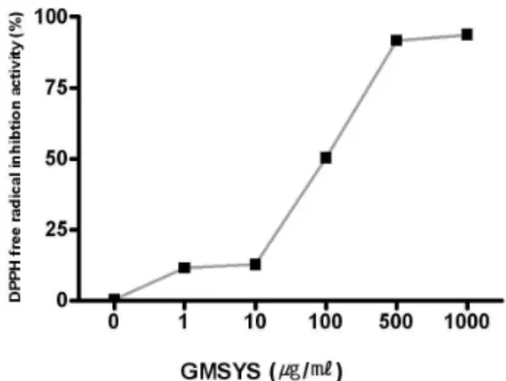
1) DPPH free radical inhibition activity 농도별 GMSYS의 DPPH free radical inhibition activity를 측정한 결과, 농도 의존적으로 증가하여 최대 활성은 1,000
㎍/㎖ GMSYS에서 93.67%로 나타났으 며, IC50은 133.60㎍/㎖이었다(Fig. 1).
2) ABTS radical cation inhibition activity
농도별 GMSYS의 ABTS radical cation
inhibition activity를 측정한 결과, 대체로
농도 의존적으로 증가하였으며 최대 활성
은 500㎍/㎖ GMSYS에서 94.67%로 나타
났으며, IC50은 106.20㎍/㎖이었다(Fig. 2).
Fig. 1. Effect of GMSYS on the generation of DPPH free radical.
Fig. 2. Effect of GMSYS on the generation of ABTS radical cation.
3) Total polyphenolic contents 10㎎/㎖ GMSYS의 total polyphenolic contents를 tannic acid 표준곡선으로부 터 구한 결과 0.78%로 나타났다.
2. SH-SY5Y細胞에 對한 保護效果 1) Cell viability
농도별 GMSYS의 SH-SY5Y cell에 대한 cell viability를 측정한 결과, 모든 농도의 GMSYS에서 통계적으로 유이한 차이가 없었다(Fig. 3).
Fig. 3. Dose-dependent effect of GMSYS treatment on the viability of SH-SY5Y cells.
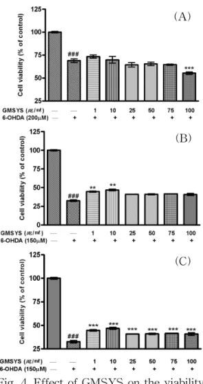
2) 6-OHDA에 對한 cell viability SH-SY5Y cell에서 농도별 GMSYS 처 리에 따른 6-OHDA에 대한 cell viability 를 측정한 결과, GMSYS pre-treatment 군의 경우 6-OHDA 200μM 단독에 의한 cell viability는 control에 비해 68.84%로 감소되었고, 10㎍/㎖ 이하의 GMSYS에서 는 6-OHDA 단독에 비하여 cell viability 가 증가되었으나 통계적으로 유의하지 않았다.
GMSYS co-treatment군의 경우 6-OHDA 150μM 단독에 의한 cell viability는 control 에 비해 37.57%로 감소되었고, 25㎍/㎖
이하의 GMSYS에서 6-OHDA 단독에 비 하여 cell viability가 통계적으로 유의하 게(p<0.01) 증가하였다.
GMSYS post-treatment군의 경우 6-
OHDA 150μM 단독에 의한 cell viability
는 control에 비해 32.59%로 감소되었고,
모든 농도의 GMSYS에서 6-OHDA 단
독에 비하여 cell viability가 통계적으로
유의하게 (p<0.001) 증가하였다(Fig. 4).
(A)
(B)
(C)
Fig. 4. Effect of GMSYS on the viability of 6-OHDA treated SH-SY5Y cells.
(A) Cell viability of GMSYS pre-treatment (B) Cell viability of GMSYS and 6-OHDA co-treatment
(C) Cell viability of GMSYS post-treatment
###; p<0.001 compared with the control
**; p<0.01, ***;p<0.001 compared with the 6-OHDA treated group
3. 胎兒中腦細胞에 對한 保護效果 1) TH-positive 細胞 保護效果
태아중뇌세포를 이용하여 GMSYS의 6-OHDA에 대한 세포보호 효과를 측정 한 결과, 6-OHDA 10μM 단독의 경우 control에 비하여 32.13%로 통계적으로 유의하게(p<0.001) 도파민 세포의 분화 및 생존을 억제시켰다.
6-OHDA와 농도별 GMSYS을 처리한
경우 6-OHDA 단독에 비하여 0.2㎍/㎖
GMSYS에서 통계적으로 유의하게(p<0.01) 도파민 세포를 증가시켰다(Fig. 5, Fig. 6).
Fig. 5. Neuroprotective effect of GMSYS in mesencephalic dopaminergic neurons against neurotoxicity induced by 6-OHDA.
###; p<0.001 compared with the control
**; p<0.01 compared with the 6-OHDA treated group
Control 6-OHDA
GMSYS 0.2㎍/㎖
+ 6-OHDA GMSYS 1㎍/㎖ + 6-OHDA Fig. 6. Neuroprotective effect of GMSYS in mesencephalic dopaminergic neurons against neurotoxicity induced by 6-OHDA (100X).
Scale bar=250㎛
2) NO inhibition 效果
태아중뇌세포를 이용하여 GMSYS의
6-OHDA 처리 후 배양액 내 NO 농도를
측정한 결과, 6-OHDA 10μM 단독의 경
우 control에 비하여 155.76%로 통계적 으로 유의하게(p<0.001) 증가되었다.
6-OHDA와 농도별 GMSYS을 처리한 경우 6-OHDA 단독에 비하여 0.2㎍/㎖
GMSYS에서 통계적으로 유의하게(p<0.01) NO 농도가 감소되었다(Fig. 7).
Fig. 7. Inhibitory effect of GMSYS on the NO production in mesencephalic dopaminergic neurons treated by 6-OHDA.
###; p<0.001 compared with the control
**; p<0.01 compared with the 6-OHDA treated group
3) TNF-α inhibition 效果
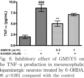
태아중뇌세포를 이용하여 GMSYS의 6-OHDA 처리 후 배양액 내 TNF-α 농 도를 측정한 결과, 6-OHDA 10μM 단독 의 경우 8.63±0.81pg/㎖로 control에 비하 여 통계적으로 유의하게(p<0.001) 증가 되었다.
6-OHDA와 농도별 GMSYS을 처리한 경우 6-OHDA 단독에 비하여 0.2㎍/㎖
GMSYS에서 통계적으로 유의하게(p<0.01) TNF-α 농도가 감소되었다(Fig. 8).
Fig. 8. Inhibitory effect of GMSYS on the TNF-α production in mesencephalic dopaminergic neurons treated by 6-OHDA.
###; p<0.001 compared with the control
**; p<0.01 compared with the 6-OHDA treated group