Protection of Primary Cultured Mouse Hepatocytes from Chemical Hypoxia- induced Injury by Hydrogen Sulfide
Min Young Lee*
Department of Molecular Physiology, College of Pharmacy, Kyungpook National University, Daegu 702-701, Korea
Received August 26, 2013 /Revised October 7, 2013 /Accepted October 14, 2013
We examined the effect of hydrogen sulfide (H
2S) in chemical hypoxia-induced injury in mouse hepatocytes. Cell viability was significantly decreased by cobalt chloride (CoCl
2), a well-known hypo- xia mimetic agent in a time- and dose- dependent manner. Sodium hydrosulfide (NaHS, a donor of H
2S) pretreatment before exposure to CoCl
2significantly attenuated the CoCl
2-induced decrease of cell viability. CoCl
2treatment resulted in an increase of intracellular ROS generation, which is inhibited by NaHS or N-acetyl-cysteine (NAC, a ROS scavenger), and p38 MAPK phosphorylation, which is al- so blocked by NaHS or NAC. The CoCl
2-induced increase of the Bax/Bcl-2 ratio was attenuated by NaHS, NAC, and SB 203580 (p38 MAPK inhibitor). The CoCl
2-induced decrease of cell viability was also attenuated by NaHS, NAC, and SB 203580 pretreatment. Additionally, NaHS inhibited the CoCl
2-induced COX-2. Similar to the effect of NaHS, NAC blocked CoCl
2-induced COX-2 expression.
Furthermore, NS-398 (a selective COX-2 inhibitor) attenuated not only the CoCl
2-induced increase of the Bax/Bcl-2 ratio, it also decreased cell viability. Taken together, H
2S protects primary cultured mouse hepatocytes against CoCl
2-induced cell injury through inhibition of the ROS-activated p38 MAPK cascade and the COX-2 pathway.
Key words : Hydrogen sulfide, cobalt chloride, chemical hypoxia, mouse hepatocytes
*Corresponding author
*Tel : +82-53-950-8577, Fax : +82-53-950-8557
*E-mail : [email protected]
This is an Open-Access article distributed under the terms of the Creative Commons Attribution Non-Commercial License (http://creativecommons.org/licenses/by-nc/3.0) which permits unrestricted non-commercial use, distribution, and reproduction in any medium, provided the original work is properly cited.
ISSN (Online) 2287-3406 Journal of Life Science 2013 Vol. 23. No. 11. 1342~1350 DOI : http://dx.doi.org/10.5352/JLS.2013.23.11.1342
Introduction
Hydrogen sulfide (H
2S), an endogenous gaseous media- tor, is produced by pyridoxal-5’-phosphate-dependent en- zymes, including cystathionine-γ-lyase, cystathionine-β- synthase and 3-mercaptopyruvate sulfurtransferase, during cysteine metabolism [6, 46]. H
2S was known as a toxic gas- eous material, but it is now a signaling gasotransmitter, which exerts physiological or pathological roles both in vivo and in vitro [36, 53]. H
2S promotes vascular smooth muscle relaxation and induces vasodilation of isolated blood vessels [9, 32]. It has been also evident that H
2S exerts their role as a potent antioxidant [27, 60]. Additionally, previous stud- ies show that H
2S shows a protective role against various stimuli-triggered injuries in many organs including heart, kidney and so forth [3, 49]. However, the mechanisms of H
2S attenuated hepatic ischemic injury remains to be elucidated.
Hepatic tissue is quite vulnerable to hypoxic injury com- pared with other organs [34]. In general, hypoxia generates reactive oxygen species (ROS) and excessive ROS generation is known to cause oxidative damage to DNA, lipids and pro- teins [5, 52]. This results in hypoxic cell injury, which is mediated by ROS-induced intracellular signaling molecules, including mitogen-activated protein kinases (MAPKs) [10].
MAPKs participate in varioius cellular functions such as cell proliferation, cell differentiation, cell motility, and cell death [37]. There are three major MAPK family subgroups: ex- tracellular signal-regulated kinase 1/2 (ERK 1/2), c-Jun N- terminal of stress-activated protein kinase (JNK), and p38 protein kinase. Especially, signal cascade involving p38 MAPK, activated by extracellular stress signals, are involved in cell differentiation and death and is known to be activated by a variety of environmental stresses or chemicals [40, 55].
Previously, it was reported that CoCl
2activates p38 MAPK,
which is involved in CoCl
2-induced apoptosis in other cell
types [65]. These findings suggest that ROS-induced p38
MAPK can be involved in CoCl
2-induced hepatic injury. It
has also been reported that ROS can stimulate cyclo-
oxygenase (COX)-2 expression in CoCl
2-induced chemical
hypoxic condition [56, 58]. At present, at least two COX iso-
enzymes, COX-1 and COX-2, have been identified [59].
or cytoprotection [4, 7, 21, 28, 50, 62]. Some studies indicated that inhibition of COX-2 activation attenuated ischemic hep- atic injury [21] while other reports showed that during ische- mic cardiotoxicity, overexpression of COX-2 play its pro- tective role [4, 28].
In the present study, we investigated the cytoprotective effect of H
2S in primary cultured mouse hepatocytes treated with cobalt chloride (CoCl
2), a widely used hypoxia mimetic agent that promotes the accumulation of hypoxia-inducible factor-1 α (HIF-1α), a critical regulator for the cellular re- sponse to hypoxia [18], and induces oxidative stress [23, 64]
and apoptosis in various cells [26, 64]. Therefore, this study assessed the effect of H
2S on hypoxic cellular injury induced by CoCl
2and its associated mechanisms in primary cultured mouse hepatocytes.
Materials and Methods Materials
Eight-week-old male ICR mice were purchased from Daehan Bio Link Co. (Incheon, Korea). All animal manage- ment procedures were conducted in accordance with the standard operation protocols established by Kyungpook National University. Cell Counting Kit-8 (CCK-8) was pur- chased from Dojindo Laboratories (Japan). CoCl
2, N-ace- tyl-l-cysteine, SB 203580, and type IV collagenase were ob- tained from Sigma-Aldrich (St. Louis, USA). The 5-(and 6)-chloromethyl-2‘,7’-dichlorodihydrofluorescein diacetate, acetyl ester (CM-H
2DCFDA) were purchased from Life Technologies (Grand Island, USA). Fetal bovine serum (FBS) was acquired from Thermo scientific (USA). The phospho- p38 MAPK antibodies were obtained from New England Biolabs (Hertfordshire, UK). The HIF-1α, Bcl-2, caspase-3, COX-1, COX-2, goat anti-rabbit IgG, and goat anti-mouse IgG antibodies were supplied by Santa Cruz Biotechnology (Santa Cruz, USA).
Isolation of mouse hepatocytes
Primary mouse hepatocytes were isolated from mouse liv- er using the two-step EDTA and collagenase perfusion method. After the mouse was anesthetized, the liver was
Krebs-Henseleit buffer without Ca and SO
4containing 0.02% collagenase and 0.1 mM CaCl
2until the liver appeared soft. The liver was then removed and gently minced, and the obtained cells were dispersed in medium (DMEM; Life Technologies, NY, USA) containing 10% FBS and 1% pen- icillin/streptomycin (Life Technologies, NY, USA). The sol- ution containing the mixed cells and debris was filtered through a 100-μm filter. Subsequently, the filtrate was centri- fuged at 50 g for 3 min at 4°C; the cells were washed with DMEM three times and then seeded in collagen-coated plates. The cells were maintained in DMEM high glucose (4.5 g/l) supplemented with 10% FBS, 1% penicillin/strep- tomycin, 1 μg/ml insulin, and 10
-12M dexamethasone for 24 h at 37°C in a humidified atmosphere (5% CO
2). The cells were incubated with fresh Williams’ E medium without FBS 24 h prior to the experiments.
Cell viability assay
Cell viability was detected using CCK-8 assay system.
Mouse hepatocytes were cultured in 96-well plates with three triplicate wells in each group. The cells were treated with conditioned medium as indicated. The CCK-8 solution was added to each well at a 1:10 dilution followed by further incubation at 37°C for 3 h. Absorbance was measured at 450 nm using a microplate reader (BioTek Instruments, Inc., VT, USA). All values are expressed as the mean (± standard er- ror, SE) of triplicate experiments. The values were converted from absolute counts to a percentage of the control.
Western blot analysis
The cell homogenates (30 μg of protein) were separated
using 10% or 12% SDS-polyacrylamide gel electrophoresis
and then transferred to polyvinylidene fluoride (PVDF)
transfer membranes. The blots were then washed with
Tris-buffered solution containing Tween-20 (TBST, 10 mM
Tris-HCl (pH 7.6), 150 mM NaCl, 0.1% Tween-20), blocked
with 5% skimmed milk powder in TBST for 1 h, and in-
cubated for 12 h with the appropriate primary antibody at
the dilutions as recommended by the supplier (1:1,000). The
membranes were then washed with TBST and incubated
with horseradish peroxidase-conjugated secondary antibody
A B
C
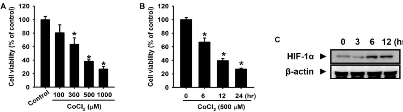
Fig. 1. Effect of CoCl2in cell viability in primary cultured mouse hepatocytes. (A) Mouse hepatocytes were incubated with CoCl2
at indicated concentrations for 12 h. (B) Cells were incubated with 500 μM CoCl2for various times (0-24 h). Cell viability was measured by CCK-8 assay. (C) Cells were incubated for various times (0-12 h), and the level of cellular HIF-1α expression was detected through western blotting. The values are expressed as the mean ± SE of three independent experiments with triplicate dishes. *
p
<0.05 vs. control.Fig. 2. Effect of H2S on CoCl2-elicited cytotoxicity. Mouse hep- atocytes were incubated with 500 μM CoCl2for 12 h in the absence or presence of pretreatment with NaHS at the indicated concentrations for 30 min. Cell viability was measured by CCK-8 assay. The values are expressed as the mean ± SE of three independent experiments with triplicate dishes. *
p
<0.05 vs. control, #p
<0.05 vs. CoCl2only.
(1:5,000) for 12 h. The bands were visualized using an en- hanced chemiluminescence detection system (Thermo Scientific, USA) according to the manufacturer’s protocols.
Detection of intracellular ROS
CM-H
2DCFDA (DCF-DA), which functions as a ROS-sen- sitive fluorophore, was used to detect the intracellular ROS.
The cells were plated on 35-mm cell culture dishes and in- cubated under the conditions as described previously. The cells were then kept in the dark and treated with 5 μM DCF-DA for 30 min at 37°C. After all the treatments were completed, the cells were washed three times with PBS and were imaged using fluorescence microscopy (100×; DM IL LED Fluo, Leica, Germany).
Statistical analysis
The results are expressed as the mean ± SE. The difference between the two mean values was analyzed by Student’s t test. A P value of <0.05 was considered significant.
Results
H
2S inhibits CoCl
2-induced cytotoxicity in primary cultured mouse hepatocytes
To investigate the effect of CoCl
2, the level of cell viability was examined with the CCK-8 assay system. As shown in Fig. 1A, CoCl
2significantly decreased the level of cell via- bility in a time-dependent manner. Fig. 1B shows that treat- ment of CoCl
2to mouse hepatocytes at concentrations rang- ing from 100 to 1,000 μM for 12 h led to a decrease in cell viability in a dose-dependent manner. Additionally, the ef- fect of CoCl
2treatment on the level of hypoxia-inducible fac-
tor-1α (HIF-1α) expression was analyzed in order to verify whether the hypoxic cell responses were induced by CoCl
2. The expression of HIF-1α was increased in a time-dependent manner (Fig. 1C). To determine the effect of hydeogen sul- fide (H
2S) on CoCl
2-induced cell injury, cells were pretreated with various concentrations of sodium hydrosulfide (NaHS, a donor of H
2S) for 30 min and then treated with 500 μM CoCl
2. NaHS significantly attenuated the decreased cell via- bility and these results indicate that NaHS pretreatment pro- tects against CoCl
2-induced hypoxic cell injury in primary cultured mouse hepatocytes (Fig. 2).
Effect of H
2S on CoCl
2-induced oxidative stress
and p38 MAPK in primary cultured mouse hepatocytes
A
B C
D E
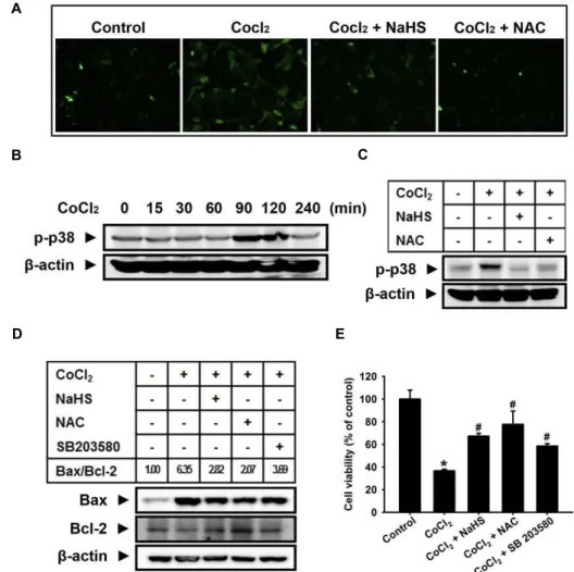
Fig. 3. Effect of H2S on the CoCl2-induced ROS generation, p38 MAPK activation, and cell injury. (A) Dichlorofluorescein (DCF)- sensitive cellular ROS was assessed. (a) Control. (b) Treatment with 500 μM CoCl2for 2 h (c) Pretreatment with 1,000 μM NaHS for 30 min before exposure to 500 μM CoCl2for 2 h (d) Pretreatment with 1,000 μM
N
-acetyl-cysteine (NAC) for 30 min before exposure to 500 μM CoCl2for 2 h. (B) Cells were incubated for various times (0-240 min) with 500 μM CoCl2. (C) Cells were pretreated with 1,000 μM NaHS or 1,000 μM NAC for 30 min and then treated with 500 μM CoCl2 for 2 h. Cell lysates were subjected to western blotting to determine the levels of phospho-p38 MAPK. (D) Cells were pretreated with 1,000 μM NaHS, 1,000 μM NAC or 1 μM SB 203580 (p38 MAPK inhibitor) for 30 min and then treated with 500 μM CoCl2for 12 h. Cell lysates were subjected to western blotting to determine the levels of Bax and Bcl-2 expression and the Bax/Bcl-2 ratio was calculated. (E) Cells were pretreated 1,000 μM NaHS, 1,000 μM NAC, or 1 μM SB 203580 for 30 min followed by incubation in the presence or absence of 500 μM CoCl2for 12 h. Cell viability was measured by CCK-8 assay. The values are expressed as the mean ± SE of three independent experiments with triplicate dishes. *p
<0.05 vs. control,#
p
<0.05 vs. CoCl2 only.N-acetyl cysteine (NAC), a common ROS scavenger (1,000 μ M) . In addition, the signaling molecule associated with CoCl
2-induced cell injury, p38 MAPK, was elevated. The maximum level of p38 MAPK activation was observed at 90-120 min after treatment with CoCl
2(Fig. 2B), and NaHS
increase of Bax/Bcl-2 ratio by CoCl
2treatment was sig- nificantly decreased by NaHS, NAC and SB 203580 (Fig. 3D).
Consistent with these results, decreased cell viability in-
duced by CoCl
2treatment was partially recovered by pre-
treatment with NaHS, NAC, and SB 203580 (Fig. 3E).
A B
C D
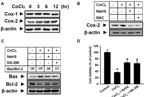
Fig. 4. Effect of H2S on CoCl2-induced COX-2 expression and cell injury. (A) Mouse hepatocytes were incubated with 500 μM CoCl2
for various times (0-24 h). (B) Cells were pretreated with 1,000 μM NaHS or 1,000 μM NAC for 30 min and then treated with 500 μM CoCl2for 12 h. Cell lysates were subjected to western blotting to determine the level of COX-2 expression.
(C) Cells were pretreated with 1,000 μM NaHS or 10 μM NS-398 (COX-2 inhibitor) for 30 min and then treated with 500 μM CoCl2for 12 h. Cell lysates were subjected to western blotting to determine the levels of Bax and Bcl-2 expression and the Bax/Bcl-2 ratio was calculated. (D) Cells were pretreated 1,000 μM NaHS or 10 μM NS-398 for 30 min followed by incubation in the presence or absence of 500 μM CoCl2for 12 h. Cell viability was measured by CCK-8 assay. The values are expressed as the mean ± SE of three independent experiments with triplicate dishes. *
p
<0.05 vs. control, #p
<0.05 vs.CoCl2 only.
Effect of H
2S on CoCl
2-induced COX-2 expression
After treatment of mosue hepatocytes with CoCl
2, COX-2 expression was significantly augmented, while COX-1 ex- pression was not significantly changed (Fig. 4A). Pretreat- ment with different concentrations of NaHS and NAC mark- edly attenuated the increased COX-2 expression by CoCl
2(Fig. 4B). We observed that pretreatment with NaHS or NS-398 (a selective COX-2 inhibitor, 10 μM) obviously in- hibited the CoCl
2-induced decrease of Bcl-2 expression and increase of Bax expression and increase of Bax/Bcl-2 ratio by CoCl
2treatment was significantly decreased by NaHS or NS-398 (Fig. 4C). In addition, decreased cell viability by CoCl
2was partially recovered by pretreatment with NaHS or NS-398 (Fig. 4D).
Discussion
Hydrogen sulfide (H
2S), an endogenous gaseous media- tor, exerts various physiological a pathophysiological effects in vivo, including anti-oxidative or anti-inflammatory effects in heart, liver, kidney and other organs [3, 13, 22, 49].
Although H
2S has long been considered as a toxic environ-
mental pollutant emerging from sewers, marshes, and vol- canic eruptions, H
2S has been recently recognized along-side nitric oxide and carbon monoxide as an endogenously pro- duced gaseous signaling molecule [22, 33, 47]. Previous stud- ies suggested that H
2S is a potent antioxidant [27, 60] and it has also been demonstrated that H
2S effectively inhibits apoptosis of a number of cell types [12, 39, 44] and this effect has been shown to promote cytoprotection. Cobalt chloride (CoCl
2) has been reported to take the place of ferrous ions in prolyl-4-hydroxylase (P4H), thereby causing a conforma- tional change in the P4H protein that consequently leads to a hypoxic condition, characterized by intranuclear accumu- lation of hypoxia inducible factor-1α (HIF-1α) [15, 42, 61].
CoCl
2has been also known to induce apoptosis in various types of cells [16, 29, 48]. In the current study, we demon- strated that chemical hypoxia-induced hepatocellular injury is markedly decreased by sodium hydrosulfide (NaHS, a do- nor of H
2S). The exposure of mouse hepatocytes to CoCl
2induced cytotoxicity, evidenced by the decreased cell via-
bility, and we observed that NaHS significantly attenuated
CoCl
2-induced decrease of cell viability. These results in-
dicate that hydrogen sulfide (H
2S) plays a role in suppress-
oxidative damage to DNA, lipids and proteins, resulting in apoptosis of cells including hepatocytes [25, 31]. In this study, exposure of mouse hepatocytes to CoCl
2increased in- tracellular ROS and CoCl
2-induced ROS generations were attenuated by pretreatment of NaHS or N-acetylcystein (NAC), a common ROS scavenger. These results show that H
2S possesses scavenging activity of ROS, which induced by CoCl
2. Indeed, Geng et al. reported that H
2S directly scav- enges superoxide anions and H
2O
2[14]. Recent studies have demonstrated that ROS are important triggers to upregulate p38 MAPK activity and that antioxidants can be con- sequently applied to inhibit p38 activation [8, 30, 65]. In hep- atocytes, the p38 signaling pathway is preferentially acti- vated by inflammatory cytokines and various stresses, such as UV light or hypoxia [2, 31, 38, 54]. ROS also can activate p38 pathway [19, 31, 63]. In accordance with previous re- ports, our results showed that NaHS or NAC attenuated CoCl
2-induced p38 MAPK activation. These results suggest that H
2S blocks p38 MAPK through its antioxidant effect.
Various studies have demonstrated that CoCl
2elicits oxida- tive stress, which constibutes to apoptosis in other cell types [30, 57, 65]. Additionally, oxidative stress-induced p38 MAPK activation was involved in hepatocyte injury [1, 20, 45]. This is supported by our results in which CoCl
2-induced cytotoxicity was prevented by NaHS, NAC, and SB 203580 (a p38 MAPK inhibitor) treatment. In addition, NaHS, NAC, and SB 203580 blocked the CoCl
2-induced increase of Bax/Bcl-2 ratio. Of the Bcl-2 family of proteins, including Bcl-2 and Bcl2-related family members such as Bcl-xL, Bad and Bax, play an important role in the regulation of apopto- sis [41]. In the present study, we showed that pretreatment of mouse hepatocytes with NaHS, NAC, and SB 203580 re- sulted in a significant decrease of increased expression of Bax, a proapoptotic Bcl-2 family member and potentiation of decreased expression of Bcl-2, an antiapoptotic Bcl-2 fam- ily member, by CoCl
2. Therefore, our results suggest that H
2S pretreatment plays a cytoprotective role via attenuation not only of CoCl
2-induced ROS, but also of p38 MAPK acti- vated by CoCl
2-induced ROS.
Our current study showed that exposure of mouse hep- atocytes to CoCl
2elevated expression of COX-2. At present,
ment with NaHS and NAC. This result indicates that COX-2 expression is enhanced by CoCl
2-induced intracellular ROS and H
2S represses COX-2 expression by its antioxidant activity. Previous studies showed that inhibition of COX-2 exerts hepatoprotective effects on liver damage [51]. COX-2 expression is upregulated by ischemia or reperfusion in the rat liver, and the inhibition of COX-2 is found to improve liver function and viability [26, 35]. Through this study, we observed that NaHS and NS-398, a selective inhibitor of COX-2, significantly attenuated CoCl
2-induced increase of Bax/Bcl-2 ratio. In addition, NaHS and NS-398 attenuated CoCl
2-induced cytotoxicity. Overall, these findings suggest that oxidative stress plays a pivotal role in hepatocellular injuries induced by CoCl
2and ROS mediate CoCl
2-induced injury through p38 MAPK and COX-2 pathway in mouse hepatocytes. In conclusion, the present study demonstrated that H
2S has a cytoprotective effect against chemical hypo- xia-induced cell injury through inhibition of the ROS, p38 MAPK, and COX-2 pathway in primary cultured mouse hepatocytes.
Acknowledgement
This research was supported by Kyungpook National University Research Fund, 2012.
References
1. Bhattacharyya, S., Pal, P. B. and Sil, P. C. 2013. A 35 kD Phyllanthus niruri protein modulates iron mediated oxida- tive impairment to hepatocytes via the inhibition of ERKs, p38 MAPKs and activation of PI3k/Akt pathway.
Food Chem Toxicol
56, 119-130.2. Bhogal, R. H., Weston, C. J., Curbishley, S. M., Adams, D.
H. and Afford, S. C. 2012. Activation of CD40 with platelet derived CD154 promotes reactive oxygen species dependent death of human hepatocytes during hypoxia and reoxygena- tion.
PLoS One
7, e30867.3. Bian, J. S., Yong, Q. C., Pan, T. T., Feng, Z. N., Ali, M. Y., Zhou, S. and Moore, P. K. 2006. Role of hydrogen sulfide in the cardioprotection caused by ischemic preconditioning in the rat heart and cardiac myocytes.
J Pharmacol Exp Ther
316, 670-678.4. Booth, E. A., Flint, R. R., Lucas, K. L., Knittel, A. K. and
Lucchesi, B. R. 2008. Estrogen protects the heart from ische- mia-reperfusion injury via COX-2-derived PGI2.
J Cardiovasc Pharmacol
52, 228-235.5. Bredesen, D. E., Rao, R. V. and Mehlen, P. 2006. Cell death in the nervous system.
Nature
443, 796-802.6. Calvert, J. W., Coetzee, W. A. and Lefer, D. J. 2010. Novel insights into hydrogen sulfide-mediated cytoprotection.
Antioxid Redox Signal
12, 1203-1217.7. Carnieto, A., Jr., Dourado, P. M., Luz, P. L. and Chagas, A. C. 2009. Selective cyclooxygenase-2 inhibition protects against myocardial damage in experimental acute ischemia.
Clinics (Sao Paulo)
64, 245-252.8. Chen, L., Liu, L. and Huang, S. 2008. Cadmium activates the mitogen-activated protein kinase (MAPK) pathway via induction of reactive oxygen species and inhibition of pro- tein phosphatases 2A and 5.
Free Radic Biol Med
45, 1035- 1044.9. Cheng, Y., Ndisang, J. F., Tang, G., Cao, K. and Wang, R.
2004. Hydrogen sulfide-induced relaxation of resistance mesenteric artery beds of rats.
Am J Physiol Heart Circ Physiol
287, H2316-2323.10. Cindrova-Davies, T., Spasic-Boskovic, O., Jauniaux, E., Charnock-Jones, D. S. and Burton, G. J. 2007. Nuclear fac- tor-kappa B, p38, and stress-activated protein kinase mi- togen-activated protein kinase signaling pathways regulate proinflammatory cytokines and apoptosis in human pla- cental explants in response to oxidative stress: effects of anti- oxidant vitamins.
Am J Pathol
170, 1511-1520.11. Dupouy, V. M., Ferre, P. J., Uro-Coste, E. and Lefebvre, H.
P. 2006. Time course of COX-1 and COX-2 expression dur- ing ischemia-reperfusion in rat skeletal muscle.
J Appl Physiol
100, 233-239.12. Elrod, J. W., Calvert, J. W., Morrison, J., Doeller, J. E., Kraus, D. W., Tao, L., Jiao, X., Scalia, R., Kiss, L., Szabo, C., Kimura, H., Chow, C. W. and Lefer, D. J. 2007. Hydrogen sulfide attenuates myocardial ischemia-reperfusion injury by pres- ervation of mitochondrial function.
Proc Natl Acad Sci USA
104, 15560-15565.13. Fiorucci, S., Antonelli, E., Mencarelli, A., Orlandi, S., Renga, B., Rizzo, G., Distrutti, E., Shah, V. and Morelli, A. 2005.
The third gas: H2S regulates perfusion pressure in both the isolated and perfused normal rat liver and in cirrhosis.
Hepatology
42, 539-548.14. Geng, B., Chang, L., Pan, C., Qi, Y., Zhao, J., Pang, Y., Du, J. and Tang, C. 2004. Endogenous hydrogen sulfide regu- lation of myocardial injury induced by isoproterenol.
Biochem Biophys Res Commun
318, 756-763.15. Goldberg, M. A., Dunning, S. P. and Bunn, H. F. 1988.
Regulation of the erythropoietin gene: evidence that the oxygen sensor is a heme protein.
Science
242, 1412-1415.16. Gotoh, M., Sano-Maeda, K., Murofushi, H. and Murakami- Murofushi, K. 2012. Protection of neuroblastoma Neuro2A cells from hypoxia-induced apoptosis by cyclic phosphatidic acid (cPA).
PLoS One
7, e51093.17. Guillemin, K. and Krasnow, M. A. 1997. The hypoxic re- sponse: huffing and HIFing.
Cell
89, 9-12.18. Guo, M., Song, L. P., Jiang, Y., Liu, W., Yu, Y. and Chen, G. Q. 2006. Hypoxia-mimetic agents desferrioxamine and cobalt chloride induce leukemic cell apoptosis through dif- ferent hypoxia-inducible factor-1alpha independent mecha- nisms.
Apoptosis
11, 67-77.19. Herrera, B., Fernandez, M., Roncero, C., Ventura, J. J., Porras, A., Valladares, A., Benito, M. and Fabregat, I. 2001.
Activation of p38 MAPK by TGF-beta in fetal rat hep- atocytes requires radical oxygen production, but is dis- pensable for cell death.
FEBS Lett
499, 225-229.20. Hyun, M. S., Hur, J. M., Mun, Y. J., Kim, D. and Woo, W.
H. 2010. BBR induces apoptosis in HepG2 cell through an Akt-ASK1-ROS-p38 MAPKs-linked cascade.
J Cell Biochem
109, 329-338.21. Ito, Y., Katagiri, H., Ishii, K., Kakita, A., Hayashi, I. and Majima, M. 2003. Effects of selective cyclooxygenase in- hibitors on ischemia/reperfusion-induced hepatic micro- circulatory dysfunction in mice.
Eur Surg Res
35, 408-416.22. Jha, S., Calvert, J. W., Duranski, M. R., Ramachandran, A.
and Lefer, D. J. 2008. Hydrogen sulfide attenuates hepatic ischemia-reperfusion injury: role of antioxidant and anti- apoptotic signaling.
Am J Physiol Heart Circ Physiol
295, H801-806.23. Jung, J. Y., Mo, H. C., Yang, K. H., Jeong, Y. J., Yoo, H.
G., Choi, N. K., Oh, W. M., Oh, H. K., Kim, S. H., Lee, J. H., Kim, H. J. and Kim, W. J. 2007. Inhibition by epi- gallocatechin gallate of CoCl2-induced apoptosis in rat PC12 cells.
Life Sci
80, 1355-1363.24. Jung, W. K., Heo, S. J., Jeon, Y. J., Lee, C. M., Park, Y. M., Byun, H. G., Choi, Y. H., Park, S. G. and Choi, I. W. 2009.
Inhibitory effects and molecular mechanism of dieckol iso- lated from marine brown alga on COX-2 and iNOS in micro- glial cells.
J Agric Food Chem
57, 4439-4446.25. Karimian, G., Buist-Homan, M., Mikus, B., Henning, R. H., Faber, K. N. and Moshage, H. 2012. Angiotensin II protects primary rat hepatocytes against bile salt-induced apoptosis.
PLoS One
7, e52647.26. Kim, H. J., Yang, S. J., Kim, Y. S. and Kim, T. U. 2003. Cobalt chloride-induced apoptosis and extracellular signal-regu- lated protein kinase activation in human cervical cancer HeLa cells.
J Biochem Mol Biol
36, 468-474.27. Kimura, Y. and Kimura, H. 2004. Hydrogen sulfide protects neurons from oxidative stress.
FASEB J
18, 1165-1167.28. Kwak, H. J., Park, K. M., Choi, H. E., Lim, H. J., Park, J.
H. and Park, H. Y. 2010. The cardioprotective effects of zi- leuton, a 5-lipoxygenase inhibitor, are mediated by COX-2 via activation of PKC delta.
Cell Signal
22, 80-87.29. Lan, A., Xu, W., Zhang, H., Hua, X., Zheng, D., Guo, R., Shen, N., Hu, F., Feng, J. and Liu, D. 2013. Inhibition of ROS-activated p38 MAPK pathway is involved in the pro- tective effect of H2S against chemical hypoxia-induced in- flammation in PC12 cells.
Neurochem Res
38, 1454-1466.30. Lan, A. P., Xiao, L. C., Yang, Z. L., Yang, C. T., Wang, X.
Y., Chen, P. X., Gu, M. F. and Feng, J. Q. 2012. Interaction between ROS and p38 MAPK contributes to chemical hypo- xia-induced injuries in PC12 cells.
Mol Med Rep
5, 250-255.sengers in cerebrovascular circulation.
J Appl Physiol
100, 1065-1076.33. Lichtman, S. N. and Lemasters, J. J. 1999. Role of cytokines and cytokine-producing cells in reperfusion injury to the liver.
Semin Liver Dis
19, 171-187.34. Nakanishi, K., Tajima, F., Nakamura, A., Yagura, S., Ookawara, T., Yamashita, H., Suzuki, K., Taniguchi, N. and Ohno, H. 1995. Effects of hy pobaric hypoxia on antioxidant enzymes in rats.
J Physiol
489, 869-876.35. Oshima, K., Yabata, Y., Yoshinari, D. and Takeyoshi, I. 2009.
The effects of cyclooxygenase (COX)-2 inhibition on ische- mia-reperfusion injury in liver transplantation.
J Invest Surg
22, 239-245.36. Pae, H. O., Lee, Y. C., Jo, E. K. and Chung, H. T. 2009.
Subtle interplay of endogenous bioactive gases (NO, CO and H(2)S) in inflammation.
Arch Pharm Res
32, 1155-1162.37. Pearson, G., Robinson, F., Beers Gibson, T., Xu, B. E., Karandikar, M., Berman, K. and Cobb, M. H. 2001. Mitogen- activated protein (MAP) kinase pathways: regulation and physiological functions.
Endocr Rev
22, 153-183.38. Quiroga, A. D., de Lujan Alvarez, M., Parody, J. P., Ronco, M. T., Carnovale, C. E. and Carrillo, M. C. 2009. Interferon- alpha2b (IFN-alpha2b)-induced apoptosis is mediated by p38 MAPK in hepatocytes from rat preneoplastic liver via activation of NADPH oxidase.
Growth Factors
27, 214-227.39. Rose, P., Moore, P. K., Ming, S. H., Nam, O. C., Armstrong, J. S. and Whiteman, M. 2005. Hydrogen sulfide protects co- lon cancer cells from chemopreventative agent beta-phenyl- ethyl isothiocyanate induced apoptosis.
World J Gastroenterol
11, 3990-3997.40. Roulston, A., Reinhard, C., Amiri, P. and Williams, L. T.
1998. Early activation of c-Jun N-terminal kinase and p38 kinase regulate cell survival in response to tumor necrosis factor alpha.
J Biol Chem
273, 10232-10239.41. Sarada, S. K., Himadri, P., Ruma, D., Sharma, S. K., Pauline, T. and Mrinalini. 2008. Selenium protects the hypoxia in- duced apoptosis in neuroblastoma cells through upregula- tion of Bcl-2.
Brain Res
1209, 29-39.42. Sharp, F. R. and Bernaudin, M. 2004. HIF1 and oxygen sens- ing in the brain.
Nat Rev Neurosci
5, 437-448.43. Smith, W. L., DeWitt, D. L. and Garavito, R. M. 2000.
Cyclooxygenases: structural, cellular, and molecular biology.
Annu Rev Biochem
69, 145-182.44. Sodha, N. R., Clements, R. T., Feng, J., Liu, Y., Bianchi, C., Horvath, E. M., Szabo, C. and Sellke, F. W. 2008. The effects of therapeutic sulfide on myocardial apoptosis in response to ischemia-reperfusion injury.
Eur J Cardiothorac Surg
33, 906-913.45. Song, M. K., Kim, Y. J., Song, M., Choi, H. S., Park, Y. K.
J Biol Chem
267, 11455-11461.47. Szabo, C. 2007. Hydrogen sulphide and its therapeutic potential.
Nat Rev Drug Discov
6, 917-935.48. Talwar, S., Jin, J., Carroll, B., Liu, A., Gillespie, M. B. and Palanisamy, V. 2011. Caspase-mediated cleavage of RNA- binding protein HuR regulates c-Myc protein expression af- ter hypoxic stress.
J Biol Chem
286, 32333-32343.49. Tripatara, P., Patel, N. S., Brancaleone, V., Renshaw, D., Rocha, J., Sepodes, B., Mota-Filipe, H., Perretti, M. and Thiemermann, C. 2009. Characterisation of cystathionine gamma-lyase/hydrogen sulphide pathway in ischaemia/re- perfusion injury of the mouse kidney: an
in vivo
study.Eur J Pharmacol
606, 205-209.50. Tu, X. K., Yang, W. Z., Shi, S. S., Wang, C. H. and Chen, C. M. 2009. Neuroprotective effect of baicalin in a rat model of permanent focal cerebral ischemia.
Neurochem Res
34, 1626-1634.51. Vadiraja, B. B., Gaikwad, N. W. and Madyastha, K. M. 1998.
Hepatoprotective effect of C-phycocyanin: protection for carbon tetrachloride and R-(+)-pulegone-mediated hep- atotoxicty in rats.
Biochem Biophys Res Commun
249, 428-431.52. Valko, M., Leibfritz, D., Moncol, J., Cronin, M. T., Mazur, M. and Telser, J. 2007. Free radicals and antioxidants in nor- mal physiological functions and human disease.
Int J Biochem Cell Biol
39, 44-84.53. Wallace, J. L. 2010. Physiological and pathophysiological roles of hydrogen sulfide in the gastrointestinal tract.
Antioxid Redox Signal
12, 1125-1133.54. Werwein, E., Dzuganova, M., Usadel, C. and Klempnauer, K. H. 2013. B-Myb switches from Cyclin/Cdk-dependent to Jnk- and p38 kinase-dependent phosphorylation and asso- ciates with SC35 bodies after UV stress.
Cell Death Dis
4, e511.55. Xia, Z., Dickens, M., Raingeaud, J., Davis, R. J. and Greenberg, M. E. 1995. Opposing effects of ERK and JNK-p38 MAP kinases on apoptosis.
Science
270, 1326-1331.56. Yang, C., Ling, H., Zhang, M., Yang, Z., Wang, X., Zeng, F., Wang, C. and Feng, J. 2011. Oxidative stress mediates chemical hypoxia-induced injury and inflammation by acti- vating NF-kappab-COX-2 pathway in HaCaT cells.
Mol Cells
31, 531-538.57. Yang, C., Yang, Z., Zhang, M., Dong, Q., Wang, X., Lan, A., Zeng, F., Chen, P., Wang, C. and Feng, J. 2011. Hydrogen sulfide protects against chemical hypoxia-induced cytotox- icity and inflammation in HaCaT cells through inhibition of ROS/NF-kappaB/COX-2 pathway.
PLoS One
6, e21971.58. Yeo, J. E., Kim, J. H. and Kang, S. K. 2008. Selenium attenu- ates ROS-mediated apoptotic cell death of injured spinal cord through prevention of mitochondria dysfunction;
in vi-
초록:화학적 허혈에 의해 손상된 마우스 간세포에 대한 hydrogen sulfide의 간세포 보호 효과 이민영*
(경북대학교 약학대학 약학과 분자생리학 연구실)
본 연구는 화학적 허혈에 의해 손상된 마우스 간세포에서 hydrogen sulfide (H
2S)의 효과를 규명하기 위해 수행 되었다. 본 연구에서 허혈 모방 화합물로 알려져 있는 cobalt chloride (CoCl
2)는 간세포 손상을 시간 및 농도 의존 적으로 유의성 있게 증가 시켰다. CoCl
2에 의한 간세포 손상은 Sodium sulfide (NaHS, H
2S 공여제)의 전처리에 의해 유의적으로 감소 되었다. CoCl
2는 세포 내 활성산소(reactive oxygen species, ROS)의 농도를 증가시켰으며, 이는 NaHS 및 N-acetyl-cysteine (NAC, a ROS 제거제)에 의해 감소하였다. 또한, CoCl
2에 의해 증가된 p38 MAPK 인산화가 NaHS 및 NAC에 의해 억제되었다. CoCl
2에 의해 증가된 Bax/Bcl-2 비율은 NaHS, NAC 및 SB 203580 (p38 MAPK 저해제)에 의해 차단되었으며, CoCl
2에 의해 유발된 간세포의 손상 또한 NaHS, NAC 및 SB 203580의 전처리에 의해 억제되었다. NaHS는 CoCl
2에 의해 증가된 COX-2의 발현을 억제하였다. 또한, NaHS의 효과와 유사하게 CoCl
2에 의해 증가된 COX-2의 발현이 NAC에 의해 억제되었다. 더욱이, NS-398 (COX-2 선택적 억제제)는 CoCl
2에 의한 Bax/Bcl-2 비율의 증가를 억제하였을 뿐 아니라, 간세포의 세포 손상 또한 억제하였다.
결론적으로, H
2S는 초대배양 된 마우스 간세포에서 CoCl
2에 의해 유발된 간세포의 손상을 ROS에 의해 유발된
p38 MAPK 및 COX-2 경로의 활성화를 억제함으로써 세포보호효과를 수행하는 것을 알 수 있었다.
tro
andin vivo
study.Cell Physiol Biochem
21, 225-238.59. Yokoyama, C. and Tanabe, T. 1989. Cloning of human gene encoding prostaglandin endoperoxide synthase and pri- mary structure of the enzyme.
Biochem Biophys Res Commun
165, 888-894.60. Yonezawa, D., Sekiguchi, F., Miyamoto, M., Taniguchi, E., Honjo, M., Masuko, T., Nishikawa, H. and Kawabata, A.
2007. A protective role of hydrogen sulfide against oxidative stress in rat gastric mucosal epithelium.
Toxicology
241, 11-18.61. Yuan, Y., Hilliard, G., Ferguson, T. and Millhorn, D. E. 2003.
Cobalt inhibits the interaction between hypoxia-inducible factor-alpha and von Hippel-Lindau protein by direct bind- ing to hypoxia-inducible factor-alpha.
J Biol Chem
278, 15911-15916.62. Yun, Y., Duan, W. G., Chen, P., Wu, H. X., Shen, Z. Q.,
Qian, Z. Y. and Wang, D. H. 2009. Down-regulation of cy- clooxygenase-2 is involved in ischemic postconditioning protection against renal ischemia reperfusion injury in rats.
Transplant Proc
41, 3585-3589.63. Zhang, Y., Qi, X., Gong, L., Li, Y., Liu, L., Xue, X., Xiao, Y., Wu, X. and Ren, J. 2008. Roles of reactive oxygen species and MAP kinases in the primary rat hepatocytes death in- duced by toosendanin.
Toxicology
249, 62-68.64. Zou, W., Yan, M., Xu, W., Huo, H., Sun, L., Zheng, Z. and Liu, X. 2001. Cobalt chloride induces PC12 cells apoptosis through reactive oxygen species and accompanied by AP-1 activation.
J Neurosci Res
64, 646-653.65. Zou, W., Zeng, J., Zhuo, M., Xu, W., Sun, L., Wang, J. and Liu, X. 2002. Involvement of caspase-3 and p38 mitogen- activated protein kinase in cobalt chloride-induced apopto- sis in PC12 cells.