15
ABBREVIATIONS: KCN, potassium cyanide; IAA, iodoacetic acid;
cPLA
2, cytosolic phospholipase A
2; ROS, reactive oxygen species;
DCFH-DA, 2',7'-dichlorofluorescein diacetate; DMTU, dimethylthiourea.
Corresponding to: Yong Keun Kim, Department of Physiology, College of Medicine, Pusan National University, Ami-dong, Suh-gu, Busan 602-739, Korea. (Tel) 82-51-240-7733, (FAX) 82-51-246-6001, (E-mail) kim430@pusan.ac.kr
Alterations in Membrane Transport Function and Cell Viability Induced by ATP Depletion in Primary Cultured Rabbit Renal Proximal Tubular Cells
Sung Ju Lee, Chae Hwa Kwon, and Yong Keun Kim
Department of Physiology, MRC for Ischemic Tissue Regeneration, College of Medicine, Pusan National University, Busan 602-739, Korea
This study was undertaken to elucidate the underlying mechanisms of ATP depletion-induced membrane transport dysfunction and cell death in renal proximal tubular cells. ATP depletion was induced by incubating cells with 2.5 mM potassium cyanide (KCN)/0.1 mM iodoacetic acid (IAA), and membrane transport function and cell viability were evaluated by measuring Na
+-dependent phosphate uptake and trypan blue exclusion, respectively. ATP depletion resulted in a decrease in Na
+-dependent phosphate uptake and cell viability in a time-dependent manner. ATP depletion inhibited Na
+-dependent phosphate uptake in cells, when treated with 2 mM ouabain, a Na
+pump-specific inhibitor, suggesting that ATP depletion impairs membrane transport functional integrity. Alterations in Na
+-dependent phosphate uptake and cell viability induced by ATP depletion were prevented by the hydrogen peroxide scavenger such as catalase and the hydroxyl radical scavengers (dimethylthiourea and thiourea), and amino acids (glycine and alanine). ATP depletion caused arachidonic acid release and increased mRNA levels of cytosolic phospholipase A
2(cPLA
2). The ATP depletion-dependent arachidonic acid release was inhibited by cPLA
2specific inhibitor AACOCF
3. ATP depletion-induced alterations in Na
+-dependent phosphate uptake and cell viability were prevented by AACOCF
3. Inhibition of Na
+-dependent phosphate uptake by ATP depletion was prevented by antipain and leupetin, serine/cysteine protease inhibitors, whereas ATP depletion-induced cell death was not altered by these agents. These results indicate that ATP depletion-induced alterations in membrane transport function and cell viability are due to reactive oxygen species generation and cPLA
2activation in renal proximal tubular cells. In addition, the present data suggest that serine/cysteine proteases play an important role in membrane transport dysfunction, but not cell death, induced by ATP depletion.
Key Words: ATP depletion, Membrane transport, cPLA
2, Serine/cysteine proteases
INTRODUCTION
Renal ischemia is a common cause of acute renal failure.
Despite numerous experimental and clinical studies on re- nal ischemia, the prognosis for patients with ischemic acute renal failure remains poor (Devarajan, 2006). Ischemia/re- perfusion injury is characterized by ATP depletion, followed by subsequent activation of a number of critical alterations in metabolism (Dagher, 2000; Devarajan et al, 2003; Nangaku
& Eckardt, 2007).
Although ischemia in vitro and in vivo has been known to induce generate reactive oxygen species (ROS) (Weinberg, 1991; Goto et al, 1999; Kim et al, 2002), the role of ROS in the pathogenesis of ischemia-induced cell injury is not fully understood. Several workers have postulated that ROS play a critical role in renal cell injury induced by hypo- xia in vitro (Paller, 1994; Kim et al, 2002) and ischemia in vivo (Paller, 1994; Devarajan, 2006), whereas, others have reported that ROS do not play any significant role
in the pathogenesis of cell injury induced by hypoxia in vitro (Borkan & Schwartz, 1989; Watabe & Nakaki, 2007) and ischemia in vivo (Joannidis et al, 1990; Kim et al, 1999;
Nistico et al, 2008). We previously observed that the role of ROS in antimycin A-induced cell death is different be- tween cultured renal proximal tubular cells and renal cort- ical slices (Kim et al, 2002). The cell death in cortical slices was prevented by antioxidants, whereas the cultured cell death was not altered.
Accumulation of unesterified fatty acids, especially arachidonic acid, occurs during ischemic or hypoxic injury in the kidney (Matthys et al, 1984; Humes et al, 1989;
Bunnachak et al, 1994). This accumulation of free fatty
acids is attributed to the degradation of membrane phos-
pholipids by activation of phospholipase A
2(PLA
2)
(Bonventre, 1993; Portilla & Creer, 1995). Although activa-
tion of PLA
2has been suggested to be an important media-
tor of ischemic or hypoxic tubular cell injury (Bonventre,
1993; Portilla et al, 1994; Choi et al, 1995; Choi et al, 1999),
the role of PLA
2activation in hypoxic cell injury remains controversial. PLA
2exerts cytoprotective effect (Zager et al, 1996) or no effect (Schnellmann et al, 1994) against hy- poxic- and antimycin A-induced proximal tubular cell injury. Most studies have been focused on the role of PLA
2activation in cell death during renal ischemia, but its role in hypoxia-mediated membrane transport dysfunction re- mains unclear.
Cysteine proteases have been implicated in ischemic and toxicant-induced cell death in various cell types including the kidney (Nicotera et al, 1989; Weinberg, 1991; Yang &
Schnellmann, 1996; Seyfried et al, 2001). However, very lit- tle has been known on the role of cysteine proteases in hy- poxia-induced membrane transport dysfunction.
The present study was, therefore, undertaken to eluci- date the role of ROS, PLA
2activation, and cysteine pro- teases in membrane transport dysfunction and cell death induced by ATP depletion, an in vitro model of ischemia, in primary cultured renal proximal tubular cells. Previous studies have shown that renal tubular cells respond with a sensitivity similar to both KCN/IAA and hypoxia (Lash et al, 1996). The data obtained herein indicated that the membrane transport dysfunction and cell death induced by ATP depletion are dependent on ROS generation and PLA
2activation. In addition, the membrane transport dysfunc- tion, but not the cell death, was prevented by cysteine pro- tease inhibitors.
METHODS
Culture of rabbit proximal tubular cells
Proximal tubular cells were isolated as previously de- scribed (Kim et al, 2002). In brief, adult New Zealand male white rabbits were killed by cervical dislocation. Their kid- neys were removed immediately, cleaned of fat and debris, and washed with sterile antibiotic-supplemented medium.
The kidneys were perfused with phosphate buffered saline (pH 7.4) through the renal artery and subsequently with DMEM/F12 containing 0.5% (wt/vol.) iron oxide until the kidneys turned a grey-black in color. The cortex was re- moved and homogenized with 4 strokes of a sterile glass homogenizer. The homogenate was passed through a series of sterile nylon mesh sieves (254 and 85 μm: TETCO, Inc., Depew, NY). Tubules and glomeruli retained on the 85-μm sieve were suspended in a tube containing DMEM/F12 me- dium and magnetic stirring bar. Glomeruli containing iron oxide were attracted to the magnetic stirring bar. The stir- ring bar was removed from the solution. The isolated prox- imal tubules were incubated briefly in DMEM/F12 medium containing 80 μg/ml of collagenase A and 0.025% soybean trypsin inhibitor. Then the dissociated tubules were washed by centrifugation, resuspended in DMEM/F12 medium, and transferred into tissue culture plates. Proximal tubule cells were grown on 24-well tissue culture plates in DMEM/F12 medium supplemented with bovine insulin (5 μg/ml), hu- man transferrin (5 μg/ml) and hydrocortisone (5×10
−8M).
The cultures were maintained in a humidified 95% air/5%
CO
2incubator at 37
oC. Culture medium was changed every 24 hr and 48 hr before each experiment. All experiments started 10∼13 days after plating when a confluent mono- layer culture was achieved.
ATP depletion
For ATP depletion, cells were treated with 2.5 mM potas- sium cyanide (KCN)/0.1 mM iodoacetic acid (IAA) in a glu- cose-free medium. The composition of the incubation me- dium was 115 mM NaCl, 5 mM KCl, 25 mM NaHCO
3, 2 mM NaH
2PO
4, 1 mM MgSO
4, and 1 mM CaCl
2(pH 7.4).
Following the incubation, phosphate uptake and cell via- bility were measured as described below. Unless otherwise stated, cells were treated with KCN/IAA for 120 min at 37
oC.
Measurement of Na
+-dependent phosphate uptake The uptake of solutes was determined in cell monolayers grown on 24 well plates. After an exposure to ATP deple- tion, the reaction buffer was removed and washed twice with the uptake buffer containing the following (in mM):
137 NaCl, 5.4 KCl, 2.8 CaCl
2, 1.2 MgSO
4, and 10 Hepes (pH 7.4). The cells were incubated for 60 min at 37
oC in the uptake buffer containing 5 μM [
32P]-phosphate. At the end of the incubation, the cells were washed three times with ice-cold uptake buffer and solubilized in 0.5 ml of 0.2%
Triton X-100. Aliquots of each sample were transferred to scintillation vials and the radioactivity was counted in a liquid scintillation counter (TRI-CARB 2100TR, Packard, USA). Protein was measured by the method of Bradford (Bradford, 1976).
Measurement of cell viability
The cells viability was determined by a trypan blue ex- clusion assay. Cells were harvested using 0.025% trypsin, incubated with 4% trypan blue solution, and were counted using a hemocytometer under light microscope. Cells failing to exclude the dye were considered nonviable, and the num- ber of nonviable cell was expressed as a percentage of the total cells.
Measurement of ROS production
The intracellular generation of ROS was measured using 2’,7’-dichlorofluorescein diacetate (DCFH-DA). The non- fluorescent ester penetrates into the cells and is hydrolyzed to DCFH by the cellular esterases. The probe (DCFH) is rapidly oxidized to the highly fluorescent compound 2’,7’-di- chlorofluorescein (DCF) in the presence of cellular perox- idase and ROS such as hydrogen peroxide or fatty acid peroxides. Cells were cultured in 35-mm tissue culture petri dishes. The culture medium was removed, and the cells were collected from flasks using trypsin-EDTA solution.
The cells were washed twice with DMEM/F12, and were suspended in glucose-free HBSS for fluorescence analysis.
The reaction was carried out in a fluorescent cuvette. The
cells were preincubated for 10 min at 37
oC in fluorescent
cuvette containing 3 ml of glucose-free HBSS with 20 μM
DCFH-DA (from a stock solution of 20 mM DCFH-DA in
ethanol). After the preincubation, the cells were treated
with KCN/IAA and incubated up to 120 min during which
the fluorescent intensity was monitored on a spectro-
fluorometer (SPEX1681, SPEX Co., USA) with excitation
wave length of 485 nm and emission wave length of 530
nm. The net increase in DCF fluorescence (arbitrary units)
was calculated by taking the difference between the values
Fig. 1. Effect of ATP depletion on Na
+-dependent phosphate uptake (A) and cell viability (B) in primary cultured renal proximal tubular cells. Cells were exposed to medium with 2.5 mM potassium cyanide/0.1 mM iodoacetic acid (KCN/IAA) or without (Control) for 0∼3 hr at 37
oC. Na
+-dependent phosphate uptake and cell viability were measured as described in ‘Materials and Methods’. Data are mean±SEM of four independent experiments performed in duplicate. *p<0.05 compared with control. (C) Effect of ATP depletion on Na
+-dependent phosphate uptake in primary cultured renal proximal tubular cells which were treated with ouabain. Cells were exposed to medium with 2.5 mM potassium cyanide/0.1 mM iodoacetic acid (KCN/IAA) or without (Control) in the presence or absence of 2 mM ouabain for 120 min at 37
oC. Na
+-dependent phosphate uptake was measured as described in ‘Materials and Methods’. Data are mean±SEM of four independent experiments performed in duplicate. *p<0.05 compared with control;
#p<0.05 compared with ouabain alone.
before and after addition of KCN/IAA.
Measurement of arachidonic acid (AA) release.
Confluent cells were incubated with
3H-AA (2 μCi/ml) in DMEM/F12 for 20 hr. After incubation, cells were gently
washed three times with HBSS (pH 7.4) to remove unbound
3
H-AA. To measure ATP depletion-induced AA release, cells were exposed to KCN/IAA for appropriate times. The me- dium was removed and centrifuged, and the radioactivity in 400 μl of supernatant was measured in a liquid scintilla- tion counter (Packard Tricarb A2100). Cells were solubi- lized with 0.1% deoxycholate and the radioactivity in 400 μl of cell lysate was measured. The amount of
3H-AA re- leased into the medium was expressed as a percent of total (cell-associated plus released).
Reverse transcriptase-polymerase chain reaction (RT-PCR) analysis
Total RNA was extracted from cells with Trizol reagent (Invitrogen, Carlsbad, CA, USA) according to the manu- facturer’s instructions. Equal amounts of total RNA were reverse transcribed by using a oligo (dT) primer and reverse transcriptase (Promega, Madison, WI, USA), and the syn- thesized cDNA was then used as a template for the PCR reaction. PCR primers were used to amplify cPLA
2(forward 5’-AACATTCAGGCAGCAGAGGA-3’ and reverse 5’-CGTA- GGTCGCACAATCCAA-3’) and GAPDH (forward 5’-GGGT- GTGAACCACGAGAAAT-3’ and reverse 5’-TTCAGCTCTG- GGATGACCTT-3’). PCR was carried out under the follow- ing conditions: the initial denaturation 95
oC for 3 min, fol- lowed by 35 cycles of denaturation at 95
oC for 30 s, anneal- ing for 30 s, extension at 72℃ for 30 s and additional ex- tension at 72
oC for 5 min. The PCR products were analyzed by 1.2% agarose gel electrophoresis.
Chemicals
Catalase, dimethylthiourea (DMTU), thiourea, antipain, leupetin, and ouabain were purchased from Sigma-Aldrich Chemical (St. Louis, MO). AACOCF
3was purchased from Calbiochem (San Diego, CA, USA). DCFH-DA was obtained from Molecular Probes (Eugene, OR, USA). All other chem- icals were of the highest commercial grade available.
Statistical analysis
Data are expressed as mean±SEM. Comparison between two groups was made using the unpaired t test. Multiple group comparisons were done using one-way analysis of variance followed by the Turkey post hoc test. p<0.05 was considered statistically significant.
RESULTS
Reduction of phosphate uptake and cell viability by ATP depletion
To determine the time course of ATP depletion-induced alterations in Na
+-dependent phosphate uptake and cell vi- ability, cells were exposed to 2.5 mM KCC/0.1 mM IAA for various times (0∼3 hr). Fig. 1A and B shows that the Na
+-dependent phosphate uptake and cell viability were de- creased in a time-dependent manner.
Since phosphate is transported across the brush border
membrane via a Na
+-dependent active mechanism in renal
proximal tubular cells, the driving force of active phosphate
uptake is the Na
+gradient across the membrane generated
by Na
+-pump. Thus, reduction of Na
+-dependent phos-
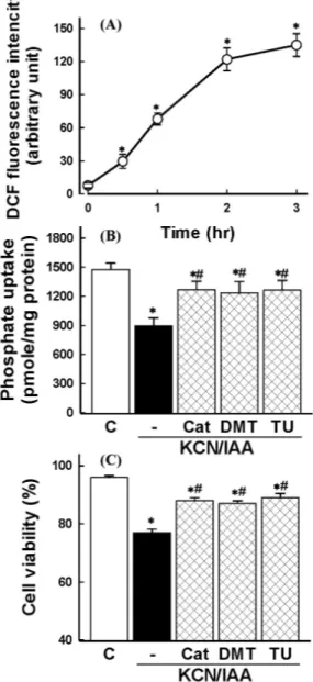
Fig. 2. (A) Time course of reactive oxygen species (ROS) generation in primary cultured renal proximal tubular cells during exposure to ATP depletion. Cells were exposed to 2.5 mM potassium cyanide/0.1 mM iodoacetic acid (KCN/IAA) for various times and ROS generation was measured. Shown is the net increase of DCF fluorescence (arbitrary units) calculated by subtracting the values for the control cells from the corresponding values for KCN/IAA-treated cells. Data are mean±SEM of three independent experiments performed in duplicate. *p<0.05 compared with zero (0) time. (B−C) Effect of radical scavengers on inhibition of phosphate uptake (B) and cell death (C) induced by ATP depletion. Cells were exposed to KCN/IAA in the presence or absence of 500 units/ml catalase (Cat), 30 mM dimethylthiourea (DMTU), or 30 mM thiourea (TU) for 3 hr at 37
oC.
Na
+-dependent phosphate uptake and cell viability were measured as described in ‘Materials and Methods’. Data are mean±SEM of four independent experiments performed in duplicate. *p<0.05 compared with control;
#p<0.05 compared with KCN/IAA alone.
phate uptake in cells exposed to KCN/IAA could be attributed to a decrease in the driving force by inhibition of Na
+-pump rather than impairment of membrane transport functional integrity. To test the possibility, the effect of ATP depletion
on Na
+-dependent phosphate uptake was examined in the absence or presence of 2 mM ouabain. As shown in Fig.
1C, treatment of primary cultured renal proximal tubular cells with ouabain partially inhibited Na
+-dependent phos- phate uptake. Treatment of the cells with KCN/IAA in the presence of ouabain significantly attenuated Na
+-depend- ent phosphate uptake further, suggesting that ATP deple- tion induces membrane transport dysfunction through Na
+-pump-independent mechanism.
Role of ROS in ATP depletion-induced alterations in membrane transport function and cell viability ROS production was examined in cells exposed to ATP depletion using DCFH-DA. ATP depletion induced an in- crease of ROS generation in a time-dependent manner (Fig. 2A).
Involvement of ROS in ATP depletion-induced alterations in Na
+-dependent phosphate uptake and cell viability was evaluated using catalase, the hydrogen peroxide scavenger, and the hydroxyl radical scavengers, such as DMTU and thiourea. As shown in Fig. 2B and C, these agents could reverse the ATP depletion-induced alterations, possibly supporting the notion that hydrogen peroxide and hydroxyl radicals are responsible for reduction in Na
+-dependent phosphate uptake and cell viability induced by ATP depletion.
Effect of amino acids on ATP depletion-induced cell injury
It has been demonstrated that several amino acids such as glycine and alanines protect cells from hypoxic injury induced by oxygen deprivation (Weinberg, 1991; Wetzels et al, 1993; Choi et al, 1999). To determine whether amino acids prevent the ATP depletion-induced cell injury, the ef- fect of glycine and alanine on the cell injury was investigated. As shown in Fig. 3, the reduction of Na
+-depen- dent phosphate uptake and cell death which were induced by ATP depletion was attenuated by these amino acids.
Role of PLA
2activation in ATP depletion-induced alterations in membrane transport function and cell viability
To determine if ATP depletion induces PLA
2activation, [
3H]AA release was measured in prelabeled cells. ATP de- pletion caused an increase of AA release in a time-depend- ent manner (Fig. 4A). The AA release by ATP depletion was inhibited by the PLA
2inhibitor AACOCF
3(Fig. 4B). Similar results were also obtained from RT-PCR of cPLA
2mRNA.
As shown in Fig. 4C, cPLA
2mRNA was increased 30 min after ATP depletion. These data suggest that ATP depletion induces cPLA
2activation in renal proximal tubular cells.
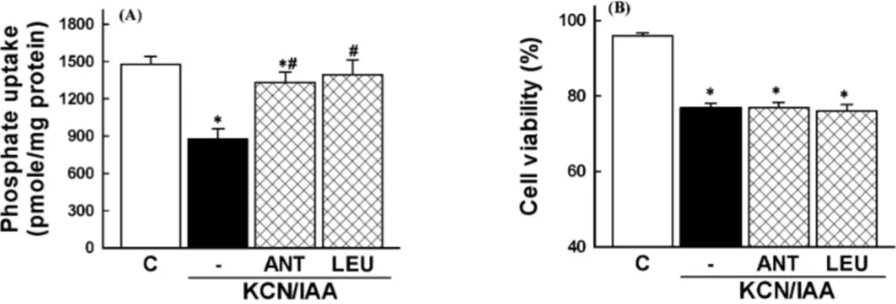
Involvement of PLA
2activation in ATP depletion-induced
membrane transport dysfunction and cell death was eval-
uated using the PLA
2inhibitor. Thus, cells were exposed
to ATP depletion in the presence of AACOCF
3, a tri-
fluoromethyl ketone analogue of arachidonyl acid which in-
hibits the 85-kD cPLA
2(Street et al, 1993). As shown in
Fig. 5, the reduction of Na
+-dependent phosphate uptake
and the cell death was attenuated by the inhibitor. These
data indicate that PLA
2is involved in ATP depletion-in-
duced alterations in membrane transport function and cell
viability.
Fig. 3. Effects of amino acids on inhibition of phosphate uptake (A) and cell death induced by ATP depletion, (B) in primary cultured renal proximal tubular cells. Cells were exposed to 2.5 mM potassium cyanide/0.1 mM iodoacetic acid (KCN/IAA) in the presence or absence of 5 mM glycine (Gly) or 5 mM alanine (Ala) for 3 hr at 37
oC. Na
+-dependent phosphate uptake and cell viability were measured as described in ‘Materials and Methods’. Data are mean±SEM of four independent experiments performed in duplicate. *p<0.05 compared with control;
#p<0.05 compared with KCN/IAA alone.
Fig. 4. Effects of ATP depletion on arachidonic acid release in primary cultured renal proximal tubular cells. Cells were prelabeled with [
3H]arachidonic acid for 20 hr, washed, and exposed to 2.5 mM potassium cyanide/0.1 mM iodoacetic acid (KCN/IAA) for various times (A) or for 3 hr in the presence or absence of 20 μM AACOCF
3(B). Data are mean±SEM of four independent experiments performed in duplicate. *p<0.05 compared with zero time (A) and control (B);
#