Protection Effects of Allylmercaptan, Metabolite of Garlic on Endothelial Cell Injury Induced by Oxidized Low Density Lipoprotein
Seung Taek Yang*
Department of Food Science and Biotechnology, Kyungsung Universtiy, Busan 608-736, Korea
Received September 27, 2010 /Accepted November 25 2010Oxidation of low density lipoprotein (LDL) has been recognized as an important role in the ini- tiation and progression of atherosclerosis. In this study, effects of allylmercaptan, a major metabolite compound of garlic, was studied on endothelial cell injury induced by oxidized low density lip- oprotein (ox-LDL). The antioxidative activity of allylmercaptan was investigated by monitoring a thi- obarbituric acid substance (TBARS). Allylmercaptan inhibited LDL oxidation induced by Cu
2+at con- centrations of 0.1, 1 and 10 mM in a dose dependent manner. Lactate dehydrogenase (LDH) release, as an index of cell injury, and intracellular glutathione levels were determined. Pulmonary artery en- dothelial cells were preincubated with allylmercaptan at 37
oC and 5% CO
2for 24 hr, washed, and then exposed to 0.1 mg/ml oxidized LDL for 24 hr. Preincubation of endothelial cells with allylmer- captan significantly prevented the LDH release and depletion of GSH. Peroxides were measured di- rectly in 24 well plates using a fluorometric assay. Allylmercaptan inhibited release of peroxides in- duced by ox-LDL in pulmonary artery endothelial cells. In a free system, allylmercaptan was shown to scavenge hydrogen peroxide. The data indicate that allylmercaptan can protect pulmonary artery endothelial cells from injury caused by oxidized LDL, and suggest that allylmercaptan may be useful for the prevention of atherosclerosis.
Key words : Allylmercaptan, oxidized low density lipoprotein (ox-LDL), atherosclerosis
*Corresponding author
*Tel:+82-51-663-4715, Fax:+82-51-622-4986
*E-mail : [email protected]
Introduction
Oxidation of low-density lipoprotein (LDL) is recognized as playing an important role in the initiation and progression of atherosclerosis [2,26]. It is well established that LDL is the major cholesterol carrier in the blood, and the elevation of LDL in plasma level is correlated with increased risk of atherosclerosis and cardiovascular disease [3,4]. LDL does not cause atherosclerosis plaques in its native form, but the oxidative modification of LDL may contribute to the pathol- ogy of atherosclerosis leading to plaque build up arteries and consequently coronary heart disease [5]. LDL can be oxi- dized by incubation of endothelial cell, smooth muscle cells [14], or macrophages monocytes [13] with a transition metal such as copper [15] of oxidized LDL (ox-LDL) induced cell injury [16,27].
Evidence in support of the oxidized LDL hypothesis also comes from studies using antioxidants. Ox-LDL is crucial to atherogenesis the potential role of antioxidants in the pre- vention of the oxidative modification of LDL assumes great
importance. Therefore, the inhibitory of LDL oxidation has been suggested as a novel approach to impede atherogenesis.
S-allylmercaptan is one of the major metabolite com- pounds in garlic and aged garlic extract [6,9,22]. Recent stud- ies indicated that allylmercaptan reduces cholesterol syn- thesis in rat hepatocytes, although its inhibitory effect ap- pears to be much less than that of allicin [29]. Another possi- ble indication bioavailability of sulfur containing com- pounds, it seemed possible to have antioxidative effects on LDL oxidation. Synthetic antioxidants, such as the drug pro- bucol, have been prescribed as an adjunct therapy along with other vasodilators [17]. However, their toxicity limits their usage and ultimately, their potential as therapeutic agent. On the other hand, many naturally occuring anti- oxidant derived from plant products offer similar protection, without the associated toxicity.
In this study, the antioxidant effects of allylmercaptan
were determined using several in vitro assay system. This
experiment now report that allylmercaptan can minimize re-
lease of lactate dehydrogenase, depletion of intracellular glu-
tathione (GSH) and the release of peroxides induced by
ox-LDL.
Materials and Methods Chemicals
Allylmercaptan 85% pure, plus 12% diallyl sulfide and 1% diallyl disulfide by HPLC analysis was provided by Nature's way products Inc. (Springville, UT, USA.). Cupric sulfate (CuSO
4 •H
2O) was obtained from Baker Chemical Co. (Phillsburg, NJ. USA). Hank's balanced salt solution (HBSS), Triton X-100, trichloroacetic acid (TCA), hydrogen peroxide (H
2O
2), ethylenediamine tetraacetic acid (EDTA), 5'5'-dithio- bis(2-nitrobenzoic acid), and phosphate buffered saline (PBS) were purchased from Sigma Chemical Company (St. Louis, MO). Horseradish peroxidase and 2,2'-azi- no-di-[3-ethylbenzthiazoline-6-sulfonic acid] (ABTS) were obtained from Boehringer Mannheim Company (Indianapolis, IN, USA). 2',7'-dichlorofluorescin diacetate was purchase from Molecular Probes (Eugene, OR, USA).
Cytotox 96
TMNonradioactive Cytotoxicity Assay Kit was supplied by, Promega Co. (Madison, WI, USA). Eagles mini- mum essential medium (EMEM), penicillin-streptomycin solution and Trypsin-EDTA solution were obtained from Mediatech Co. (Washington, DC, USA). Bovine calf serum (BCS) and fetal calf serum (FCS) were obtained from Hyclone Laboratory (Logun, UT. USA).
Cell culture
Pulmonary artery endothelial cells (PAECs) was obtained from American Type Culture Cellection (Rockville, MD, USA). PAECs were grown in EMEM with 20% BCS and 200 µ/ml penicillin-streptomycin solution. All cells were in- cubated at 37
oC in a 5% humidified CO
2atmosphere for at least 3~4 days before use. Throughout the experiments, cell viability was always greater than 95% as determined by try- pan blue exclusion.
Preparation of ox-LDL
Ox-LDL was prepared by the method of Heinecke [12].
Protein content was determined using the method of Lowry et al. [13], while the presence of ox-LDL was confirmed via agarose gel electrophoresis [10].
TBARS measurement
Various concentration of allylmercaptan in 0.1 ml vol- umes and 0.1 ml of LDL (0.2 mg protein/ml) were added to 0.5 ml of 0.5 µM CuSO
4and incubated at 37
oC for 60 min. The reaction was stopped by adding 0.1 ml of 10 mM
EDTA. The extent of peroxidation was determined by meas- uring thiobarbituric acid reactive substance (TBARS) [7].
Lactate dehydrogenase release
The PACEs (8×10⁴cells/well) in 24-well plates were pre- incubated with 0.1, 1, and 10 mM of allylmercaptan for 24 hr, washed with HBBS, and then incubated with 0.1 mg/ml Ox-LDL in HBBS for 24 hr. The supernatant was collected from each well and stored at 4
oC. Cell monolayers were treated with 0.2 ml of 0.8% Triton X-100 for 30 min at room temperature to disrupt cell membranes. The lysates were then collected. Lactate dehydrogenase (LDH) activity was measured in both the supernatant and the cell lysate frac- tions using the Cyto-Tox96 Nonradioactive Cytotoxicity Assay Kit following the manufacturer's instruction. This as- say is based on the conversion of a tetrazolium salt into red formazan product. The intensity of color is proportional to LDH activity. Absorbance was determined at 492 nm with a 96-well plate enzyme-linked immunosorbent assay (ELISA) reader (400 AT ETA, Whittaker Bioproducts, Walkersville, MD, USA). Percentage LDH released from the cells was determined using the formula
Percent release = LDH activity in supernatant / (LDH ac- tivity in supernatant + LDH activity in cell lysate)
Determination of intracellular glutathione (GSH)
Intracellular GSH was determined according to the meth- od of Sedlak and Lindsay [25]. The PACEs (4.0×10⁶cells) in 75 cm
2flask were preincubated with different concentrations of allylmercaptan for 24 hr, washed with HBSS, and then incubated with 0.1 mg/ml ox-LDL in HBSS for 24 hr. After cell monolayers were washed with Ham's HBSS to remove ox-LDL, they were treated with 0.2 ml of 0.8%-Triton X-100 for 20 min at room temperature to lyse cell membranes. To measure the intracellular GSH, 0.2 ml of the lysates were added to 0.2 ml of 10%-TCA, and the mixture was centri- fuged at 15,000 rpm for 10 min at 4
oC. To the supernatant was added 0.5 ml of 0.4 M Tris-HCl buffer (pH 8.9) and 20 ml of 10 mM DTNB-methanol solution. The absorbance was then measured at 412 nm. The GSH level was compared with that of endothelial cells without exposure to ox-LDL and expressed as percentage of control.
Determination of peroxides
Released peroxides were measured by flurometric assay
using 2',7'-dichlorofluorescin diacetate (DCFH-DA) as a
probe [28]. Upon exposure to peroxides, non-fluorescent DCFH-DA becomes deacetylated and is transformed into 2',7'-dichloroflurescein (DCF), which exhibit a characteristic absorbance and emissions at 485 nm and 550 nm. PACEs were grown to confluency on 24-well plates. Allylmercaptan at concentrations of 0.1, 1, 10 and 20 mM was added to the medium 24 hr before ox-LDL stimulation. Cells were washed twice with HBSS, and 10 ml of 0.5 mM DCFH-DA were add- ed to each well prior to stimulation, Time course studies were then performed using ox-LDL. Fluorescein intensity was measured every 20 min for 3 hr using a 7620 microplate fluorometer (Cambridge Tee. Watertown. MH. USA).
H
2O
2scavenging assay
The scavenging effect of allylmercaptan on H
2O
2was de- termined according to the method of Okamoto, Hayase et al. [24] One-tenth ml of 50 nM H
2O
2, 0.1 ml of different con- centrations of allylmercaptan of HBSS, 0.6 ml of 10 U/ml peroxidase, and 0.6 ml of 0.1% ABTS were added to 1.8 ml of 0.1 M phosphate buffer (pH 6.0). The solution was then incubated at 37
oC for 15 min. Absorbance at 414 nm was measured using the spectrophotometer.
Statistical analysis
Data were analyzed using a one-way analysis of variance (ANOVA) followed by Turky's multiple range test for sig- nificant difference, and results were expressed as the mean±SE. A p value of less than 0.05 was considered significant. All statistical procedures were performed with statgraphics software version 5.0 (STSC. Inc. Rockville, MD.
USA).
Results and Discussion Inhibitory effects of allylmercaptan on Ox-LDL
LDL oxidation in vitro previously upon incubation with metal ions or with activated endothelial cells gas been dem- onstrated (Parthasarathy et al. 1985,). Moreover, our labo- ratory gas demonstrated that ox-LDL induces membrane damage in bovine enSdothelial cells as measured by lactate dehydrogenase release [16].
Antioxidative effects of sulfur containing compounds, al- lylmercaptan on the oxidation of LDL, as measured by pro- duction of TBARS. Cu
2+ions was found to be effective at ini- tiating the oxidation of LDL as measured by TBARS. To ob- tain the ox-LDL, LDL was oxidized by Cu
2+in the time de-
pendent manner and the production of TBARS reached a plateau after 60 min of incubation. Each experiment was re- peated at least three times. With consistent results indicating that the assays used in this study were highly reproducible LDL incubation with CuSO
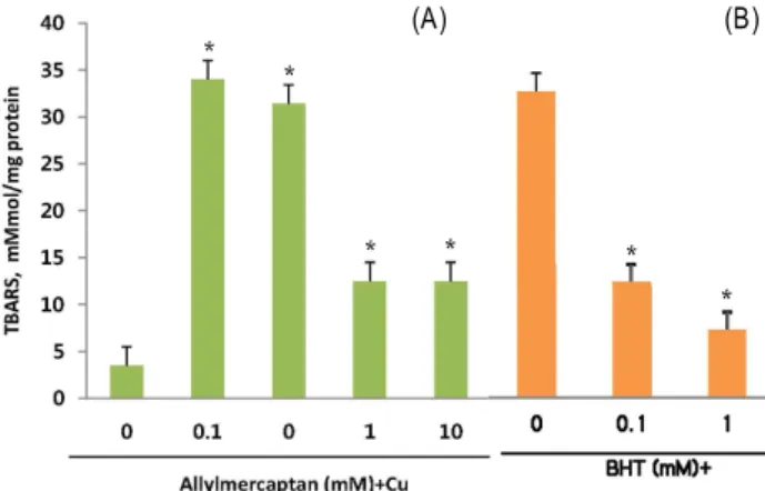
4resulted in a significant increase TBARS. Upon coincubation with varying concentration of allylmercaptan, its effect on TBARS formation was most no- ticeable at 1 and 10 mM (Fig. 1A). The known antioxidant butylated hydroxytoluene (BHT) was used as a positive con- trol (Fig. 1B).
Addition of allylmercaptan resulted in dose-dependent decrease in LDL oxidation, as measured by reduced TBARS formation. Likewise, the effect of the known antioxidant, BHT, on Cu
2+-induced TBARS formation was examined and compared to the effect of allylmercaptan. These results in- dicate allylmercaptan has substancial antioxidant properties and may inhibit LDL oxidation via antioxidant mechanisms.
Studies involving chemical analysis of garlic suggest that or- ganosulfur compounds are responsible for the bioactive [23].
Particles are innocuous, possessing lipophilic antioxidants(α -toccopherol and β-carotene) to protect against oxidant at- tack [25,31].
Allylmercaptan possess a reducing sulfur center attached to organic side groups, enabling stabilization of their negative charge. This combination in chemical structure thus confers strong antioxidant properties upon some of those compounds [1]. Allylmercaptan is recognized as a significant allyl sulfur compound occuring in garlic preparations [29].
Among these, our laboratory has placed emphasis on
* *
* * *
*
(A) (B)
Fig. 1. The effect of allylmercaptan on Cu2+-induced LDL oxida- tion (A) and BHT (B) as control. Data represents mean±SE of triplicate samples. *Significant differences (
p
<0.05) compared with samples exposed to ox-LDL but without pretreatment with allylmercaptan.allylmercaptan. Antioxidants have been the focus of recent studies involving the atherosclerotic process, primarily be- cause of the oxidant-dependent nature of atherogenic initiation.
Effects of LDH release and GSH level
Low-density lipoprotein oxidation has been recognized as playing an important role in the intiation and progression of atherosclerosis [5,28]. LDL has been shown to be oxidized by cultured cells such as macrophages and enothelial and smooth muscle cells with transition metals. Ox-LDL appears ti ubitiate vascular dysfunction by directly promoting cytitixicity. Effects of allylmercaptan on LDH release (histogram) and GSH level (line graph) when cells were ex- posed to 0.1 mg/ml ox-LDL (Fig. 2). Ox-LDL caused an in- crease in LDH release and a decrease in intracellular GSH compared with cells not exposed to ox-LDL. Pretreatment of cells with allylmercaptan resulted in a dose-dependent inhibition of LDH release and intracellular GSH depletion.
Ox-LDL appears to initiate vascular function by directly pro- moting cell cytotoxicity. It can alter the composition and per- meability of the endothelial barrier and it thus cytotoxic for endothelial cell [11]. Previously some reports indicated that garlic compounds inhibit Cu
2+induced LDL oxidation [15]
and protect vascular endothelial cells from ox-LDL induced cell injury [16].
In the present study, LDL release was measured as an index of cell injury. LDH is an intracellular enzyme that leaks from cells when their membranes are damaged.
Ox-LDL caused an increase in LDH release, indicating cell injury. This detail indicated that preincubation with allyl- mercaptan significantly inhibited the increase in LDH re- lease induced by ox-LDL, showing the protective effects of
Fig. 2. Effects of allylmercaptan on LDH release and intracellular GSH. Histogram depicts LDH release.
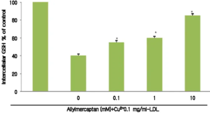
Fig. 3. Effect of allylmercaptan on intracellular GSH. Data repre- sent means±SE of triplicate samples. Untreated indicates that cell were not exposed to ox-LDL.*Significant differ- ence (
p
<0.05) compared with samples exposed to ox-LDL but without pretreatment with allylmercaptan.its compound on the cell membranes. Allylmercaptan ex- hibited a dose dependent inhibition of ox-LDL induced LDL release.
The effect of allylmercaptan on GSH level when cells were exposed to 0.1 mg/ml ox-LDL. Ox-LDL caused a decrease in intracellular GSH compared with cells not exposed to ox-LDL. Pretreatnebt if cekks with allylmercaptan resulted in dose independent of intracellular GSH delpletion (Fig. 3) Ox-LDL has been shown to deplets intracellar GSH in cultured endothelial cells. Intracellular GSH depletion can lead to increased endothelial cell susceptibility to injury caused by ox-LDL [19]. GSH is the most abundant low molecular weight thiol compound in the cell and plays an important role in antioxidant defense and detoxification.
GSH depletion compromises cell defences against oxida- tive damage and may lead to cell death [20]. Although the mechanism of allylmercaptan is not clearly under- stood, it has been suggested that allylmercaptan could in- terfere with cellular sulfur hydryl (SH) homeostasis by af- fecting the glutathione (GSH) by the sulfoxide group. It has been shown that allylmercaptan inactivates GSH de- pletion by reacting with the SH group of the enzyme and formation of the mixed disulfide. The depletion of GSH results in protein-SH oxidation, peroxidation and dis- turbances of cellular metabolism, which may lead to cell death.
This data indicate that incubation of PAECs with ox-LDL
for 24 hr caused a decrease of total GSH. Preincubation of
PAECs with allylmercaptan prevented GSH depletion, sug-
gesting that its compound can be a potent protective agent
against ox-LDL induced cytotoxicity.
Effects of allylmercaptan on peroxide
Under oxidant-stressed conditions, peroxides such as H
2O
2and lipid peroxides change cell function and inter- action with surrounding cells. For unstance, H
2O
2serves as an important second messenger in the activation of the tran- scriotion factor, NF-
kB, which is associated with expression of cell adhesion factors, vascular cell adhesion molecule-1 (VCAM-1), and intercellular adhesion molecule-1 (ICAM=1) [8,27]. Furthermore, H
2O
2damages cell membranes, reduces cell viability, and induces lipid peroxidation [32]. H
2O
2also yields hydrixyl radicals (OH) by reacting with a transition metal such as Fe
2+or Cu
2+. Generation of these hydroxyl radicals results in DNA damage and lipid peroxidation, leading to cell dysfunction and death. Lipid peroxides in en- dothelial cells change the permeability of cell membranes and ion efflux.
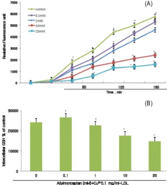
Peroxide release was measured using a fluorometric using a fluorometer assay. ox-LDL concentrations at 0.1 mg/ml were used to stimulate the PACEs (Fig. 3) and DCF for- mation was measured. Incubation of PACEs with ox-LDL caused an increase of fluorescence, indicating release of peroxides. Allylmercaptan inhibited this release in a dose-dependent manner.
Moreover, exposure of PACEs to oxidized LDL resulted in a significant release of peroxides. Allylmercaptan demon- strated a similar dose-dependent decrease in DCF formation abeit to a much lesser extent (Fig. 4A). Even after 24 hr of coincubation, allylmercaptan still exerted its inhibition at concentrations of 10 and 20 mM (Fig. 4B). The results estab- lish significant allylmercaptan inhibition of peroxide per- metion in PACEs cell line.
The results was induced upon stimulation of PACEs cell lines. Once LDL oxidative modification has occurred, the ox-LDL particle can exert a myriad of pathogenic effects in atherosclerotic progression. With its native LDL receptor do- main cleaved during oxidative modification, ox-LDL moves across the cell membrane via an alternate scavenge receptor abundant on the cell surfaces of endothelial cells [20]. Such event may accelerate the formation of atherogenic lesions and cell death.
In a times course study, allylmercaptan inhibited release of peroxide in endothelial cells dose-dependently while still retaining its inhibitory effect 24 hr later. It also observed the direct scavenging effect of allylmercaptan on peroxides.
Although several beneficial antioxidative properties of allyl- mercaptan have been reported [8], this study presents a novel
Fig. 4. Effects of allylmercaptan on release of peroxides. PACES were incubated with 0.1 mg/ml ox-LDL and various con- centrations of allylmercaptan. 2' 7'-Dichlorofluorerescein fluorescence, indicating release of peroxides was moni- tored every 30 min for 3 hr (Fig. 3A). Release of per- oxides was also measured at 24 hr (Fig. 3B). Data repre- sent mean±SE of triplicate samples. *Significant differ- ence (
p
<0.05) compared with control exposed to ox-LDL without allylmercaptan.activity, suggesting that allylmercaptan may reduce the per- oxides and may thus be useful for pathologies associated with oxygen species.
Scavenging effects of hydrogen peroxide
Table 1 shows the scavenging effect of directly allylmer- captan on hydrogen peroxide. Decreases of hydrogen per- oxide reflecting scavenging by allylmercaptan were noted,
Table 1. Scavenging effect of allylmercaptan on hydrogen per- oxide
Allylmercaptan (mM) H2O2 (nmol±SE) Scavenging (%) 0
0.11 1020
5.00 4.90±0.08* 4.03±0.08* 0.98±0.06* 1.02±0.06*
- 19.42.0 80.479.6 Data represent mean±SE of triplicate samples.
*Significant difference compared with samples without allyl- mercaptan (
p
<0.05).(A)
(B)
with significant activity observed at the concentration of 0.1, 1, 10 and 20 mM of allylmercaptan.
Under the oxidative stress conditions, peroxides such as hydrogen peroxides and lipid peroxides change cell function and interaction with surrounding endothelial cell. For in- stance, hydrogen peroxide serves as an important second messenger in the activation of the transcription factor, NF-kB which is associated with expression of cell adhesion factors, vascular cell adhesion molecule, and intracellular adhesion molecule[26]. Furthermore, H
2O
2damages cell membranes, reduces cell viability, and induce lipid peroxidation [30].
H
2O
2yields hydroxyl radical (•OH) by reacting with a tran- sition metal such as Cu
2+and Fe
2+. Generation of these hy- droxyl radicals results in DNA damage and lipid perox- idation, leading to cell dysfunction and death.
This results observed the direct scavenging effect of allyl- mercaptan on hydrogen peroxide. This data thus suggest that allylmercaptan can protect endothelial cells from oxi- dant injury by removing the intracellular peroxides such as hydrogen peroxide and lipid peroxide.
The dependence of the atherogenic process on oxidation and peroxide generation makes antioxidant intervention a feasible therapentic measure. In recent years, probucol has been employed as a therapeutic adjunct with other anti- arteriosclerosis drugs with great success [30]. Although orig- inally intended as a cholesterol-lowering agent, its anti- oxidant properties were soon recognized and put on clinical use. Synthetic antioxidants, such as the drug probucol [18], have been prescribed as an adjunct therapy along with other vasodilators [30]. The lipophilic properties of probucol allow it to incorporate evenly in cellular membranes in a fashion similar to many of the tocopherol antioxidants. However, as with many lipophilic compounds, a limit to beneficial ef- fects is reached as increased concentration membrane chemistry. Therefore, hydrophilic antioxidants such as SAC become increasingly significant oxidant scavengers, as limits to the intake of lipophilic antioxidants are attained. The hy- rophilic nature of SAC allows it to not only react with oxi- dant molecules imbedded in cellular membranes (i.e., lipid perooxides), but also to scavenge the extracellular medium for more hydrophilic ROs such as superoxdied anions and hydroxyl radicals. Therefore, SAC exerts a synergistic effect in reducing oxidant load by scavenging extracellular and membrane-bound oxidants, while concomitantly regenerat- ing lipid-soluble antioxidants. However, their toxicity limits their usage and ultimately, their potential as therapeutic
agents. On the other hand, many naturally occuring anti- oxidants [17] derived from natural products such as allyl- mercaptan offer similar protection.
In conclusion, the data from this in vitro study indicate that allylmercaptan may protect endothelial cells from ox-LDL induced injury by preventing depletion of GSH and removing peroxides, and suggest that allylmercaptan may be useful in the prevention of atheriosclerosis.
Acknowledgement
This research was supported by Kyungsung University Research Grants in 2010.
References
1. Block, E. 1992. The organosulfur chemistry of the genus
Allium
implications for the organic chemistry of sulfur.Angew Chem. Int. Ed. Engl.
31, 1135-1178.2. Bonetti, P. O., L. O. Lerman, and A. Lerman. 2003.
Endothelial dysfunction. A marker of atherosclerotic risk.
Artherioscler. Thromb. Uasc. Biol.
23, 188-175.3. Cai, H. and D. G. Harrison. 2001. Endlothelial dysfunction in cardiovascular diseases: the role of oxidant stress.
Crit.
Res.
87, 840-844.4. Cathcart, M. K., D. W. Morel, and G. M. Chisolm. 1985.
Monocytes and neutrophiles oxidized low density lip- oprotein making it cytotoxic.
J. Leukocyte Biol
. 38, 341-350.5. Cox, D. A. and M. L. Cohen. 1996. Effects of oxidized low density lipoprotein on vascular contraction and relaxation:
clinical and pharmacological implications in atherosclerosis.
Pharmacol. Rev.
48, 3-19.6. Egon-Schmind, C., R. Eckardm, and E. H. Kemper. 1992.
Metabolism of garlic constituents in the isolated perfused rat liver.
Planta Med
. 58, 301-305.7. Esterbauer, H., G. Strigel, H. Puhl, and M. Rotheneder. 1989.
Continuous monitoring of
in vitro
oxidation of human low density lipoprotein.Free Radic. Res. Commun
. 6, 67-75.8. Geng, Z., Y. Rong, and B. H. S. Lau. 1997. S-Allylcysteine inhibits activation of nuclear factor kappa B in human T cells.
Free Radic. Biol. Med
. 23, 345-3509. Gebhardt, R. and H. Beck. 1996. Differential inhibitory effects of garlic-derived organosulfur compounds on cholesterol bi- osynthesis in primary rat hepatocyte cultures.
Lipids
31, 1269-1276.10. Greenspan, P. and R. L. Gutman. 1993. Detection by nile red of agarose fell electrophoresed native and modified low density lipoprotein.
Electrophoresis
14, 65-68.11. Guretzk, H. J., K. D. Gerbitz, B. Olgemoller, and E.
Schleicher. 1994. Atherogenic levels of low density lip- oprotein alter the permeability and composition of the endo- thelial barrier.
Aherosclerosis
107, 15-24.12. Heinecke, J. W. 1987. Free radical modification of low-den- sity lipoprotein: Mechanisms and biological consequences.
Free Rad. Biol. Med
. 3, 65-73.13. Henriksen, T., E. M. Mahoney, and D. Steinberg. 1981.
Enhanced macrophage degradation of biologically modified low density lipoprotein.
Atherosclerosis
3, 149-159.14. Henriksen, T., E. M. Mahoney, and D. Steinberg. 1981.
Enhanced macrophage degradation of low density lip- oprotein previously incubated with cultured endothelial cells: Recognition by receptors for acetylated low density lipoprotein.
Proc. Natl. Acad. Sci. USA
78, 6449-6504.15. Ide, N., A. B. Nelson, and B. H. S. Lau. 1997. Aged garlic extract and its constituents inhibit Cu2+-induced oxidative modification of low density lipoprotein.
Planta Med
. 63, 263-264.16. Ide, N. and B. H. S. Lau. 1997. Garlic compounds protect vascular endothelial cells from oxidized low density lip- oprotein-induced injury.
J. Pharm. Pharmacol.
49, 908-911.17. Parthasarathy, S., U. P. Steinbrecher, J. Barnett, J. L. Witzum, and D. Steinberg. 1985. Essential role of phospholipase A2
activity in endothelial cell-induced modification of low-den- sity lipoprotein.
Proc. Natl. Acad. Sci. USA
82, 3000-3004.18. Kuzuya, M. and F. Kuzuya. 1993. Probucol as an antioxidant and antiatherogenic drug.
Free Radic. Biol. Med
. 14, 67-77.19. Kuzuya, M., M. Naito, C. Funaki, T. Hayashi, K. Asai, and F. Kuzuya. 1989. Protective role of intracellular glutathione against oxidized low density lipoprotein in cultured endo- thelial cells.
Biochem. Biophys. Res. Commun
. 163, 1466-1472.20. Kuzuya, M., M. Naito, C. Funaki, T. Hayashi, K. Asai, and F. Kuzuya. 1991. Lipid peroxide and transition metals are reqired for the toxicity of oxidized low density lipoprotein to cultured endothelial cells.
Biochim. Biophys. Acta.
1096, 155-161.21. Lowry, O. H., N. J. Rosebrough, A. L. Farr, and R. J. Randall.
1951. Protein measurement with the Folin phenol reagent.
J. Biol. Chem
. 193, 265-275.22. Lawson, L. D. 1993. Bioactive organosulfur compounds of garlic and garlic products: Their role in reducing blood
lipids. In human medicinal agents from plants. In Kinghorn, A. D. and M. F. Balandrin (eds.), pp. 306-330, American Chemical Society. Washington, DC, USA.
23. Lawson, L. D. 1996. The composition and chemistry of garlic cloves and processed garlic, in garlic: The science and ther- apeutic application of allium sativum L. and related species.
2nd eds. In Koch, H. P. and L. D. Lawson (eds.), pp. 37-107, Williams and Wilkins, Baltimore.
24. Okamoto, G., F. Hayase, and H. Kato. 1992. Scavenging of active oxygen species by glycated protein.
Biosci. Biotech.
Biochem.
56, 928-931.25. Ryu, B. H., J. W. Jeung, L. G. Robert, and P. Greenspan.
1990. Antioxidative activity for human low density lip- oprotein oxidation by a novel compound purified from ma- rine microbial origin.
J. Marine Biotech.
81, 175-182.26. Sedlak, J. and R. H. Lindsay. 1968. Estimation of total, pro- tein-bound, and nonprotein sulfhydryl groups in tissue with Ellman's reagent.
Anal. Biochem
. 25, 192-205.27. Sen, C. K. and L. Packer. 1996. Antioxidant and redox regu- lation of gene transcription.
FASEB
10, 709-720.28. Steinberg, D., S. Parthasarathy, T. E. Carew, J. C. Khoo, and J. L. Witztum. 1989. Beyond cholesterol: Modification of low-density lipoprotein that increase its atherogenicity.
N.
Engl. J. Med
. 320, 915-924.29. Wan, C. P., E. Myung, and B. H. S. Lau. 1993. An automated micro-fluorometric assay for monitoring oxidative burst ac- tivity of phagocytes.
J. Immunol.
159, 131-138.30. Xu, S. and B. H. Simon Cho. 1999. Allyl mercaptan, a major metabolite of garlic compounds, reduces cellular cholesterol synthesis and its secretion in Hep-G2 cells.
J. Nutr. Biochem
. 10, 654-659.31. Yang, S. T. 2007. Antioxidative activity of extracts of aged black garlic on oxidation of human low density lipoprotein
J. Life Sci.
17. 1330-1335.32. Yamasaki, T., L. Li, and B. H. S. Lau. 1994. Garlic compounds protect vascular endothelial cells from hydrogen per- oxide-induced oxidant injury.