Kor. J. Hort. Sci. Technol. 29(6):623-632, 2011
배추에서 항암물질 phenylethylisothiocyanate의 다량 합성을 위한 myrosinase와 glutathione S-transferase 유전자 분리 및 이를 이용한
형질전환체 육성
박지현1ㆍ이수진1ㆍ김보령1ㆍ우은택2ㆍ이지선1ㆍ한은향1ㆍ이윤형1ㆍ박영두1*
1경희대학교 생명과학대학 원예생명공학과, 2캐로톱씨드 육종연구소
Isolation of Myrosinase and Glutathione S-transferase Genes and Transformation of These Genes to Develop Phenylethylisothiocyanate
Enriching Chinese Cabbage
Ji-Hyun Park
1, Su-Jin Lee
1, Bo-Ryung Kim
1, Eun-Teak Woo
2, Ji-Sun Lee
1, Eun-hyang Han
1, Youn-Hyung Lee
1, and Young-Doo Park
1*1Department of Horticultural Biotechnology, Kyunghee University, Yongin 446-701, Korea
2Breeding Research Institute, Carrotop Seed Co., Anseong 456-882, Korea
Abstract. To increase the anti-carcinogens phenylethylisothiocyanate (PEITC), myrosinase (MYR), and glutathione S-transferase (GST), genes related to PEITC pathway were isolated and the gene expressions were regulated by Agrobacterium transformation. Isolated cDNAs, MYR, and GST genes were 1,647 bp and 624 bp, respectively, and the protein expression was confirmed through pET system. Thereafter, we constructed a sense-oriented over-expressing myrosinase (pBMY) and RNAi down-regulated GST (pJJGST) binary vectors for the Chinese cabbage transformation. After the transformation, thirteen over-expressing transgenic Chinese cabbage plants (IMS) with pBMY and five down-regulated ones (IGA) with pJJGST were selected by PCR analysis. Selected T
0transgenic plants were generated to T
1plants by self-pollination. Based on the Southern blot analysis on these T
1transgenic plants, 1-4 copies of T-DNA were transferred to Chinese cabbage genome.
Thereafter, RNA expression level of myrosinase gene or GST gene was analyzed through real-time RT PCR of IMS, IGA, and non-transgenic inbred lines. In case of IMS lines, myrosinase gene was increased 1.03-4.25 fold and, in IGA lines, GST gene was decreased by 26.42-42.22 fold compared to non-transgenic ones, respectively. Analysis of PEITC concentrations using GC-MS it showed that some IMS lines and some IGA lines increased concentrations of PEITC up to 4.86 fold and up to 3.89 fold respectively compared to wild type. Finally in this study IMS 1, 3, 5, 12, and 15 and IGA 1, 2, and 4 were selected as developed transgenic lines with increasing quantities of anti-carcinogen PEITC.
Additional key words: down regulation vector, over-expression vector
*Corresponding author: [email protected]
※ Received 17 August 2011; Accepted 27 September 2011. 본 연구는 농촌진흥청에서 차세대 바이오그린21사업(과제 번호: PJ0080762011)의 지원에 의해 이루어진 것임.
서 언
채소와 과일의 섭취가 많을수록, 특히 배추과 채소를 섭 취할수록 항암효과에 도움을 준다는 연구결과가 지속적으 로 발표되고 있다(Fahey et al., 2001; Lam et al., 2009;
Langouet et al., 2000; Thornalley, 2002). 배추과 작물은 높은
함량의 glucosinolate를 가지고 있어 생체 내 화학적 변화로
인한 2차 대사산물들을 생성한다. 그 종류로는 isothiocynate
(ITC)와 indol 화합물 등이 있으며 이는 화학적 방어에 효과
가 있다고 밝혀졌다(Lampe and Peterson, 2002). Glucisonolate
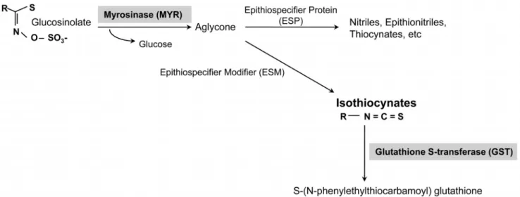
Fig. 1. Biosynthesis pathway of phenethyl isothiocyanate and related genes.
의 2차 대사산물의 일종인 phenylethylisothiocyanate(PEITC) 는 ITC의 한 종류로 glucosinolate가 myrosinase(MYR)에 의해 가수분해 되면서 생성되며 해충저항성(Zhang et al., 2006)과 항암효과를 보이는 물질이다. Glucosinolate가 MYR 효소에 의해 중간물질인 algycone을 형성하고, algycone은 epithiospecifier protein(ESP)유전자와 epithiospecifier modifier (ESM)유전자에 의해 상보적으로 nitriles과 thiocyanate로 분해되거나 isothiocyanate로 분해된다(Halkier and Gershenzon, 2006; Lambrix et al., 2001; Zhang et al., 2006). 한편 생성 된 ITC는 미토콘드리아에 존재하는 glutathione S-transferase (GST)에 의해 촉매되어 S-(N-pheylethio-carbamoyl)-glutathione 으로 전환된다(Eklind et al., 1990)(Fig. 1).
ITC는 배추에서 PEITC, benzyl-isothiocyanate 및 3-phenyl- propyl-isothiocyanate등의 형태로 존재하며 그 중 PEITC는 암의 발달을 억제하는 항암물질로써 전립선암, 유방암, 자궁 경부암, 대장암, 폐암 및 식도암 등에 효과가 있음이 보고되 었다(Doerr-O'Rourke et al., 1991; Lai et al., 2010; Lam et al., 2009; Stoner et al., 1991; Tseng et al., 2004; Xu et al., 2006; Yu et al., 1996).
Glucosinolate가 분해되는 과정에서 MYR는 유용물질인 PEITC의 생성을 유도하는 매개자로써 중요한 역할을 한다.
MYR 효소의 기능이 활발할수록 glucosinolate로부터 생성 되는 PEITC의 양이 증가할 것으로 기대된다. 또한 생성된 PEITC는 mercapturic acid pathway로 유입되는데(Wu et al., 2009) 이를 조절하는 첫 번째 단계 효소인 GST를 억제 시키면 분해되는 PEITC의 양이 상대적으로 적어 생체 내 존재하는 PEITC 양은 증가할 것으로 기대된다.
따라서 본 연구에서는 배추에서 항암효과가 있는 유용물 질인 PEITC의 합성 과정에 관여하는 주요 유전자인 MYR
과 GST를 코딩하는 유전자를 분리하였고 유전자의 특성을 분석한 후 pET system을 통하여 단백질 발현을 확인하였다.
분리된 유전자는 PEITC 물질을 다량으로 유도하기 위하여 MYR 유전자는 과발현(over-expression)을 유도하는 vector를 제작하였고 GST유전자는 발현 억제(down-regulation)를 유도 할 수 있는 vector를 제작하였다. 완성된 각각의 vector로 배 추 형질전환을 실시하였고 형질전환체 선발 후 Southern 분 석으로 T-DNA copy 수를 확인하였으며 선발된 형질전환체 에서의 MYR 유전자와 GST 유전자의 발현량을 real-time RT PCR 방법으로 분석하였다. 최종적으로는 형질전환체에 서 생성되는 PEITC 함량 분석을 실시하고 함량이 증가된 형질전환 계통을 선발・육성하였다.
재료 및 방법
Reverse transcription(RT)-PCR과 5’RACE 방법을 이용한 MYR와 GST 유전자의 분리
실험에 사용된 배추는 inbred line인 CT001(Carrotop seed company, An-Sung)이며 이를 파종하여 한 달 후에 어린잎 을 채취하여 RNA 추출에 사용하였다. RNA 추출은 RNeasy Kit(QIAGEN, USA)를 이용하였고 방법은 제조회 사에서 제공한 방법에 따라 수행하였다.
PEITC 대사과정에 관련된 유전자 GST는 reverse transcription
(RT)-PCR 방법, MYR는 5’RACE 방법을 이용하여 완전장
의 cDNA를 분리하였다. RT-PCR 방법은 one-step RT-PCR
kit(ABgene, UK)를 이용하였으며 각각의 유전자마다 specific
primer를 작성하여 1μg의 RNA를 49℃에서 30분 동안
cDNA를 합성한 후 94℃에서 30초, 55℃에서 45초, 72℃에
서 90초를 1cycle로 하여 30cycle을 반응시키고 마지막 단계에
Table 1. Primer sequences for isolation of glutathione S-transferase (GST) and myrosinase (MYR) genes.
Primer name Sequence
GST Sense primer 5'-CCT TCA CAT TCA ACA TTA ATT AAA C-3'
Antisense primer 5'-GTA CTG AGA AAT TGG GCA ATG A-3'
MYR GeneRacerTM 5' primer 5'- CGA CTG GAG CAC GAG GAC ACT GA -3'
Antisense primer 5'- TTT TTC ATT AGT AAC TGA TCA TCT TT- 3'
서 72℃, 5분 처리를 하였다. 유전자를 분리하기 위하여 작 성된 primer는 Table 1에 제시하였다. 5’RACE 방법을 이용 한 MYR 유전자의 완전장 cDNA 분리는 GeneRacer
TMKit (invitrogen, USA)을 사용하였다. 분리한 RNA에 GeneRacer
TMRNA oligo를 oligo dT primer와 역전사효소를 이용하여 cDNA를 합성한 뒤 GeneRacer
TMRNA oligo로부터 유래된 GeneRacer
TM5'primer와 MYR유래의 antisense primer를 (Table 1) 첨가하고 PCR을 실시하여 분리하였다. cDNA합성 프로그램 조건은 전 처리된 1μg의 RNA에 oligo dTprimer를 넣고 70℃에서 RNA 이차구조를 제거한 뒤 42℃에서 30분 간 역전사시키고 역전사효소를 불활성화시키기 위해 75℃
에서 10분간 유지하였다. 그 후 합성된 cDNA 1μg에 MYR antisense primer, GeneRacer
TM5'primer, Tag polymerase를 첨가하여 PCR을 실시하였고 프로그램은 96℃에서 2분, 9 6℃에서 30초, 55℃에서 30초, 72℃에서 1분으로 40cycle 수행하고 마지막으로 72℃에서 10분간 반응시켰다. 각각의 primer들로 증폭된 유전자는 1.0%의 agarose gel에 전기영 동으로 확인하였고, 염기서열 분석을 위해 pGEM-T easy vector(promega,USA)에 삽입하고 T7과 SP6 primer를 이용 하여 염기서열을 분석하였다. 분리된 유전자의 단백질 domain 분석은 NCBI에서 제공하는 conserved domains search 프로 그램을 이용하였다.
pET system을 이용한 유전자의 단백질 발현 확인
MYR와 GST 유전자의 단백질 발현여부를 확인하기 위하 여 E. coli 발현시스템인 pET 벡터를 이용하였는데 GST 유 전자는 pET-15b를, MYR 유전자는 pET-17b를 이용하였다.
각각의 유전자를 발현벡터에 삽입하기 위해 coding region에 GST 유전자는 NdeI과 BamHI, MYR는 HindIII과 SpeI의 제한효소 site를 결합시켜주고 PCR로 증폭하였다. 최종적으 로 PCR 산물과 pET 벡터를 동시에 각각의 제한효소로 처 리하여 삽입하였고 E. coli 균주로는 BL21을 사용하여 37℃에 서 배양하였다. 단백질 발현은 Western 분석으로 확인하였다.
형질전환을 위한 각 유전자의 식물 발현 vector 작성 배추에서 PEITC의 다량 합성을 위하여 MYR와 GST 유전
자의 형질전환용 식물발현 vector를 제작하였다. 발현 조절을 위하여 MYR 유전자는 과발현이 되도록 over-expression 벡 터(pBMY)를 작성하였고 GST 유전자는 그 기능을 억제시 키기 위하여 RNAi 벡터(pJJGST)를 작성하였다. 벡터 작성 의 자세한 내용은 결과 및 고찰에서 제시하였다.
Agrobacterium을 이용한 배추 형질전환
배추종자를 ethanol과 sodium hypochlorite(NaOCl)로 소독 한 후 , 파종하여 기내생장시킨 개체에서 하배축을 절단하여 형질전환에 이용하였다 . 절단된 하배축은 pBMY와 pJJGST vector를 운반하는 각각의 Agrobacterium LBA4404 strain 용액에 접종하고, 공동배양배지[Murashige & Skoog(MS) 기본배지, 3% sucrose, 0.8% plant agar, NAA 1mg・L
-1, BA 4mg・L
-1, AgNO
34mg・L
-1, acetosyringone 10mg・L
-1, 0.8%
plant agar, pH 5.8]에 치상하였다. 21 ± 2℃, 암조건에서 3 일 동안 공동배양한 절편체는 cefotaxime 200mg・L
-1이 포 함된 MS 기본액체배지에서 3회 반복하여 Agrobacterium을 제거해 주고 pJJGST는 hygromycin 5mg・L
-1가 그리고 pBMY는 phosphinotricin 20mg・L
-1가 첨가된 선발배지(MS 기본배지 , 3% sucrose, NAA 1mg・L
-1, BA 4mg・L
-1, AgNO
34mg・L
-1, acetosyringone 10mg・L
-1, cefotaxime 200mg・L
-1, 0.8% bacto-agar, pH 5.8)에 치상하였다. 절편체에서 발생된 callus를 동일한 조건의 선발배지에 옮겨 신초를 유도하였 고 , 유기된 신초는 발근배지(MS 기본배지, 3% sucrose, 0.8%
plant agar, pH 5.8)에 옮겨 발근시켰다. pBMY가 삽입된 형 질전환체는 IMS라 명명하였고 pJJGST가 삽입된 형질전환 체를 IGA로 명명하였다.
PCR을 통한 형질전환체 선발과 Southern 분석을 통한 copy 수 분석
형질전환된 배추의 선발을 위해 T
0세대에서 pBMY와
pJJGST의 선발마커인 bar 유전자와 hygromycin저항성 유
전자를 목표로 프라이머를 작성하고 이를 이용해 선발한 후
T
1세대로 증식시켰다. 자가수분을 통하여 확보한 T
1세대의 형
질전환체 역시 형질전환 여부를 확인하기 위해 PCR검정을 실
시하였고 삽입된 T-DNA copy수를 확인하기 위해 Southern
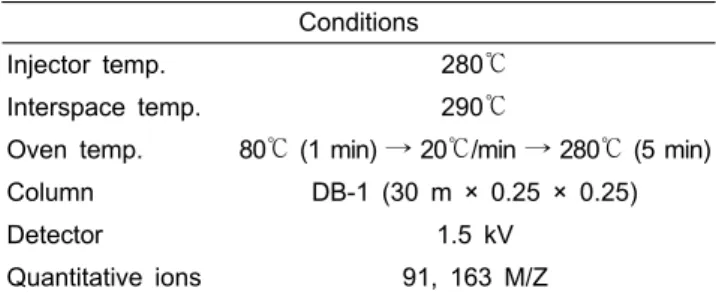
Table 2. GC/MS analysis conditions for phenylethylisothiocyanate (PEITC) concentration measurement.
Conditions
Injector temp. 280℃
Interspace temp. 290℃
Oven temp. 80℃ (1 min) → 20℃/min → 280℃ (5 min)
Column DB-1 (30 m × 0.25 × 0.25)
Detector 1.5 kV
Quantitative ions 91, 163 M/Z
분석을 실시하였다. 실험을 위한 genomic DNA 분리는 Cho et al.(1994)의 방법에 따라 수행하였다. PCR 반응은 IMS 형질전환체 선발을 위한 ppt 프라이머(F: 5’-GGT CTG CAC CAT CGT CAA CC-3’, R: 5’-TCA AAT CTC GGT GAC GGG CA-3’)와 IGA 형질전환체 선발을 위한 hpt 프 라이머(F: 5'-TTT CCA CTA TGC GCG AGT AC-3', R:
5'-TGT CGA GAA GTT TCT GAT CGA -3')를 사용하여 수 행하였으며 Southern 분석은 IMS와 IGA에서 분리된 genomic DNA 10μg을 제한효소 HindIII를 처리하여 37℃에서 18시간 동 안 반응시킨 후 McCouch et al.(1988)의 방법에 따라 수행하였다.
Real-time RT PCR을 통한 형질전환체의 유전자 발현분석 형질전환체로 판명된 IMS와 IGA의 T
1세대 개체에서 유 전자의 발현 정도를 알아보기 위해 real-time RT PCR을 이 용하여 정량적 분석을 하였다. Real-time RT PCR은 각각의 형질전환체에서 RNeasy
®Kit(QIAGEN, Germany)를 이용하여 total RNA를 분리하였으며, SensiMix One-Step Kit(Quantace) 에서 제공하는 방법에 따라 Rotor-Gene
TM6000(Corbett, Australia) 을 이용하여 PCR 반응을 수행하였다. 각 유전자를 특이적으 로 인식하는 real-time RT PCR용 primer로는 IMS는 MYRF: 5'- GTT GCA TCT GCT TAC CA -3', MYRR: 5'- TGT AGC CAG TAG CAT TGA GT -3'과 IGA는 GSTF:
5'- GGC AGG TAT CAA AGT TTT CG- 3', GSTR: 5'- TTG ATT CGA AGA GCT TGA GG -3'와 같으며, 대조 유 전자로는 housekeeping 유전자인 actin을 이용하여 비교 분석 하였다.
PEITC 물질량 측정
IMS와 IGA에서의 PEITC 함량을 wild type의 배추에 대 비하여 측정하기 위해 GC-MS 분석을 수행하였다. 실험에 사용된 시료는 줄기 기부로부터 3cm를 제거하고 그로부터 5cm 상단까지 채취하였다. 준비된 시료는 액체질소에서 마 쇄한 후 1g을 측정하여 5mL의 물에서 30℃ 20분간 추출한 후 3mL의 용액을 2mL의 dichloromethane로 2번 분배한 다 음 진공 농축한 후 GC-MS(VF-5MS, Varian, USA)로 분석 하였다. 함량 정량은 PEITC 표준시료를 사용하여 검량곡선 을 작성한 후 시료 내 PEITC 정량에 사용하였다. 자세한 PETIC 분석조건은 Table 2에 제시하였다.
결과 및 고찰
GST와 MYR 유전자의 분리
Table 1에 제시한 각 유전자의 primer sequence들을 사용
하여 RT-PCR과 5’RACE 방법으로 분리된 유전자는 개시 코돈과 종결코돈을 포함하고 있었으며 1% agarose gel에 전 기영동한 사진은 Fig. 2A에 제시하였다. 분리된 각 유전자 의 개시코돈을 시작으로 종결코돈까지의 cDNA크기가 GST 유전자(특허등록번호; 제10-0744612호, GenBank accession No. AY567976)는 642bp이며 MYR 유전자(특허등록번호:
제054493호, GenBank accession No. AY567974)의 cDNA 는 1647bp이었다. 각각의 유전자의 염기 서열을 NCBI BLAST search 프로그램을 이용하여 상동성을 비교한 결과 GST의 경우 Brassica juncea와 98%의 상동성을 보였으며 MYR는
Brassica napus와 98%의 상동성을 보였다. 또한 유전자들의 염기서열을 아미노산 서열로 번역한 결과 GST는 214개, MYR는 549개의 아미노산으로 구성되어 있으며 각 유전자 의 아미노산 서열을 Figs. 2D와 2F에 제시하였다.
MYR 유전자는 B. napus와 A. thaliana에서 3개의 유전자
가 family로 구성되어 있는 것으로 밝혀졌고(Lenman et al.,
1993; Thangstad et al., 1993; Xue et al., 1995; Zhang et
al., 2002) 3개의 MYR 유전자중 하나의 유전자는 기능이
없는 위유전자 (pseueogene)이며 기능을 가진 유전자는 12
개의 exon과 11개의 intron으로 구성되어 있다(Thangstad et
al., 1993; Zhang et al., 2002). MYR는 모든 조직에서 어느
정도의 활성을 보이고 생장초기에는 각 조직마다 그 활성이
다르게 나타나지만(Bones, 1990) 생장하면서 조직간의 활
성의 차이는 거의 보이지 않는다. GST 유전자는 대부분의
식물체에 존재하는 유전자로써 세포질 내에 용해되어 존재
하며(Edwards et al., 2000; Marrs,1996) 구조적으로 GSH와
결합하는 active site와 독성물질 등의 화합물과 결합하는
substrate binding site가 존재한다(Atmstrong, 1997; Sheehan
et al., 2001). 보고된 바에 의하면 GST 유전자는 여러 그룹
이 존재하며 식물체에는 대표적으로 phi, zeta, tau, theta,
lambda의 5 그룹이 존재하는데 이중 phi와 tau 그룹이 식물
특이적으로 존재한다 . 배추에서 분리한 GST는 phi 그룹에 속
해있으며 아라비돕시스에서 phi 그룹의 GSTs는 herbicide-
detoxifying활성을 가지며 tau 그룹의 GSTs는 auxin-induced
Fig. 2. Characteristics of isolated glutathione S-transferase (GST) and myrosinase (MYR) genes in Chinese cabbage. (A) RT-PCR products of GST and MYR genes. M; 100 bp DNA ladder, Lane 1: GST-800 bp, 2: MYR-1796 bp. (B) Protein expression analysis of GST gene in E.coli by western blot using His Ab (1:5000). (C) Protein expression analysis of MYR gene in E.coli by western blot using His Ab (1:5000). (D) Amino acid sequences of GST protein and analysis of protein domain using NCBI Conserved Domains search program. Gray background is GST-N-phi subfamily domain and black is GST-C family superfamily domain.
(E) Amino acid sequences of MYR protein and analysis of protein domain using NCBI Conserved Domains search program.
Gray background is glycol-hydro-1 superfamily domain. The stop codon is marked as *.
GSTs가 주기능이며 2개의 exon으로 이루어져 있다고 보고 되었다(Dixon et al., 2002, 2009; Edwards et al., 2000).
pET system을 이용한 유전자의 단백질 발현확인과 단백질 domain 분석
분리한 유전자들의 DNA 염기서열을 바탕으로 triplet
codon에 의한 단백질 발현여부를 확인하기 위하여 pET
system을 이용한 발현실험을 수행하였다. 각 유전자는 6X
histidine이 tagging된 벡터에 삽입되었으며 발현된 단백질
의 크기는 GST 유전자는 pET-15b에서 약 29 KDa의 단백
질을, MYR은 pET-17b 벡터로부터 약 65 KDa의 단백질을
확인할 수 있었다(Figs. 2B and 2C). GST 유전자는 발현여
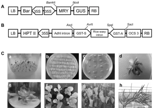
Fig. 3. T-DNA constructs and procedures for Agrobacterium–mediated Chinese cabbage transformation. (A) Over-expression vector (pBMY) of myrosinase gene for Agrobacterium–mediated Chinese cabbage transformation. (B) RNAi vector (pJJGST) of glutathione S-transferase gene for Agrobacterium–mediated Chinese cabbage transformation. (C) Chinese cabbage transformation. a, sowing onto medium in vitro; b, co-cultivation of hypocotyl explants; c, callus induction onto selection medium; d, shoot induction onto selection medium; e, root induction in rooting medium; f, acclimation in pots; g, flowering and self-pollination in greenhouse;
h, obtaining of T0 seeds.
부 확인 시 어려움은 없었으나 MYR의 경우 단백질이 불용 해성으로 발현되어 변성 조건에서 Ni-NTA affinity 결합 검 정을 진행하였다.
배추로부터 분리된 각 유전자들의 domain을 NCBI에서 제공하는 conserved domains search 프로그램을 통하여 분 석한 결과(Figs. 2D and 2F) GST유전자는 phi계통의 유전 자로써 항해충성의 효과를 보이는 그룹에 속해 있었다.
GST C family와 N family는 식물에만 특이적으로 존재하는 그룹인 phi 계열의 subfamily며 C family에는 GSH binding site가 존재하며 N family는 substrate binding site가 존재한 다. 이는 곰팡이, 세균의 단백질과 세포내의 발암유발물질, 환경적 독소등을 glutathione(GSH)에 전달하여 세포질내의 독성제거 역할을 하기도 하며(Dixon et al., 1998) PEITC물 질을 GSH에 전달하여 PEITC-SH complex가 세포막 단백 질인 multidrug resistance protein(MRP) channel을 통해 배 출되면 세포질 내에 GSH가 감소하면서 PEITC가 항암작용 과 관련된 protein receptor에 결합하고 signal을 전달하여 암 세포의 감소를 야기하는 역할을 하기도 한다(Thornalley, 2002). MYR 유전자는 Glycosyl hydrolase family 1(GH1) 이 존재하는 것으로 분석되었는데 glycoside hydrolase는 아 미노산 서열을 바탕으로 2개 또는 2개 이상의 탄수화물 사 이의 glycosidic결합이나 한 개의 탄수화물과 비탄수화물의
결합을 분해하는 역할을 한다(Henrissat, 1991; Henrissat and Davies, 1997). 여러 진핵생물 또는 고세균들을 포함한 미생물들은 GH family 1을 가지고 있으며 전형적으로 G-O-X 또는 G-S-X구조를 가지고 있다. G는 β-linked glucosyl, galactosyl, mannosyl, fucosyl, 6-phosphoglucosyl, 6-phosphogalactosyl, 또는 glucuronide residue가 대표적이고, X는 다른 glycosyl residue 또는 non-carbohydrate 그룹(예, aglycone)이 대표적 이다(Barrett et al., 1995; Burmeister et al., 1997; Czjzek et al., 2000). 따라서 myrosinase 유전자가 존재하는 식물체 는 외부의 충격이 가해졌을 때나 해충으로부터 피해를 입었 을 때 glycosyl hydrolase 기능을 띄며 glucosinolate를 분해 하는 화학적 반응이 진행되는 것으로 분석되었다.
형질전환을 위한 각 유전자의 형질전환용 벡터작성
배추에서 PEITC 물질을 효과적으로 다량 유도하기 위하여
각 유전자의 형질전환 벡터를 제작하였다. MYR 유전자는
glucosinolate의 분해를 촉진시키기 위하여 과발현되도록 작성
하여 중간산물인 aglycone을 더 많이 생성할 수 있도록 하였
다 . MYR 유전자 5’개시코돈 앞쪽에 NcoI을 결합하고 3’종결
코돈 뒤쪽에 BamHI 제한효소 site를 결합하여 pCAMBIA3301
(CAMBIA, Australia)에 삽입하여 과발현 벡터인 pBMY를
작성하였다(Fig. 3A). GST유전자는 발현을 억제시키도록 하
는 RNAi 벡터를 제작하였다. RNAi 벡터 제작시 효과적인 down regulation을 위해서 목표 유전자의 conserved domain 분석을 기반으로 디자인한다면 효과적으로 식물체 내 모든 유전자 copy를 억제할 수 있다. 대표적인 예로 벼(Oryza
sativa, Miki et al., 2005), 까마중(Solanum americanum, Sinet al., 2006) 및 콩(Glycine max, Tuteja et al., 2009) 등에서 conserved된 유전자 지역을 blocking하여 유전자군(gene family) 전체를 조절하여 목적 대사 경로를 조절하였다. GST RNAi 벡터 역시 효과적인 down regulation을 위해 GST의 conserved domain 분석을 하여 GST의 주요 기능인 GSH와 결합하는 위치와 substrate binding pocket을 목적으로 하는 부분이 포 함되도록 GST유전자의 단편 570bp를 RNAi 벡터인 JJ374 에 정방향과 역방향으로 삽입하였다. GST 단편을 정방향으 로 삽입하기 위해 AscI과 AvrII의 제한효소 site를 결합시켰 으며 역방향인 유전자 단편에는
SpeI과 SacI을 결합시켜GST RNAi 벡터인 JJGST를 작성하였다(Fig. 3B).
한편 작성된 RNAi 벡터가 small RNA가 제대로 형성되는 지는 웹사이트 http://rna.tbi.univie.ac.at/cgi-bin/RNAfold.cgi 를 통하여 RNAi vector 제작시 효과적으로 헤어핀 구조를 형성하는 것을 시뮬레이션하여 확인하였다(자료 미제시).
시뮬레이션은 RNAi 벡터에 삽입된 정방향의 GST 염기서 열, 헤어핀을 이루는 rice waxy intron 1과 역방향으로 삽입 된 GST 염기서열을 분석한 것으로 정방향과 역방향으로 삽 입된 염기서열은 -1015.40kal・mol
-1의 매우 낮은 자유에너 지를 갖는 것으로 보아 결합이 매우 잘 됨을 보여주었다. 또 한 헤어핀을 구성하는 intron 부위 역시 자유에너지가 높아 그 결합이 이루어지지 않아 올바른 헤어핀 구조를 보이고 있다고 판단되었다.
Agrobacterium을 이용한 형질전환 배추의 선발과 T-DNA copy수 분석
pBMY와 pJJGST vector를 Agrobacterium tumefaciens 매개로 inbred 배추 품종에 형질전환하였다(Fig. 3C). pBMY 로 형질전환된 배추는 IMS라 명명하였고 pJJGST로 형질전 환된 개체는 IGA라 명명하였다. 각각의 벡터로 형질전환되 어 순화된 개체들은 IMS가 15개체, IGA가 6개체이었으며 IMS의 경우 선발마커인 bar 유전자를 목표로 하여 ppt 프라 이머로 490bp PCR 산물의 유무로 형질전환체 선발을 하였 고 IGA는 hygromycin 저항성 유전자를 목표로 하여 hpt 프 라이머로 1149bp PCR 산물의 유무로 형질전환체 선발을 하였다. PCR 검정을 통해 순화된 개체들 중 형질전환체로 판단된 개체는 IMS가 13개체, IGA가 4개체이었다. 형질전 환체로 선별된 T
0의 각 개체를 계통으로 하여 자가수분으로
T
1세대의 종자를 확보하였고 T
1세대 분석을 위해 획득한 종자를 파종을 하여 T
0세대와 같은 방법으로 계통별 1개체씩 형질전환체를 선발하였다. 선발된 T
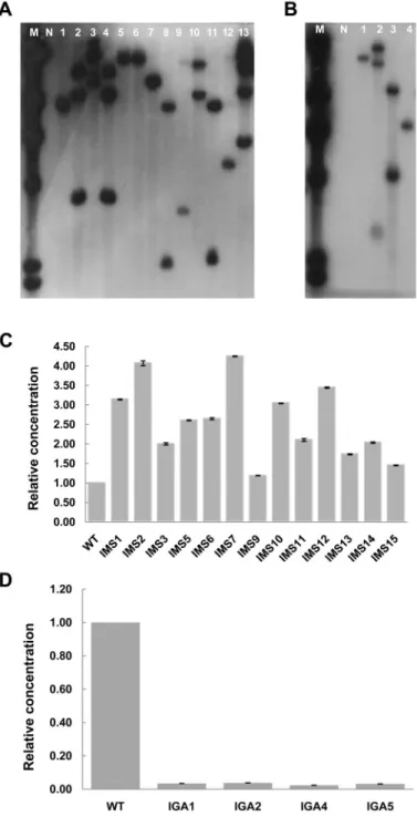
1세대에서 배추 genome 내로 삽입된 T-DNA의 copy수를 Southern 분석으로 확인하 였다. IMS 13계통과 IGA4라인을 각각 제한효소 HindIII를 처리하여 분석한 결과 1 copy로 밝혀진 계통은 IMS의 경우 6 계통, IGA의 경우 2 계통으로 밝혀졌고, 2 copy인 계통은 각각 4 계통, 1 계통이었으며 3 copy인 계통은 각각 2 계통, 1 계통으로 밝혀졌고 4 copy로 밝혀진 계통은 IMS에서 1계 통이었다(Figs. 4A and 4B).
배추형질전환체의 MYR와 GST 유전자 발현분석
T-DNA의 삽입이 확인된 T
1형질전환계통 IMS와 IGA에 서 각각의 유전자가 발현된 수준을 정량적으로 확인하기 위 하여 real-time RT PCR을 실시하였다. 실험은 총 3반복으로 실시하였으며, 각 실험의 결과는 delta-delta C
Tvalues 법을 이용하여 유전자 발현량을 도출해 내었으며, melting analysis 법을 이용하여 증폭된 산물 결과의 신뢰성을 검증 하였다(Livak and Schmittgen, 2001). IMS 형질전환 계통에 서의 MYR 유전자 발현 정도는 wild type에 비하여 1.03-4.25 배 가량 증가하여 발현됨을 확인하였으며, IGA 형질전환 계 통에서의 GST 유전자가 wild type에 비하여 26.42-42.22배 가량 발현량이 감소된 것을 확인하였다(Figs. 4C and 4D).
PEITC 물질량 측정
Real-time RT PCR을 이용해 배추에 형질전환된 벡터 pBMY와 pJJGST가 목적에 맞게 발현이 조절된 것을 확인 하였으며 IMS와 IGA에서의 발현량 증가와 감소가 실제로 PEITC 생성에 영향을 주었는지 확인하기 위하여 GC-MS방 법으로 PEITC 물질량 분석을 하였다. 형질전환된 배추에서 물질양을 조사하기 위해 시료를 준비하기 전 성숙모본에서 의 각각의 형질전환체는 표현형으로 나타나는 원예적 형질 [식물체(키), 바깥잎(크기, 모양, 잎면의 요철 등) 등]에 차이 를 보이지 않았다. PEITC 물질분석을 위하여 표준시료를 사용하여 검량곡선을 작성한 결과 PEITC는 표준시료로부 터 retention time이 7.06min임을 알 수 있었으며 표준검량 곡선을 이용한 결과 함량이 0.02μg・g
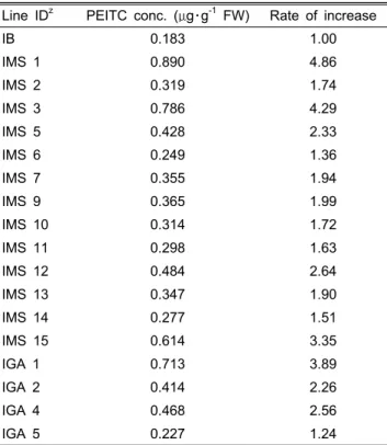
-1F.W.(분자량 163)로 정량되었다. 물질분석은 각 계통마다 오차를 줄이기 위해 3 반복으로 실시하였으며 물질량은 평균치를 제시하였다(Table 3). 그 결과 IMS 13개 계통과 IGA 4개의 계통에서 wild type의 배추에 비해 1.24배에서 4.86배까지 대부분 PEITC 물질의 증가를 확인할 수 있었다.
특정 물질과 관련된 대사과정에서 물질량을 조절하기 위
Fig. 4. Southern hybridization and real-time RT PCR analysis of T1 transgenic Chinese cabbage plants. (A) Southern hybridization of DNA from over-expressed T1 transgenic Chinese cabbage plants (IMS) with pBMY vector. (B) Southern hybridization of DNA from down-regulated T1 transgenic Chinese cabbage plants (IGA) with pJJGST vector. Genomic DNA was digested with HindIII, and probed with a [32P]-labeled 500bp of bar gene and 1.1 kb of hpt gene from pBMY and pJJGST plasmid DNA, respectively.
M: λ Hind III marker, N: non-transgenic plant, lane 1-13 (A) and lane 1-4 (B): transgenic plants. (C) The quantified analysis of mRNA accumulation of IMS. (D) The quantified analysis of mRNA accumulation of IGA. Quantified analysis was conducted by real-time RT PCR using delta-delta CT values method. The vertical T-bars indicate standard deviation.
해 T-DNA 삽입을 통하여 유전자를 조절하고 그에 따른 결 과로 물질량을 조사한 연구는 지속적으로 이어지고 있다. 감 자에서 저장되는 전분의 양을 증가시키기 위하여 amylose와 amylopectin의 경쟁적인 대사경로를 조절하였고(Kok-Jacon et al., 2003), potato sucrose transporter를 과발현시켜 녹말의 양을 증가시킨 사례가 있다(Rosche et al., 2002). 또한 담배
에서 화색관련 유전자인 DFR을 과발현시켜 안토시아닌의 증가로 인한 화색이 짙은 담배꽃을 확인한 사례도 있다(Lee et al., 2008).
본 연구를 통하여 PETIC 대사과정에 관련된 MYR 유전
자와 GST 유전자의 조절을 통한 항암물질 PEITC량의 증가
를 각각의 유전자가 조절된 형질전환개체에서 확인하였으
Table 3. Phenylethylisothiocyanate (PEITC) concentrations of transgenic IMS and IGA lines.
Line IDz PEITC conc. (μg・g-1 FW) Rate of increase
IB 0.183 1.00
IMS 1 0.890 4.86
IMS 2 0.319 1.74
IMS 3 0.786 4.29
IMS 5 0.428 2.33
IMS 6 0.249 1.36
IMS 7 0.355 1.94
IMS 9 0.365 1.99
IMS 10 0.314 1.72
IMS 11 0.298 1.63
IMS 12 0.484 2.64
IMS 13 0.347 1.90
IMS 14 0.277 1.51
IMS 15 0.614 3.35
IGA 1 0.713 3.89
IGA 2 0.414 2.26
IGA 4 0.468 2.56
IGA 5 0.227 1.24
zIB: inbred line, IMS: over-expressed transgenic Chinese cabbage plants with pBMY vector, IGA: down-regulated transgenic Chinese cabbage plants with pJJGST vector.
며 증가량이 대조개체(비형질전환체)에 비해 2배 이상 증가 한 형질전환계통 IMS 1, 3, 5, 12, 15 및 IGA 1, 2, 4를 선발 하였다. 선발된 형질전환계통은 PEITC대사과정에서 하나 의 경로만 조절한 결과이지만 합성 경로에서 두 곳을 동시 에 조절한다면 PEITC 물질양의 증가률이 더욱 높아질 것이 라 기대되며 추후 MYR 유전자와 GST 유전자를 집적시키 기 위해 IMS와 IGA의 교배를 실시할 예정이다.
초 록
본 연구는 배추에서 항암물질 PEITC의 함량을 높이기 위 하여 PEITC 대사과정에서 관련 유전자인 myrosinase (MYR)와 Glutathione S-transferase(GST) 유전자를 분리하 고 Agrobacterium tumefacien 형질전환 방법을 통하여 유전 자 발현을 조절하였다. 분리된 MYR과 GST의 cDNA는 각 각 1647bp와 624bp임을 확인하였고 pET system으로 단백 질의 발현을 확인하였다. 형질전환을 위해서 MYR-과발현 벡터와 GST-발현억제 벡터를 제작하였으며 이를 이용하여 배추에 형질전환한 후 PCR 검정을 통해 MYR-과발현 벡터 로 형질전환된 개체(IMS) 13개체를 GST-발현억제 벡터로 형질전환된 개체(IGA) 5개체를 선발하였다. 선발된 T
0개체
는 T
1세대로 진전시켰으며 T
1형질전환 계통의 서던분석 결 과 배추 genome내로 1-4 copy의 T-DNA가 삽입된 것을 확 인하였다. 유전자 발현양을 real-time RT PCR로 조사한 결과 IMS는 발현량이 1.03-4.25배 증가하였고 IGA는 26.42-42.22 배 감소하였다. IMS와 IGA의 각 계통에서 PEITC의 농도를 GC-MS 방법을 이용하여 확인한 결과 IMS는 PEITC 함량 이 형질전환이 되지 않은 대조군에 비해 최대 4.86배까지 증가한 계통을 확인하였고 IGA는 최대 3.89배까지 증가된 계통을 확인하였다. 최종적으로 본 연구를 통하여 항암물질 PEITC량의 증가를 보인 형질전환계통 IMS 1, 3, 5, 12, 15 및 IGA 1, 2, 4를 선발하였다.
추가 주요어 : 발현 억제 벡터, 과발현 벡터
인용문헌
Armstrong, R.N. 1997. Structure, catalytic mechanism, and evolution of the glutathione transferases. Chem. Res. Toxicol. 10:2-18.
Barrett, T., C.G. Suresh, S.P. Tolley, E.J. Dodson, and M.A.
Hughes. 1995. The crystal structure of a cyanogenic β -glucosidase from white sweet clover, a family 1 glycosyl hydrolase. Structure 3:951-960.
Bones, A.M. 1990. Distribution of β-thioglucosidase activity in intact plants, cell and tissue cultures and regenerant plants of Brassica napus L. J. Exp. Bot. 41:737-744.
Burmeister, W.P., S. Cottaz, H. Driguez, R. Iori, S. Palmieri, and B. Henrissat. 1997. The crystal structure of Sinapis alba myrosinase and a covalent glycosyl-enzyme intermediate provide insights into the substrate recognition and activesite machinery of an S-glycosidase. Structure 5:663-675.
Cho, Y.G., M.Y. Eun, S.R. Mccouch, and Y.A. Chae. 1994. The semidwarf gene, sd-1, of rice (Oryza sativa L.) II molecular mapping and marker-assisted selection. Theor. Appl. Genet.
89:54-59.
Czjzek, M., M. Cicek, V. Zamboni, D.R. Bevan, B. Henrissat, and A. Esen. 2000. The mechanism of substrate (aglycone) specificity in β-glucosidases is revealed by crystal structures of mutant maize β-glucosidase-DIMBOA, -DIMBOAGlc, and -dhurrin complexes. Proc. Natl. Acad. Sci. USA 97:13555–
13560.
Dixon, D.P., L. Adrian, and E. Robert. 2002. Plant glutathione transferases. Genome Biol. 3: Reviews 3004.1-3004.10.
Dixon, D.P., L. Cummins, D.J. Cole, and R. Edwards. 1998.
Glutathione-mediated detoxification systems in plants. Curr.
Opin. Plant Biol. 1:258-266.
Dixon, D.P., M. Skipsey, and R. Edwards. 2009. Goles for glutathione transferases in plant secondary metabolism.
Phytochemistry 71:338-350.
Doerr-O'Rourke, K., N. Trushin, S.S. Hecht, and G.D. Stoner.
1991. Effect of phenethyl isothiocyanate on the metabolism of the tobacco-specific nitrosamine 4-(methylnitrosamino)-1- (3-pyridyl)-1-butanone by cultured rat lung tissue. Carcinogenesis
12:1029-1034.
Edwards, R., D.P. Dixon, and V. Walbot. 2000. Plant glutathione S-transferases: Enzymes with multiple functions in sickness and in health. Trends Plant Sci. 5:193-198.
Eklind, K.L., M.A. Morse, and F.I. Chung. 1990. Distribution and metabolism of the natural anticarcinogen phenylethyl isothiocyanate in A/J mice. Carcinogenesis 11:2033-2036.
Fahey, J.W., A.T. Zalcmann, and P. Talalay. 2001. The chemical diversity and distribution of glucosinolates and isothiocynates among plants. Phytochemistry 56:5-51.
Halkier, B.A. and J. Gershenzon. 2006. Biology and biochemistry of glucosinolates. Ann. Rev. Plant Biol. 57:303-333.
Henrissat, B. 1991. A classification of glycosyl hydrolases based on amino acid sequence similarities. Biochem. J. 280:309-316.
Henrissat, B. and G.J. Davies. 1997. Structural and sequence based classification of glycosyl hydrolases. Curr. Opin. Struct. Biol.
7:637-644.
Kok-Jacon, G.A., Q. Ji, J.P. Vincken, and R.G. Visser. 2003.
Towards a more versatile alpha-glucan biosynthesis in plants.
J. Plant Physiol. 160:765-777.
Lai, K.C., S.C. Hsu, C.L. Kuo, S.W. IP, J.S. Yang, Y.M. Hsu, H.Y. Huang, S.H. Wu, and J.G. Chung. 2010. Phenethyl isothiocyanate inhibited tumor migration and invasion via suppressing multiple signal transduction pathways in human colon cancer HT29 cells. J. Agric. Food Chem. 58:11148-11155.
Lam, T.K., L. Gallicchio, L. Lindsley, M. Shiels, E. Hammond, X.G. Tao, L. Chen, K.A. Robinson, L.E. Caulfield, J.G.
Herman, E. Guallar, and A.J. Alberg. 2009. Cruciferous vegetable consumption and lung cancer risk: A systematic review. Cancer Epidemiol. Biomarkers Prev. 18:184-195.
Lambrix, V., M. Reichelt, T. Mitchell-Olds, D.J. Kliebenstein, and J. Gershenzon. 2001. The Arabidopsis epithiospecifier protein promotes the hydrolysis of glucosinolates to nitriles and influences Trichoplusia in herbivory. Plant Cell 13:2793-2807.
Lampe, J.W. and S. Peterson. 2002. Brassica, biotransformation and cancer risk: genetic polymorphisms alter the preventive effects of cruciferous vegetables. J. Nutr. 132:2991-2994.
Langouet, S., L.L. Furge, N. Kerriguy, K. Nakamura, A.
Guillouzo, and F.P. Guengerich. 2000. Inhibition of human cytochrome P450 enzymes by 1,2-dithiole-3-thione, oltipraz and its derivatives, and sulforaphane. Chem. Res. Toxicol.
13:245-252.
Lee, W.S., J.A. You, H. Chung, Y.H. Lee, N.I. Baek, J.S. Yoo, and Y.D. Park. 2008. Molecular cloning and biochemical analysis of dihydroflavonol 4-reductase (DFR) from Brassica rapa ssp. pekinesis (Chinese cabbage) using a heterologous system. J. Plant Biol. 51:42-47.
Lenman, M., A. Falk, J. Xue, and L. Rask. 1993. Characterization of a Brassica napus myrosinase pseudogene: Myrosinases are members of the BGA family of β-glycosidases. Plant Mol.
Biol. 21:463-474.
Livak, K.J. and T.D. Schmittgen. 2001. Analysis of relative gene expression data using real-time quantitative PCR and the 2(-Delta DeltaC(T))method. Methods 25:402-408.
Marrs, K.A. 1996. The functions and regulation of glutathione S-transferases in plants. Plant Mol. Biol. 47:127-158.
Mccouch, S.R., G. Kochert, Z.H. Yu, Z.Y. Wang, G.S. Khush, W.R. Coffman, and S.D. Tanksley. 1988. Molecular mapping of rice chromosomes. Theor. Appl. Genet. 76:815-829.
Miki, D., R. Itoh, and K. Shimamoto. 2005. RNA silencing of single and multiple members in a gene family of rice. Plant Physiol. 138:1903-1913.
Rosche, E., D. Blackmore, M. Tegeder, T. Richardson, H.
Schroeder, T.J. Higgins, W.B. Frommer, C.E. Offler, and J.W.
Patrick. 2002. Seed-specific overexpression of a potato sucrose transporter increases sucrose uptake and growth rates of developing pea cotyledons. Plant J. 30:165-175.
Sheehan, D., G. Meade, V.M. Foley, and C.A. Dowd. 2001.
Structure, function and evolution of glutathione transferases:
implications for classification of non-mammalian members of an ancient enzyme superfamily. Biochem. J. 360:1-16.
Sin, S.F., E.C. Yeung, and M.L. Chye. 2006. Downregulation of Solanum americanum genes encoding proteinase inhibitor II causes defective seed development. Plant J. 45:58-70.
Stoner, G.D., D.T. Morrissey, Y.H. Heur, E.M. Daniel, A.J. Galati, and S.A. Wagner. 1991. Inhibitory effects of phenetyl isothiocyanate on N-nitrosobenzylmethylamine carcinogenesis in the rat esophagus. Cancer Res. 51:2063-2068.
Thangstad, O.P., P. Winge, H. Husebye, and A. Bones. 1993.
The myrosinase (thioglucoside glucohydrolase) gene family in Brassicaceae. Plant Mol. Biol. 23:511-524.
Thornalley, P.J. 2002. Isothiocyanates: Mechanism of cancer chemopreventive action. Anti-cancer Drugs 13:331-338.
Tseng, E., E.A. Scott-Ramsay, and M.E. Morris. 2004. Dietary organic isothiocyanates are cytotoxic in human breast cancer MCF-7 and mammary epithelial MCF-12A cell lines. Exp.
Biol. Med. 229:835-842.
Tuteja, J.H., G. Zabala, K. Varala, M. Hudson, and L.O. Vodkin.
2009. Endogenous, tissue-specific short interfering RNAs silence the chalcone synthase gene family in glycine max seed coats. Plant Cell 21:3063-3077.
Wu, X., O.H. Zhou, and K. Xu. 2009. Are isothiocyanates potential anti-cancer drugs? Acta Pharmacol. Sin. 30:501-512.
Xu, C., G. Shen, X. Yuan, J.H. Kim, A. Gopalkrishnan, Y.S.
Keum, S. Nair, and A.N. Kong. 2006. ERK and JNK signaling pathways are involved in the regulation of activator protein 1 and cell death elicited by three isothiocyanates in human prostate cancer PC-3 cells. Epub. 27:437-445.
Xue, J., M. Jorgensen, U. Philgren, and L. Rask. 1995. The myrosinase gene family in Arabidopsis thaliana: gene organization, expression and evolution. Plant Mol. Biol. 27:911-922.
Yu, R., J.J. Jiao, J.L. Duh, T.H. Tan, and A.N. Kong. 1996.
Phenethyl isothiocyanate, a natural chemopreventive agent, activates c-Jun N-terminal kinase 1. Cancer Res. 56:2954-2959.
Zhang, J., B. Pontoppidan, J. Xue, L. Rask, and J. Meijer. 2002.
The third myrosinase gene TGG3 in Arabidopsis thaliana is a pseudogene specificially expressed in stamen and petal.
Physiol. Plant 115:25-34.
Zhang, Z., J.A. Ober, and D.J. Kliebenstein. 2006. The gene controlling the quantitative trait locus epithiospecifer modifer 1 alters glucosinolate hydrolysis and insect resistance in Arabidopsis. Plant Cell 18:1524-1536.