Pretreatment with GPR88 Agonist Attenuates Postischemic Brain Injury in a Stroke Mouse Model
Seo-Yeon Lee
1*, Jung Hwa Park
2,3,4, Min Jae Kim
2,3,4, Byung Tae Choi
2,3,4and Hwa Kyoung Shin
2,3,4*
1Department of Pharmacology, Wonkwang University School of Medicine, Iksan, Jeonbuk 54538, Korea
2Department of Korean Medical Science, School of Korean Medicine, Pusan National University, Yangsan, Gyeongnam 50612, Korea
3Korean Medical Science Research Center for Healthy-Aging, Pusan National University, Yangsan, Gyeongnam 50612, Korea
4Graduate Training Program of Korean Medicine for Healthy-Aging, Pusan National University, Yangsan, Gyeongnam 50612, Korea Received July 9, 2020 /Revised August 4, 2020 /Accepted October 5, 2020
Stroke is one of the leading causes of neurological disability worldwide and stroke patients exhibit a range of motor, cognitive, and psychiatric impairments. GPR88 is an orphan G protein-coupled re- ceptor (GPCR) that is highly expressed in striatal medium spiny neurons; its deletion results in poor motor coordination and motor learning. There are currently no studies on the involvement of GPR88 in stroke or in post-stroke brain function recovery. In this study, we found a decrease in GPR88 pro- tein and mRNA expression levels in an ischemic mouse model using Western blot and real-time PCR, respectively. In addition, we observed that, among the three types of cells derived from the brain (brain microvascular endothelial cells, BV2 microglial cells, and HT22 hippocampal neuronal cells), the expression of GPR88 was highest in HT22 neuronal cells, and that GPR88 expression was down- regulated in HT22 cells under oxygen-glucose deprivation (OGD) conditions. Moreover, pretreatment with RTI- 13951-33 (10 mg/kg), a brain-penetrant GPR88 agonist, ameliorated brain injury following ischemia, as evidenced by improvements in infarct volume, vestibular-motor function, and neuro- logical score. Collectively, our results suggest that GPR88 could be a potential drug target for the treatment of central nervous system (CNS) diseases, including ischemic stroke.
Key words : Cerebral ischemia, GPCR, oxygen glucose deprivation, stroke
*Corresponding authors
*Tel : +82-51-510-8476, Fax : +82-51-510-8437
*E-mail : [email protected] (Hwa Kyoung Shin) [email protected] (Seo-Yeon Lee)
This is an Open-Access article distributed under the terms of the Creative Commons Attribution Non-Commercial License (http://creativecommons.org/licenses/by-nc/3.0) which permits unrestricted non-commercial use, distribution, and reproduction in any medium, provided the original work is properly cited.
Introduction
Stroke is the leading cause of long-term disability and cog- nitive impairment worldwide; thus, it demands enormous resources from healthcare systems and incurs substantial so- cial cost. Remarkable advances have been made in acute vas- cular treatments to reduce infarct size and improve neuro- logical outcomes [10, 13]. Traditionally, the focus of stroke treatments has been on decreasing ischemic cell death, which has proven to be effective, given the progressive de- cline of stroke mortality in the past few decades [4].
However, stroke patients display motor, cognitive, and psy- chiatric impairments even after recovery [4]. The FDA-ap- proved treatment for ischemic stroke is thrombolysis with tissue plasminogen activator (tPA) up to 4.5 hr after occlu-
sion [1]. Although acute tPA treatment is beneficial against ischemic stroke, a more effective method of stroke treatment must be developed to further improve stroke recovery.
GPR88 is an orphan G protein-coupled receptor (GPCR) that is highly expressed in both the dorsal and ventral areas of the striatum [22], which is a major component of the basal ganglia circuitry. GPR88 expression has also been observed in other brain regions, including the cerebral cortex, amyg- dala, and hypothalamus [6, 15]. GPR88 is highly expressed in both D1 dopamine receptor (D1R)- and D2R-expressing medium spiny neurons (MSNs) [14]. Many basal ganglia functions are controlled by MSN networks, including volun- tary movement, motor learning, motor planning, and deci- sion making. The pathophysiology of neurological disorders, such as Parkinson's disease, Huntington's disease, bipolar disorder, schizophrenia, and attention deficit hyperactivity disorder (ADHD), are closely associated with the altered neural circuitry involving striatal MSN [21].
A study by Quintana et al reported that the lack of GPR88
in MSNs results in increased glutamatergic excitation and
reduced GABAergic inhibition, which consequently enhan-
ces their excitability. GPR88 knockout mice exhibit behav-
ioral hyperactivity, deficits in motor coordination, and im- paired acquisition and integration of visual or auditory cues, leading to poor cue-based learning [19]. Reduced expression of GPR88 affects dopamine depletion, as observed in a uni- lateral 6-hydroxydopamine (6-OHDA)-lesioned rat model of Parkinson's disease [14]. In aged BACHD mice, a Hunting- ton's disease model expressing the full-length human mu- tant huntingtin (mHTT) gene, dendritic spine loss was ob- served and this is attributable to the reduction in expression of the striatum-specific GPR88 [20]. GPR88 expression is not only confined to the striatum, but also expressed in the cere- bral cortex, amygdala, and hypothalamus [6, 15]. GPR88 knockout mice thus acquired striatal deficits (hyperactivity, stereotypies, and motor impairment) and exhibited reduced anxiety-like behaviors [17]. Thus, the therapeutic potential of GPR88 extends to cognitive and anxiety disorders [17].
In addition, GPR88 knockout mice showed enhanced moti- vation for voluntary alcohol-drinking and -seeking behav- iors [3]. However, there are currently no studies on the in- volvement of GPR88 in stroke, as well as in the post-stroke brain function recovery.
In this study, we investigated the possible role of GPR88 in ischemic stroke by determining the expression levels of GPR88 in ischemic mice and neural cells. We then used a GPR88 agonist, RTI-13951-33 [8, 9], and examined its effects on ischemic brain damage, particularly in terms of infarct volume, neurological deficits, and motor function, using a focal cerebral ischemia mouse model.
Materials and Methods
Animals
C57BL/6 mice (six-week-old, male) were purchased from Nara Biotechnology (Seoul, Korea). Mice were housed under a 12-hr light/dark cycle and allowed ad libitum access to food and water. All animal experiments were conducted in compliance with the guidelines set by the Pusan National University-Institutional Animal Care and Use Committee (PNU-IACUC; approval number PNU-2019-2167). GPR88 ag- onist (RTI-13951-33 hydrochloride: 5 and 10 mg/kg/total volume 100 μl, respectively) was intraperitoneally injected 30 min prior to ischemic brain injury [9].
Focal cerebral ischemia experiments
For the cerebral ischemia and reperfusion model, middle cerebral artery occlusion (MCAO) using the intraluminal fil-
ament technique was performed. The creation of the MCAO model was performed by inserting a 7-0 monofilament (Doccol Corporation, Redlands, CA) coated with silicone in- to the internal carotid artery to occlude the middle cerebral artery. The monofilament was withdrawn 60 min after occlu- sion to achieve reperfusion. The mice were sacrificed, and their brains were removed 4 hr, 24 hr, and 48 hr after occlusion. For the permanent ischemic occlusion model, fo- cal cerebral ischemia was induced by photothrombosis of the cortical microvessels, as previously described [12].
Briefly, mice received an intraperitoneal (i.p.) injection of Rose Bengal (Sigma-Aldrich, St. Louis, MO; 0.1 ml of 10 mg/ml in 0.9% saline) 5 min prior to illumination. A fi- ber-optic bundle containing a CL 6000 LED cold light source (Carl Zeiss, Jena, Germany) was positioned over the sensor- imotor cortex of the exposed skull (2.4 mm lateral from the bregma) and was used to illuminate the samples for 15 min.
The mice were then returned to their home cages to recover under a heating lamp.
Measurements of infarct volume
The brains were removed 24 hr after ischemic injury, and the infarct size was determined by 2,3,5-triphenyltetrazolium chloride (TTC) staining of 2 mm-thick brain sections. Infarct size was quantified using the i-Solution software (Image &
Microscope Technology, Vancouver, Canada). Measurements of the direct infarct volume included areas of the ipsilateral side that sustained direct damage. The indirect infarct vol- ume was calculated according to the following formula: con- tralateral hemisphere (mm
3) - undamaged ipsilateral hemi- sphere (mm
3).
Behavior test
Neurological score and wire-grip tests were performed for
brain functionality studies. Neurological Score. Neurological
deficits were evaluated 24 hr after ischemic injury using the
following scoring system: 1 = turning in the direction of the
ipsilateral (nondamaged) side when held by the tail, 2 =
turning in the direction of the contralateral (damaged) side
and difficulty bearing weight, 3 = unable to bear weight on
the contralateral side, and 4 = no spontaneous movement
[11]. Wire-Grip Test. Vestibular-motor function was evaluated
24 hr after the ischemic injury using the wire-grip test. Each
mouse was suspended on a metal wire and forced to hang
with both forepaws. Wire grip was scored as follows: 1 =
not holding onto the wire; 2 = holding onto the wire using
both forepaws and hind paws but not the tail; 3 = holding onto the wire using both forepaws and hind paws as well as the tail, without movement; 4 = moving on the wire using both forepaws, both hind paws, and tail; and 5 = moving well on the wire [11].
Cell cultures
Mouse primary brain microvascular endothelial cells (mEC) were purchased from Cell Biologics (C57-6023, Chicago, IL, USA) and cultured in EGM-2-MV complete medium (CC-3202, Lonza, Basel, Switzerland). HT22 cells (murine hippocampal neuronal cells) and BV2 cells (murine micro- glial cells) were cultured in Dulbecco’s Modified Eagle Medium (DMEM; 11965, Thermo Fisher Scientific, Waltham, MA, USA) supplemented with 10% fetal bovine serum (FBS;
16000, Thermo Fisher Scientific, Waltham, MA, USA), 100 U/ml penicillin, and 100 mg/ml streptomycin (1% of P/S;
15140, Thermo Fisher Scientific, Waltham, MA, USA). Cells were maintained at 37℃ in a humidified chamber under 5%
CO
2. For oxygen glucose deprivation (OGD) studies, HT22 cells (2×10
5cells/6 wells) were cultured in 10% DMEM/FBS for one day, washed twice with PBS (SH30264.01, Hyclone Laboratories, Logan, UT, USA), and resuspended in glucose- free DMEM (11966-025, Thermo Fisher Scientific, Waltham, MA, USA) containing 3% FBS. Cells were then incubated for 24 hr in a hypoxic chamber (SMA-30D, ASTEC, Fukuoka, Japan) filled with 1% O
2, 5% CO
2, and balanced with N
2.
RNA isolation and real-time PCR
Total RNA from brain tissues and cells was prepared us- ing TRIzol
TMReagent (15596, Thermo Fisher Scientific, Wal- tham, MA, USA) following the manufacturer’s protocol. Two micrograms of total RNA were converted to cDNA using the PrimeScript
TM1st strand cDNA Synthesis Kit (6110, Takara, Shiga, Japan). PCR was performed with the Roter- Gene SYBR Green PCR kit (204074, Qiagen, Hilden, Ger- many), and PCR samples were run on a Rotor-Gene Q PCR system (Qiagen, Hilden, Germany). The primer sequences used were as follows: mouse GPR88 (Forward 5'–GCCCCA AATCAAGCAGGCAA–3', Reverse 5'–CCACGATTCTTC TTCCTCGCA–3') and m18S (Forward 5'–GTAACCCGTT GAACCCCATT–3, Reverse 5–CCATCCAATCGGTAGTA GCG–3').
Cell lysis and Western blot analysis
Brain tissues and cells were lysed using RIPA buffer
(9806, Cell signaling, Beverly, MA, USA) supplemented with a protease inhibitor mixture (P3100, Genedepot, Katy, TX, USA) and a phosphatase inhibitor mixture (P3200, Genede- pot, Katy, TX, USA). The total protein contents (~25 μg) were separated in a 10% SDS-PAGE, transferred onto a nitro- cellulose membrane (0.2 µm; 10600004, Amersham, Little Chalfort, UK), and immunoblotted with GPR88 antibody (ab110760, Abcam, Cambridge, UK). β-actin (A5316, Sigma- Aldrich, St. Louis, MO, USA) was used as an internal control.
Chemiluminescence was quantified using an ImageQuant LAS 4000 apparatus (GE Healthcare Life Sciences, Uppsala, Sweden), and band intensity was analyzed using Image J software (NIH, Bethesda, MD. USA).
MTT cell viability assay
Cell viability was evaluated using an MTT assay kit (Sigma-Aldrich, St. Louis, MO, USA), following manufactur- er's instructions. HT22 cells were seeded on a 96-well plate (1×10
4cells/well) and incubated overnight for cell attach- ment. The cells were then washed twice with PBS, re- suspended in glucose-free DMEM containing 3% FBS, and incubated for 24 hr in a hypoxic chamber (SMA-30D, ASTEC, Fukuoka, Japan) for OGD conditions. Subsequently, the MTT solution (10 μl per well) was added, and the plate was in- cubated at 37℃ for an additional 3 hr. Absorbance was measured at 570 nm using a SpectraMax 190 spectropho- tometer (Molecular Devices, Sunnyvale, CA, USA).
Statistical Analysis
All data were expressed as the mean ± standard error of the mean (SEM). The statistical differences between two groups were compared using a Student's t-test. Results with
p<0.05 were considered statistically significant.Results
GPR88 downregulation after focal cerebral ischemia and in neuron under OGD
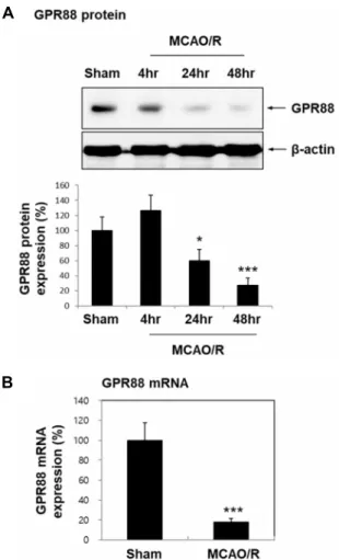
To investigate the potential involvement of GPR88 in is- chemic brain damage, we determined whether the expre- ssion of GPR88 in brain tissue is altered in an ischemic stroke model. GPR88 expression was significantly downregulated in ischemic brains at both the protein (24 hr; 59.66%, p<0.05;
48 hr; 27.24%, p<0.001) and mRNA levels (56.28%, p<0.001)
(Fig. 1). To identify which type of cells among brain cells
exhibits high GPR88 expression, we examined BV2 micro-
A
B C
Fig. 2. GPR88 expression in three types of brain cells and under OGD conditions. (A) Western blots showing GPR88 ex- pression in BV2 microglial cells, HT22 neuronal cells, and brain microvascular endothelial cells (mEC). Data was presented as mean ± SEM (n=4). **p<0.01 versus BV2 cells. (B, C) HT22 cells under OGD conditions for 24 hr were analyzed for GPR88 mRNA expression (B) by RT-PCR (n=3) and protein expression (C) by Western blot (n=3). Data was presented as mean ± SEM. *p<0.05 and ***p<0.001 versus control group.
A
B
Fig. 3. Cell viability reduction in HT22 under OGD conditions.
(A) HT22 cells were cultured under OGD conditions for 24 hr. Magnification: ×200; scale bar=200 μm. (B) Cell viability was determined via MTT assay. Three inde- pendent experiments with triplicates were performed.
Data are presented as mean ± SEM. ***p<0.001 versus the control group.
A
B
Fig. 1. Downregulation of GPR88 after transient MCAO in C57 BL/6 mice. Six-week-old C57BL/6 mice underwent tran- sient focal cerebral ischemia by middle cerebral artery occlusion (MCAO) for 60 min, followed by reperfusion for 4 hr, 24 hr, and 48 hr (MCAO/R). (A) Ipsilateral brain tissues were lysed and analyzed for GPR88 expression by Western blotting (n=5 each). Quantification graph was presented as mean ± SEM. *p<0.05 and ***p<0.001 versus sham group. (B) GPR88 mRNA expression at 60 min MCAO/24 hr reperfusion by real-time PCR. Data was presented as mean ± SEM (n=4). ***p<0.001 versus sham group.
glial cells, HT22 neuronal cells, and brain microvascular en- dothelial cells (mEC) (Fig. 2). GPR88 protein expression was found to be high in HT22 hippocampal neuronal cells (p<
0.01 versus BV2 cells) (Fig. 2A). We confirmed that GPR88 was significantly downregulated in HT22 cells under oxygen glucose deprivation (OGD)-like ischemic conditions (mRNA 33.47%, p<0.001; protein 19.97%, p<0.05) (Fig. 2B, Fig. 2C).
We then investigated the viability of HT22 cells under OGD
conditions (Fig. 3). In comparison with control cells, OGD
cells exhibited reduced confluence (Fig. 3A) and significantly
lower levels of cell viability (32.26%, p<0.001) (Fig. 3B). These
A
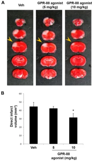
B
Fig. 4. Effect of GPR88 agonist on brain injury. (A) Mice were intraperitoneally injected with the GPR88 agonist RTI- 13951-33 hydrochloride (5 or 10 mg/kg, n=5 each) or PBS (vehicle group, n=5) 30 min prior to ischemic insult.
Representative photographs of brain sections stained with TTC. (B) Quantification of direct infarct volume 24 hr postischemia. *p<0.05 versus vehicle group.
A B
Fig. 5. Effect of GPR88 agonist on brain function and behavior following ischemic brain injury. Neu- rological score (A) and wire-grip tests (B) were performed to evaluate the recovery of neuro- logical deficits and vestibular motor functions after ischemic brain injury. Data was presented as mean ± SEM (n=5~7 each). *p<0.05, ***p<
0.001 versus vehicle group.
results suggest the possible involvement of GPR88 in cell survival, i.e., GPR88 reduction affects cell death.
Effect of GPR88 activation using GPR88 agonist on ischemic brain damage
To determine whether GPR88 activation reduces ische- mia-induced brain damage, mice were intraperitoneally in- jected with RTI-13951-33 hydrochloride (5 or 10 mg/kg), a strong GPR88 agonist, 30 min prior to ischemic injury (Fig.
4, Fig. 5). A significant reduction in infarct volume was ob- served in mice pretreated with 10 mg/kg of GPR88 agonist (31.57±4.67, p<0.05) (Fig. 4). We then investigated the effect of the GPR88 agonist on neurological deficits and motor function. Significant improvements in neurological score (1.43±0.2, p<0.001) and in wire-grip (3±0.2, p<0.05) were ob- served in mice pretreated with 10 mg/kg of GPR88 agonist, relative to those observed in the vehicle group (Fig. 5). These results suggest that GPR88 activation via an agonist may contribute to the amelioration of brain damage following ischemia.
Discussion
In this study, we found that GPR88 expression was de- creased in ischemic brains and in neurons under OGD conditions. GPR88 activation with a GPR88 agonist amelio- rated brain injury following ischemia, as evidenced by im- provements in infarct volume, vestibular-motor function, and movement.
Motor disorders that manifest after a stroke are primarily associated with lesions in the basal ganglia and the thalamus.
The basal ganglia consists of the principal subcortical com-
ponent of the circuits that link the cerebral cortex and the
thalamus [7]. GPR88 is highly expressed in striatal MSN,
which is a major component of the basal ganglia circuitry
striatum [22]. In the absence of GPR88, MSN exhibited en-
hanced excitability, resulting in hyperactivity, poor motor
coordination, and impaired cue-based learning in mice.
Striatal GPR88 re-expression in GPR88
cre/cremice rescued their molecular and electrophysiological abnormalities and led to normalized behavior, suggesting the importance of GPR88 function in the regulation of MSN excitability in neu- rological and psychiatric diseases [19]. We observed that both GPR88 mRNA and protein levels were downregulated following ischemic stroke (Fig. 1). Neurons highly expressed GPR88, but OGD insult led to GPR88 downregulation (Fig.
2). Furthermore, administration of the GPR88 agonist RTI- 13951-33 ameliorated ischemia-induced brain functional de- fects affecting vestibular-motor function and movement (Fig.
5). Therefore, these results suggest that the development of movement disorders after infarction could possibly be due to a decrease in GPR88 expression in the neurons.
The importance of GPR88 in the regulation of striatal function has been reported, and GPR88 has been suggested as a promising drug target for basal ganglia-associated disorders. Transcriptional profiling experiments have re- vealed that GPR88 gene expression is modulated either by treatment or conditions related to schizophrenia [16], bipolar disorder [18], depression [5], and drug addiction [2]. We also found that pretreatment with RTI-13951-33, a GPR88 agonist, reduced infarct volume and restored brain function after is- chemic stroke (Fig. 4, Fig. 5). Jin C et al previously reported RTI-13951-33 as a potent, selective, and brain-penetrant GPR88 agonist, which has the advantage of having a higher aqueous solubility compared to 2-PCCA [2-(Pyridin-2-yl)cy- clopropanecarboxylic acid], another pre-characterized GPR88 agonist [8, 9]. Taken together, although we cannot rule out the possibility that GPR88 has additional roles in brain re- gions other than the striatum, the behavioral restoration ob- served in the treatment groups subjected to administration with a GPR88 agonist following ischemia suggests that GPR88 may be a promising drug target for ischemic stroke recovery.
Data Availability
The dataset supporting the conclusions of this article is included within the article. All data generated or analyzed during this study are included in this published article or are available from the corresponding author upon reason- able request.
Acknowledgments
This work was supported by a 2-Year Research Grant of
Pusan National University. Conceived and designed the ex- periments: S.-Y.L. and H.K.S. Performed the experiments:
J.H.P. and M.J.K. Analyzed the data: J.H.P., M.J.K., B.T.C., S.-Y.L., and H.K.S. Wrote the manuscript: S.-Y.L. and H.K.S.
All authors contributed extensively to this work and ap- proved the final manuscript.
The Conflict of Interest Statement
The authors declare that they have no conflicts of interest with the contents of this article.
References
1. Alper, B. S., Malone-Moses, M., McLellan, J. S., Prasad, K.
and Manheimer, E. 2015. Thrombolysis in acute ischaemic stroke: Time for a rethink? BMJ. 350, h1075.
2. Befort, K., Filliol, D., Ghate, A., Darcq, E., Matifas, A., Muller, J., Lardenois, A., Thibault, C., Dembele, D., Le Merrer, J., Becker, J. A., Poch, O. and Kieffer, B. L. 2008. Mu-opioid receptor activation induces transcriptional plasticity in the central extended amygdala. Eur. J. Neurosci. 27, 2973-2984.
3. Ben Hamida, S., Mendonca-Netto, S., Arefin, T. M., Nasseef, M. T., Boulos, L. J., McNicholas, M., Ehrlich, A. T., Clarke, E., Moquin, L., Gratton, A., Darcq, E., Harsan, L. A., Maldo- nado, R. and Kieffer, B. L. 2018. Increased alcohol seeking in mice lacking gpr88 involves dysfunctional mesocortico- limbic networks. Biol. Psychiatry 84, 202-212.
4. Benjamin, E. J., Blaha, M. J., Chiuve, S. E., Cushman, M., Das, S. R., Deo, R., de Ferranti, S. D., Floyd, J., Fornage, M., Gillespie, C., Isasi, C. R., Jimenez, M. C., Jordan, L. C., Judd, S. E., Lackland, D., Lichtman, J. H., Lisabeth, L., Liu, S., Longenecker, C. T., Mackey, R. H., Matsushita, K., Mozaffar- ian, D., Mussolino, M. E., Nasir, K., Neumar, R. W., Pala- niappan, L., Pandey, D. K., Thiagarajan, R. R., Reeves, M.
J., Ritchey, M., Rodriguez, C. J., Roth, G. A., Rosamond, W.
D., Sasson, C., Towfighi, A., Tsao, C. W., Turner, M. B., Virani, S. S., Voeks, J. H., Willey, J. Z., Wilkins, J. T., Wu, J. H., Alger, H. M., Wong, S. S., Muntner, P., American Heart Association Statistics, C. and Stroke Statistics, S. 2017.
Heart disease and stroke statistics-2017 update: A report from the american heart association. Circulation 135, e146- e603.
5. Conti, B., Maier, R., Barr, A. M., Morale, M. C., Lu, X., Sanna, P. P., Bilbe, G., Hoyer, D. and Bartfai, T. 2007. Region-specif- ic transcriptional changes following the three antidepressant treatments electro convulsive therapy, sleep deprivation and fluoxetine. Mol. Psychiatry 12, 167-189.
6. Ehrlich, A. T., Semache, M., Bailly, J., Wojcik, S., Arefin, T.
M., Colley, C., Le Gouill, C., Gross, F., Lukasheva, V., Hogue, M., Darcq, E., Harsan, L. A., Bouvier, M. and Kieffer, B.
L. 2018. Mapping gpr88-venus illuminates a novel role for gpr88 in sensory processing. Brain Struct. Funct. 223, 1275-
1296.
7. Ghika-Schmid, F., Ghika, J., Regli, F. and Bogousslavsky, J. 1997. Hyperkinetic movement disorders during and after acute stroke: The lausanne stroke registry. J. Neurol. Sci. 146, 109-116.
8. Jin, C., Decker, A. M., Huang, X. P., Gilmour, B. P., Blough, B. E., Roth, B. L., Hu, Y., Gill, J. B. and Zhang, X. P. 2014.
Synthesis, pharmacological characterization, and structure- activity relationship studies of small molecular agonists for the orphan gpr88 receptor. ACS. Chem. Neurosci. 5, 576-587.
9. Jin, C., Decker, A. M., Makhijani, V. H., Besheer, J., Darcq, E., Kieffer, B. L. and Maitra, R. 2018. Discovery of a potent, selective, and brain-penetrant small molecule that activates the orphan receptor gpr88 and reduces alcohol intake. J.
Med. Chem. 61, 6748-6758.
10. Kalaria, R. N., Akinyemi, R. and Ihara, M. 2016. Stroke in- jury, cognitive impairment and vascular dementia. Biochim.
Biophys. Acta 1862, 915-925.
11. Kim, M. J., Lee, S. Y., Hwang, J. Y., Kim, H., Ha, K. T., Choi, B. T., Baek, J. U. and Shin, H. K. 2018. Pretreatment with shuanghe-tang extract attenuates postischemic brain injury and edema in a mouse model of stroke: An analysis of medicinal herbs listed in dongui bogam. Oxid. Med. Cell.
Longev. 2018, 2479602.
12. Kim, M. J., Park, K. H., Lee, J. Y., Ha, K. T., Choi, B. T., Baek, J. U., Yun, Y. J., Lee, S. Y. and Shin, H. K. 2019.
Weisheng-tang ameliorates acute ischemic brain damage in mice by maintaining blood-brain barrier integrity. Oxid.
Med. Cell. Longev. 2019, 4379732.
13. Lin, D. J., Finklestein, S. P. and Cramer, S. C. 2018. New directions in treatments targeting stroke recovery. Stroke 49, 3107-3114.
14. Massart, R., Guilloux, J. P., Mignon, V., Sokoloff, P. and Diaz, J. 2009. Striatal gpr88 expression is confined to the whole projection neuron population and is regulated by dopaminergic and glutamatergic afferents. Eur. J. Neurosci.
30, 397-414.
15. Massart, R., Mignon, V., Stanic, J., Munoz-Tello, P., Becker,
J. A., Kieffer, B. L., Darmon, M., Sokoloff, P. and Diaz, J.
2016. Developmental and adult expression patterns of the g-protein-coupled receptor gpr88 in the rat: Establishment of a dual nuclear-cytoplasmic localization. J. Comp. Neurol.
524, 2776-2802.
16. Matsuoka, T., Tsunoda, M., Sumiyoshi, T., Takasaki, I., Tabuchi, Y., Seo, T., Tanaka, K., Uehara, T., Itoh, H., Suzuki, M. and Kurachi, M. 2008. Effect of mk-801 on gene expre- ssions in the amygdala of rats. Synapse 62, 1-7.
17. Meirsman, A. C., Le Merrer, J., Pellissier, L. P., Diaz, J., Clesse, D., Kieffer, B. L. and Becker, J. A. 2016. Mice lacking gpr88 show motor deficit, improved spatial learning, and low anxiety reversed by delta opioid antagonist. Biol. Psyc- hiatry 79, 917-927.
18. Ogden, C. A., Rich, M. E., Schork, N. J., Paulus, M. P., Geyer, M. A., Lohr, J. B., Kuczenski, R. and Niculescu, A. B. 2004.
Candidate genes, pathways and mechanisms for bipolar (manic-depressive) and related disorders: An expanded con- vergent functional genomics approach. Mol. Psychiatry 9, 1007-1029.
19. Quintana, A., Sanz, E., Wang, W., Storey, G. P., Guler, A.
D., Wanat, M. J., Roller, B. A., La Torre, A., Amieux, P. S., McKnight, G. S., Bamford, N. S. and Palmiter, R. D. 2012.
Lack of gpr88 enhances medium spiny neuron activity and alters motor- and cue-dependent behaviors. Nat. Neurosci.
15, 1547-1555.
20. Rocher, A. B., Gubellini, P., Merienne, N., Boussicault, L., Petit, F., Gipchtein, P., Jan, C., Hantraye, P., Brouillet, E.
and Bonvento, G. 2016. Synaptic scaling up in medium spi- ny neurons of aged bachd mice: A slow-progression model of huntington's disease. Neurobiol. Dis. 86, 131-139.
21. Sesack, S. R. and Grace, A. A. 2010. Cortico-basal ganglia reward network: Microcircuitry. Neuropsychopharmacology 35, 27-47.
22. Van Waes, V., Tseng, K. Y. and Steiner, H. 2011. Gpr88 - a putative signaling molecule predominantly expressed in the striatum: Cellular localization and developmental regu- lation. Basal Ganglia 1, 83-89.
초록:
GPR88 효현제의 전처리에 의한 뇌졸중후 뇌손상 감소효과 연구이서연
1*․박정화
2,3,4․김민재
2,3,4․최병태
2,3,4․신화경
2,3,4*
(1원광대학교 의과대학 약리학교실, 2부산대학교 한의학전문대학원 한의과학과, 3부산대학교 건강노화 한의과학
연구센터, 4부산대학교 건강노화 한의전문인력양성팀)