- 287 -
Inhibitory Effects of Lycopene on the Expression of Pro-inflammatory Genes in Human Vascular Endothelial Cells
Tae Hoon Kim
1and Jong-Sup Bae
2†1
Department of Herbal Medicinal Pharmacology, Daegu Haany University, Gyeongsan 712-715, Korea
2
College of Pharmacy, Research Institute of Pharmaceutical Sciences, Kyungpook National University, Daegu 702-701, Korea
혈관내피세포에서 라이코펜이 염증유전자 발현에 미치는 영향
김태훈
1․배종섭
2†1
대구한의대학교 한약재약리학과,
2경북대학교 약학과, 약학연구소
Abstract
Lycopene, found in tomatoes and tomato products, has antioxidant, anticancer, and anti-inflammatory effects.
High-mobility-group box 1 (HMGB1) mediates the pro-inflammatory responses in several inflammatory diseases.
In this study, the potential roles of lycopene in the HMGB1-mediated pro-inflammatory gene expressions in the primary human-umbilical-vein endothelial cells (HUVECs) were investigated. The data showed that HMGB1 upregulated the expressions of monocyte chemotactic protein 1 (MCP-1), interleukin-6 (IL-6), secretory phospholipase A2 (sPLA2)-IIA, and prostaglandin E2 (PGE2). Lycopene pre-incubation for 6 h decreased the HMGB1-mediated induction of MCP-1, IL-6, sPLA2-IIA, and PGE2. Further study revealed that the inhibitory effects of lycopene on the HMGB-1 induced expression of pro-inflammatory genes were mediated by the inhibition of two important inflammatory cytokines: tumor necrosis factor (TNF)-α and nuclear factor (NF)-κB. These results suggest that HMGB1 upregulated the expression of pro-inflammatory genes and lycopene inhibited HMGB-1-induced pro-inflammatory genes by inhibiting TNF-α and NF-κB. This finding will serve as an important evidence in the development of a new medicine for the treatment of inflammatory diseases.
Key words :lycopene; HMGB1; inflammation; endothelium
서 론
1)
토마토에 함유되어 있는 라이코펜은 항산화효과, 항암효 과와 항염증효과가 있다고 알려져 있다(1). 그리고 토마토 또는 토마토를 재료로 한 음식을 섭취하는 것은 만성혈관질 환, 염증질환 및 암을 예방한다(1). 최근에 라이코펜이 혈관 내피세포의 투과성, 세포부착단백질의 발현 및 백혈구의 혈관내피세포에 대한 부착과 이동을 억제하고 그 기전으로 써 염증을 유발하는데 중요한 두 가지 사이토카인 tumor necrosis factor-α (TNF-α)와 nuclear factor-κB (NF-κB)의 발 현을 억제하는 것이 보고되었다(2).
High mobility group box 1 (HMGB1)은 비히스톤 DNA
†
Corresponding author. E-mail:[email protected] Phone:82-53-950-8570, Fax:82-53-950-8557
결합물질로서, 뉴크레오좀 형성을 안정화(3), 유전자 발현 을 촉진(4), 스테로이드 호르몬 수용체를 조절(5)하는 단백 질이다. HMGB1은 인체내에서 흉선, 림프절, 고환, 태아의 간 등에서 높은 농도로 존재하며, 간세포와 뇌세포를 제외 하면 대부분의 세포에서는 주로 핵 내에 존재하지만, 염증 을 유발하는 물질에 의해 세포나 조직이 활성화되면 세포 밖으로 분비된다(6,7). HMGB1은 염증을 유발하는 사이토 카인이고, HMGB1의 길항제가 항염증물질로 효과가 있다 는 것이 보고되었다(8).
염증반응 초기에 다양한 종류의 물질 예를 들어 각종 사이토카인들이 만들어져 염증을 더욱더 악화시킨다(9).
이러한 염증반응 매개물질에는 케모카인, 사이토카인, 혈 장효소단백질, 지용성 염증매개물질 등을 들을 수 있다.
케모카인은 약 100여개의 아미노산으로 구성된 작은 단백
질들의 집단으로, 선택적으로 백혈구 소집단의 세포접합,
화학유인작용, 활성화 등을 조절하는 사이토카인을 말한다 (10). 이런 케모카인 중 하나인 MCP-1 (monocyte chemotactic protein 1)은 염증성세포들 중 단핵세포, 기억 T세포 및 혈관 내피세포에 작용하는 중요한 화학주성 인자로 염증에 의해 발현이 증가되는 대표적인 물질이다(11). 염증반응을 촉진 하는 사이토카인들은 급성 및 만성 염증반응에서 중요한 역할을 한다. 특히 인터루킨-8 (IL-8)은 염증반응에서 2차 매개단백질 (secondary mediator)로 작용하여 염증세포들을 활성화하고, 그들을 염증부위로 유인하는 화학유인인자 (chemotatic factor, chemokines)의 작용을 가지고 있다(11).
뿐만 아니라, TNF-α는 그람음성세균 감염에 의해서 만들어 지는데, 세균의 세포막에 있는 내독소 (bacterial endotoxin) 인 lipopolysaccharide (LPS)에 의해 활성화된 림프구에 의 해서 만들어진다(12). 중증 염증질환인 패혈증과 같이 LPS 가 다량 존재하게 되면 TNF-α가 많이 만들어져 조직의 손상 이나 전신혈관응고와 같은 심각한 결과를 초래할 수 있다 (12). Nuclear factor-κB (NF-κB) 또한 혈관염증을 매개하는 물질로 잘 알려져 있는데, NF-κB의 자극으로 특정 유전자 발현 속도가 증가하면 관련 효소 및 단백질 생산이 늘어나 서 염증성 프로스타글란딘 및 기타 에이코사노이드 생산을 비정상적으로 증가시킨다(12,13,14,15). NF-κB 활성이 증 가하면 면역계가 과잉 활성 되어 다양한 자가 면역 질환 및 염증 반응이 악화된다(12,13,14,15). 지용성 염증매개물 질인 프로스타글란딘 E2 (PGE2)는 염증에서 아주 중요한 역할을 한다고 알려져 있다(16). PGE2는 phospholipase A2 (PLA2), cyclooxygenase (COX), 프로스타글란딘 E synthase (PGES)등과 같은 효소의 작용에 의해 phospholipid로부터 생성이 된다(16). PGE2는 홍반이나 발열, 통증, 부종을 유발 하여 염증을 유도한다(16). 또 다른 염증매개물질인 분비성 IIA형 phospholipase A2 (secretory phospholipase A2-IIA, sPLA2-IIA)는 염증액과 염증관련 질환의 혈장에서 발견되 며, 염증과 조직손상에 있어서 중요한 역할을 한다고 알려 져 있다(17).
본 연구에서는 HMGB1에 의해서 유도되는 염증을 촉진 하는 물질 (MCP-1, IL-8, sPLA2-IIA, PGE2)이 증가함을 확인하였고, 라이코펜은 HMGB1에 의해 증가되는 물질의 발현을 감소시켰다. 특히 그 기전으로 HMGB1에 의해 증가 하는 NF-κB와 TNF-α의 발현이 라이코펜에 의해 저해됨을 확인하였고, 그 결과를 보고하고자 한다.
재료 및 방법
실험재료
라이코펜과 MTT (3-(4,5-dimethyl-2-yl)-2,5-diphenyltetrazolium bromide)은 Sigma (St Louis, MO, USA)에서, HMGB1은 Abnova (Taipei City, Twiwan)에서 구입하여 사용하였다.
라이코펜은 DMSO에 녹여서 사용하였다.
세포배양
인간제대정맥내피세포 (HUVEC)는 Cambrex Bio Science Inc. (Charles City, IA, USA)에서 구입하여 사용하였고, 세 포의 배양은 이전에 방법대로 배양하였다 (18). HUVEC은 계대배양을 하였고, passage number는 3번에서 5번사이의 세포를 사용하였다. 라이코펜의 처리 농도는 0 - 20 µM로 1회 처리하였다.
세포독성실험
라이코펜이 가지는 세포독성을 측정하기 위해 MTT를 사용하였다. 혈관내피세포 (5 x 10
3개/well)을 하루동안 배양한 후, 농도별 라이코펜을 처리하였다. 48시간 후, 세포 를 씻은 후, 100 µl의 MTT (1 mg/mL)을 넣어주고 4시간 배양하였다. 100% DMSO 3 mL를 첨가하여 세포에 흡수된 MTT formazan을 녹여내고 96-well 배양접시에 100µl 씩분 주하여 540 nm에서 흡광도를 측정하였다.
MCP-1, IL-8, sPLA2-IIA, PGE2, NF-κB 그리고 TNF-α 농도측정을 위한 ELISA 방법
라이코펜을 6시간 처리한 후, HMGB1 (1 µg/ml)을 16시 간 처리한 후, 세포배양액 또는 nuclear lysates을 따로 모은 후, -80℃에 보관하였다. 세포로부터 분비 또는 세포핵에 있는 각종 염증관련 단백질의 농도를 측정하기 위한 ELISA Kit MCP-1 (R&D System, Minneapolis), IL-8 (R&D System, Minneapolis), sPLA2-IIA (Cayman Chemical, Ann Arbor, MI, USA), PGE2 (Cayman Chemical, Ann Arbor, MI, USA), NF-κB (Cell Signaling Technology, Inc, Danvers, MA, USA) 그리고 TNF-α (R&D System, Minneapolis)을 구입하였고, 실험방법은 제조사의 guideline에 따라 진행하였다. MCP-1, IL-8, sPLA2-IIA, PGE2와 TNF-α 농도측정은 세포배양액으 로부터 측정하였고, NF-κB는 nuclear extract로부터 측정하 였다.
웨스턴 브로팅
라이코펜을 6시간 처리한 후, HMGB1 (1 µg/ml)을 16시 간 처리한 후, 세포배양액 또는 nuclear lysates을 따로 모은 후, -80℃에 보관하였다. 정량된 단백질 10 µg을 각 웰에 넣고 10% SDS-PAGE에서 전기영동 하여 polyvinylidene fluoride (PVDF) membrane에 옮겨, blocking 용액 (10%
far-free milk and 0.05% Tween 20), 1차항체 (anti-
phopho-NF-kB 또는 anti-TNF-α, Cell Signaling, MA, USA),
2차항체 (Anti-rabbit IgG-HRP, Cell Signaling, MA, USA)를
차례로 처리한 후 암실에서 enhanced chemiluminescence
(Amersham ECL Plus, NJ, USA)를 이용하여 단백질을 증폭
시켰다. Intra control로써는 actin을 사용하였다.
통계처리
각 실험은 최소 3번 이상 검정하였고, 실험 결과는 평균
± 표준오차로 표시하였고 Student’s t test로 검정하여 p값이 5% 미만일 때 통계적으로 유의 하다고 간주하였다.
결과 및 고찰
라이코펜이 MCP-1과 IL-8의 발현에 미치는 영향
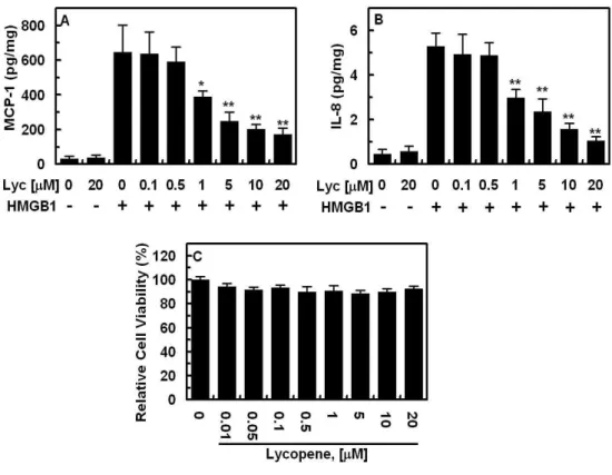
본 연구에서는 인간제대정맥내피세포 (HUVEC)를 사용 하였다. 인간제대정맥내피세포는 염증을 유발하는 각종물 질에 민감하게 반응하기 때문에 혈관염증을 연구하는데 많이 사용된다. MCP-1와 IL-8은 혈관내피세포에서 분비되 며 염증과정에서 중요하게 작용을 하고, 염증자체를 유발 한다고 알려져 있다. 염증을 유발하는 물질인 HMGB1 (1 µg/ml)을 HUVEC에 16시간 전 처리했을 경우, MCP-1와 IL-8의 발현이 증가하였으며, 라이코펜만 처리했을 경우에 는 MCP-1와 IL-8의 발현이 변화가 없었다(Fig. 1). 라이코펜 을 전 처리했을 경우에는 HMGB1에 의해 증가되는 MCP-1 와 IL-8의 발현이 농도의존적으로 감소하였다(Fig. 1). 이와 같은 결과가 라이코펜이 가지는 세포독성 때문에 기인하는 것을 배제하기 위해 라이코펜이 가지는 세포독성을 측정하
Fig. 1. Effect of lycopene on HMGB1-mediated MCP-1 or IL-8 expression in HUVECs.
HUVECs were stimulated with HMGB1 (1 µg/ml, B, C) for 16 h after treating the cell monolayer with the indicated concentrations of lycopene for 6 h. The release of MCP-1 (A) or IL-8 (B) was measured by ELISA as described under “Materials and methods”. (C) effect of lycopene on cellular viability as described under “Materials and Methods”.
All results are shown as mean ± SD of three different experiments. All results are shown as the means ± SD of different three experiments. * p<0.05 and **p<0.01 as compared to HMGB1 alone.
였다(Fig. 1C). 그 결과 라이코펜은 세포에 아무런 독성을 가지지 않는다. 따라서 라이코펜은 세포에 아무런 독성을 가지지 않으면서 혈관염증조건에서 발현이 증가하는 단백 질인MCP-1과 HMGB1의 발현을 저해한다.
라이코펜이 sPLA2-IIA과 PGE2의 발현에 미치는 영향
sPLA2-IIA는 패혈증, 위장염증질환, 급성 췌장염, 류머 티즘성 관절염 등 각종 염증질환과 밀접한 관련이 있다(19).
그리고 패혈증과 패혈쇼크를 포함하는 혈관염증질환 환자 의 혈액샘플에서 많은 양의 sPLA2-IIA이 발견되었다 (20,21,22). 이것은 염증질환에서 sPLA2-IIA의 중요한 역할 을 시사한다.
HMGB1 (1 µg/ml)을 HUVEC에 16시간 전 처리했을 경 우, sPLA2-IIA이 분비되는 양은 증가하였으며, 라이코펜만 처리했을 경우에는 sPLA2-IIA의 분비량의 변화는 없었다 (Fig. 2,A). 라이코펜을 전 처리했을 경우에는 HMGB1에 의해 증가되는 sPLA2-IIA의 양이 농도의존적으로 감소하 였다 (Fig. 2A).
sPLA2-IIA의 활성도는 PGE2의 생성에 상당히 중요하다
(23). HMGB1은 sPLA2-IIA의 분비량을 촉진하였기 때문
에, PGE2의 생성량에 영향을 줄 것이다. 이것을 검증하기
위하여, HMGB1 (1 µg/ml)을 HUVEC에 16시간 전 처리하
Fig. 2. Effect of lycopene on HMGB1-mediated sPLA2-IIA or PGE2 expressions in HUVECs.
HUVECs were stimulated with HMGB1 (1 µg/ml, B, C) for 16 h after treating the cell monolayer with the indicated concentrations of lycopene for 6 h. The release of sPLA2-IIA (A) or PGE2 (B) was measured by ELISA as described under “Materials and methods”. All results are shown as the means ± SD of different three experiments. * p<0.05 and **p<0.01 as compared to HMGB1 alone.
고 세포배양액에서 PGE2의 양을 측정하였다. HMGB1 은 충분히 PGE2의 생성량을 촉진시켰다(Fig. 2B). 다음으 로 라이코펜이 HMGB1에 의해 증가되는 PGE2의 양에 미 치는 영향을 알아보기 위해, HMGB1을 처리하기 전에 라이 코펜을 처리하였다. 라이코펜만 처리했을 경우에는 PGE2 의 생성양에는 아무런 영향을 미치지 못했으며(Fig. 2B), 라이코펜은 HMGB1에 의해 증가되는 PGE2의 양은 농도의 존적으로 감소하였다(Fig. 2B). 이것은 라이코펜이 sPLA-2IIA와 PGE2의 유전자 발현을 감소시키는 작용으로 라이코펜의 항염증기전을 지지해주는 결과이다.
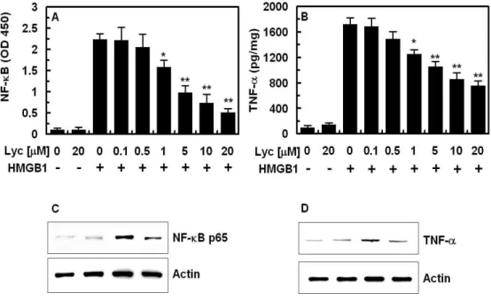
Fig. 3. Effect of lycopene on HMGB1-mediated NF-κB or TNF-α expressions in HUVECs.
HUVECs were stimulated with HMGB1 (1 µg/ml, B, C) for 16 h after treating the cell monolayer with the indicated concentrations of lycopene for 6 h. The activation of NF-κB (A, C) or TNF-α (B, D) was measured by ELISA (A, B) or westernn blotting (B, D) as described under “Materials and methods”. All results are shown as the means
± SD of different three experiments. * p<0.05 and **p<0.01 as compared to HMGB1 alone.
라이코펜이 NF-κB의 활성과 TNF-α의 발현에 미치는 영향
NF-κB는 염증을 유발하는 염증성 전자인자로 잘 알려져 있다(13,14,15). 염증반응에서 필수적인 요소인 NF-κB의 활성을 HMGB1을 처리한 HUVEC에서 측정하였다.
HMGB1은 NF-κB의 활성도를 충분히 증가시켰다 (Fig.
3A). 라이코펜이 HMGB1에 의해 증가되는 NF-κB의 활성
에 미치는 영향을 알아보기 위해 HMGB1을 처리하기 전에
라이코펜을 처리하였다. 라이코펜만 처리했을 경우에는
NF-κB의 활성에는 아무런 영향을 미치지 못했으며, 라이코
펜은 HMGB1에 의해 증가되는 NF-κB의 활성을 농도의존
적으로 감소하였다(Fig. 3A).
TNF-α는 NF-κB에 의해 발현이 증가하여 염증반응을 유 발시키고, 동맥경화증을 포함하는 혈관염증에서 중요한 역 할을 한다(24,25,26). 그래서 HMGB1을 처리한 HUVEC에 서 TNF-α의 발현양을 측정하였다. 그 결과 HMGB1은 TNF- α 의 발현양을 충분히 증가시켰다(Fig. 3B). 라이코펜이 HMGB1에 의해 증가되는 TNF-α의 발현양에 미치는 영향 을 알아보기 위해, HMGB1을 처리하기 전에 라이코펜을 처리하였다. 라이코펜만 처리했을 경우에는 TNF-α의 발현 양에는 아무런 영향을 미치지 못했으며, 라이코펜은 HMGB1에 의해 증가되는 TNF-α의 발현양을 농도의존적 으로 감소하였다(Fig. 3B). 이것은 라이코펜이 HMGB1에 의해 증가하는 각종 염증관련 물질들의 발현을 저해하는 기전으로 NF-κB와 TNF-α의 발현을 저해함으로써 이루어 진다는 것을 시사한다.
1999년도에 HMGB1이 염증을 유발하며, HMGB1의 길 항제가 패혈증에서 치료효과를 보인다는 사실이 보고되었 다(8). HMGB1의 수용체로는 RAGE (receptor for advanced glycation end-products), Toll like receptor 2와 4(TLR2와 TLR4)가 알려져 있다(27,28). HMGB1과 수용체와의 결합 은 NF-κB를 활성화시키며, TNF-α, IL-6 그리고 IL-8같은 cytokine의 분비를 촉진시키고 결국 염증반응을 확대 증폭 시킨다(29). 이러한 작용기전을 고려하면 HMGB1이 패혈 증과 같은 전신 염증질환에서 중요한 역할을 시행할 가능성 이 높다. 기존에 알려진 염증매개 사이토카인인 TNF-α나 IL-1β등은 패혈증의 초기에 최고치에 이른 뒤에 바로 혈중 치가 정상화되기 때문에 TNF-α나 IL-1β 등에 대한 길항제 를 패혈증의 치료에 사용하려는 시도들은 치료시기의 범위 가 너무 좁기 때문에 모두 실패하였다. 반면에 은 패혈증의 후기에 분비가 최고조에 달하기 때문에 치료시기의 범위가 넓은 장점이 있어서 HMGB1에 대한 길항제가 패혈증의 치료제로서의 유용성이 높을것으로 예상된다(8,30). 따라 서 본 연구에서 HMGB1에 의해 증가되는 각종 염증관련물 질이 라이코펜에 의해 감소되는 것은 향후 라이코펜이 혈관 염증질환을 치료하는 후보물질이 될 수 있음을 시사한다.
뿐만 아니라, 토마토 또는 토마토를 재료한 음식을 섭취하 는 것은 혈관염증질환을 예방하는 효과도 기대할 수 있다.
요 약
본 연구에서는 HMGB1 의해 증가되는 각종 염증관련 물질에 대해 라이코펜이 가지는 저해 역할을 규명하고 하 였다.
라이코펜은 HMGB1에 의해 증가되는 MCP-1, IL-8, sPLA2-IIA, PGE2의 발현을 NF-κB 그리고 TNF-α의 활성를 저해함으로써 감소시켰다. 특히, 1 mM에서 그 효능이 통계
적으로 유효하였다. 결론적으로 HMGB1에 의해서 발생하 는 각종 혈관염증질환에서 라이코펜은 증가하는 각종 염증 관련물질을 저해하였고, 결국 라이코펜이 패혈증을 포함하 는 염증질환을 효과적으로 치료할 수 있는 방법에 있어 많은 방향성을 제시할 것으로 기대한다.
감사의 글
이 논문은 2011학년도 경북대학교 신임교수정착연구비 에 의하여 연구되었음.
참고문헌
1. Heber D, Lu QY (2002) Overview of mechanisms of action of lycopene. Exp Biol Med (Maywood), 227, 920-923
2. Bae JW, Bae JS (2011) Barrier protective effects of lycopene in human endothelial cells. Inflamm Res, 60, 751-758
3. Goodwin GH, Sanders C, Johns EW (1973) A new group of chromatin-associated proteins with a high content of acidic and basic amino acids. Eur J Biochem, 38, 14-19 4. Bustin M, Reeves R (1996) High-mobility-group chromosomal proteins: architectural components that facilitate chromatin function. Prog Nucleic Acid Res Mol Biol, 54, 35-100
5. Boonyaratanakornkit V, Melvin V, Prendergast P, Altmann M, Ronfani L, Bianchi ME, Taraseviciene L, Nordeen SK, Allegretto EA, Edwards DP (1998) High-mobility group chromatin proteins 1 and 2 functionally interact with steroid hormone receptors to enhance their DNA binding in vitro and transcriptional activity in mammalian cells. Mol Cell Biol, 18, 4471-4487
6. Mosevitsky MI, Novitskaya VA, Iogannsen MG, Zabezhinsky MA (1989) Tissue specificity of nucleo-cytoplasmic distribution of HMG1 and HMG2 proteins and their probable functions. Eur J Biochem, 185, 303-310
7. Andersson U, Tracey KJ (2011) HMGB1 is a therapeutic target for sterile inflammation and infection. Annu Rev Immuno, l29, 139-162
8. Wang H, Bloom O, Zhang M, Vishnubhakat JM,
Ombrellino M, Che J, Frazier A, Yang H, Ivanova S,
Borovikova L, Manogue KR, Faist E, Abraham E,
Andersson J, Andersson U, Molina PE, Abumrad NN, Sama A, Tracey KJ (1999) HMG-1 as a late mediator of endotoxin lethality in mice. Science, 285, 248-251 9. Esmon CT, Fukudome K, Mather T, Bode W, Regan LM, Stearns-Kurosawa DJ, Kurosawa S (1999) Inflammation, sepsis, and coagulation. Haematologica, 84, 254-259
10. Alon R, Shulman Z (2011) Chemokine triggered integrin activation and actin remodeling events guiding lymphocyte migration across vascular barriers. Exp Cell Res, 317, 632-641
11. Castellani ML, De Lutiis MA, Toniato E, Conti F, Felaco P, Fulcheri M, Theoharides T C, Caraffa A, Antinolfi P, Conti P, Cuccurullo C, Ciampoli C, Felaco M, Orso C, Salini V, Cerulli G, Kempuraj D, Tete S, Shaik B (2010) Impact of RANTES, MCP-1 and IL-8 in mast cells. J Biol Regu lHomeost Agents, 24, 1-6
12. Bradle JR (2008) TNF-mediated inflammatory disease.
J Pathol, 214, 149-160
13. Javaid K, Rahman A, Anwar KN, Frey RS, Minshall RD, Malik AB (2003) Tumor necrosis factor-alpha induces early-onset endothelial adhesivity by protein kinase Czeta-dependent activation of intercellular adhesion molecule-1. Circ Res, 92, 1089-1097.
14. Lockyer JM, Colladay JS, Alperin-Lea WL, Hammond T, Buda AJ (1998) Inhibition of nuclear factor-kappaB-mediated adhesion molecule expression in human endothelial cells. Circ Res, 82, 314-320 15. Marui N, Offermann MK, Swerlick R, Kunsch C, Rosen
CA, Ahmad M, Alexander R W, Medford RM (1993) Vascular cell adhesion molecule-1 (VCAM-1) gene transcription and expression are regulated through an antioxidant-sensitive mechanism in human vascular endothelial cells. J Clin Invest, 92, 1866-1874 16. Jaulmes A, Thierry S, Janvier B, Raymondjean M,
Marechal V (2006) Activation of sPLA2-IIA and PGE2 production by high mobility group protein B1 in vascular smooth muscle cells sensitized by IL-1beta. FASEB J, 20, 1727-1729
17. Vadas P, Pruzanski W (1986) Role of secretory phospholipases A2 in the pathobiology of disease. Lab Invest, 55, 391-404
18. Bae JS, Rezaie AR (2008) Protease activated receptor 1 (PAR-1) activation by thrombin is protective in human pulmonary artery endothelial cells if endothelial protein C receptor is occupied by its natural ligand. Thromb Haemost, 100, 101-109
19. Dennis EA (1997) The growing phospholipase A2 superfamily of signal transduction enzymes. Trends Biochem Sci, 22, 1-2
20. Nakano T, Ohara O, Teraoka H, Arita H (1990) Group II phospholipase A2 mRNA synthesis is stimulated by two distinct mechanisms in rat vascular smooth muscle cells. FEBS Lett, 261, 171-174
21. Oka S, Arita H (1991) Inflammatory factors stimulate expression of group II phospholipase A2 in rat cultured astrocytes. Two distinct pathways of the gene expression.
J Biol Chem, 266, 9956-9960
22. Menschikowski M, Hagelgans A, Siegert G (2006) Secretory phospholipase A2 of group IIA: is it an offensive or a defensive player during atherosclerosis and other inflammatory diseases? Prostaglandins Other Lipid Mediat, 79, 1-33
23. Hajjar DP, Pomerantz KB (1992) Signal transduction in atherosclerosis: integration of cytokines and the eicosanoid network. FASEB J, 6, 2933-2941
24. Branen L, Hovgaard L, Nitulescu M, Bengtsson E, Nilsson J, Jovinge S (2004) Inhibition of tumor necrosis factor-alpha reduces atherosclerosis in apolipoprotein E knockout mice. Arterioscler Thromb Vasc Biol, 24, 2137-2142
25. Li Y, Schwabe RF, DeVries-Seimon T, Yao PM, Gerbod-Giannone MC., Tall AR, Davis RJ, Flavell R, Brenner DA, Tabas I (2005) Free cholesterol-loaded macrophages are an abundant source of tumor necrosis factor-alpha and interleukin-6: model of NF-kappaB- and map kinase-dependent inflammation in advanced atherosclerosis. J Biol Chem, 280, 21763-21772 26. Stoll LL, Denning GM, Weintraub NL (2006) Endotoxin,
TLR4 signaling and vascular inflammation: potential therapeutic targets in cardiovascular disease. Curr Pharm Des, 12, 4229-4245
27. Bonaldi T, Talamo F, Scaffidi P, Ferrera D, Porto A, Bachi A, Rubartelli A, Agresti A, Bianchi ME (2003) Monocytic cells hyperacetylate chromatin protein HMGB1 to redirect it towards secretion. EMBO J, 22, 5551-5560
28. Par, JS, Svetkauskaite D, He Q, Kim JY, Strassheim D, Ishizaka A, Abraham, E (2004) Involvement of toll-like receptors 2 and 4 in cellular activation by high mobility group box 1 protein. J Biol Chem, 279, 7370-7377 29. Andersson U, Wang H, Palmblad K, Aveberger AC,
Bloom O, Erlandsson-Harris H, Janson A, Kokkola R,
Zhang M, Yang H, Tracey KJ (2000) High mobility group
1 protein (HMG-1) stimulates proinflammatory cytokine synthesis in human monocytes. J Exp Med, 192, 565-570 30. Wang H, Yang H, Czura CJ, Sama AE, Tracey KJ (2001)
HMGB1 as a late mediator of lethal systemic inflammation. Am J Respir Crit Care Med, 164, 1768-1773
(접수 2011년 11월 26일 수정 2012년 2월 3일 채택 2012년 2월 17일)