Coil-Protected Embolization Technique for a Branch-Incorporated Aneurysm
Yon-Kwon Ihn, MD
1, Byung Moon Kim, MD, PhD
2, Sang Hyun Suh, MD
3, Dong Joon Kim, MD
2, Dong Ik Kim, MD
21Department of Radiology, The Catholic University of Korea College of Medicine, St. Vincent’s Hospital, Suwon 442-723, Korea; 2Department of Radiology, Yonsei University College of Medicine, Severance Hospital, Seoul 120-752, Korea; 3Department of Radiology, Yonsei University College of Medicine, Gangnam Severance Hospital, Seoul 135-720, Korea
Objective: A small branch-incorporated aneurysm is an aneurysm with a small branch incorporated into the sac or the neck.
It is one of the most difficult aneurysms to treat with coil embolization. The aim of this study was to evaluate the safety and effectiveness of the coil-protected embolization technique for small-branch incorporated aneurysm.
Materials and Methods: Fourteen aneurysms (2 ruptured and 12 unruptured) in 12 patients (mean age, 56 years, range, 40-73 years; 6 men and 6 women) were treated with the coil-protected embolization technique during the period between February 2007 and October 2011. Clinical and angiographic outcomes were retrospectively evaluated.
Results: All aneurysms were successfully treated without any complications during the procedure. Immediate post- treatment angiographies demonstrated complete or near complete occlusion in 12 and incomplete occlusion in 2 patients.
Two patients had a delayed small embolic infarction in the relevant posterior circulation territory and middle cerebral artery territory 10 days and 14 days later, respectively, but both recovered completely or almost completely (modified Rankin scale score [mRS score], 0 and 1, respectively). During the clinical follow-up period (mean, 21 months; range: 2-58 months), all patients reported an mRS score of 0 (n = 10) or 1 (n = 2). Vascular imaging follow-up (catheter angiography: n
= 3 and MR angiography: n = 8) was available in 11 aneurysms at 6-12 months. All 11 aneurysms showed complete occlusion except for 1 minor neck recurrence that did not require further treatment.
Conclusion: In this series of cases, the coil-protected embolization technique seems to be feasible and effective in the treatment of small-branch incorporated aneurysms.
Index terms: Intracranial aneurysm; Coil embolization; Coil protection
Received May 1, 2012; accepted after revision August 8, 2012.
This study was supported by a grant (No. A085136) of the Korea Healthcare Technology R&D Project, Ministry for Health, Welfare &
Family Affairs, Republic of Korea.
Corresponding author: Byung Moon Kim, MD, PhD, Department of Radiology, Yonsei University College of Medicine, Severance Hospital, 50 Yonsei-ro, Seodaemun-gu, Seoul 120-752, Korea.
• Tel: (822) 2228-7400 • Fax: (822) 393-3035
• E-mail: [email protected]
This is an Open Access article distributed under the terms of the Creative Commons Attribution Non-Commercial License (http://creativecommons.org/licenses/by-nc/3.0) which permits unrestricted non-commercial use, distribution, and reproduction in any medium, provided the original work is properly cited.
Korean J Radiol 2013;14(2):329-336
INTRODUCTION
Coil embolization has been increasingly used to treat intracranial ruptured or unruptured aneurysms. The rapid development of devices and embolization techniques have allowed endovascular treatment to be applied to more complex aneurysms that were previously impossible to be treated via endovascular methods. However, a small branch-incorporated aneurysm still remains a challenge for endovascular treatment (1, 2). Although a balloon or stent is very useful in treating wide-necked aneurysms, sometimes it cannot protect the incorporated small branch into the
pISSN 1229-6929 · eISSN 2005-8330
size, 4.1 mm; range, 2.5-7 mm) were treated with the coil- protected embolization technique in 2 referral hospitals during the period between February 2007 and October 2011.
A small branch (≤ 1 mm in diameter)-incorporated aneurysm is an aneurysm with a small branch incorporated into the sac or neck (1, 2). The locations of aneurysms under study were in the middle cerebral artery (MCA) bifurcation and M1 portion in 8, anterior communicating artery in 2, vertebral artery - posterior inferior cerebellar artery (PICA) origin in 2, anterior cerebral artery in the A2 portion in 1, and basilar artery - superior cerebellar artery (SCA) origin in 1 (Table 1).
Patients with unruptured small aneurysm were decided to treat after discussing with the responsible neurosurgeons for probable increased risk of aneurysm rupture (past history or family history of subarachnoid hemorrhage or simultaneous coil embolization of multiple intracranial aneurysms).
Medical records and imaging studies were reviewed to obtain the relevant clinical and radiographic information.
microcatheter for coiling. While a small-sized coil (helical or 3D shape, 1.5 or 2 mm in diameter and 2-6 cm in length) was deployed through the catheter facing the incorporated branch origin for protection, the initial coil frame was made with another catheter and coil (Fig. 1C). Just after making the initial coil frame, the protected coil was carefully retrieved (Fig. 1D), and angiography was performed to confirm the preservation of the incorporated branch and stability of the coil basket (Fig. 1E). Once the stability of the coil frame and preservation of the incorporated branch were confirmed, further coil embolization was completed by repeating the procedure.
RESULTS
All aneurysms were successfully embolized without any treatment-related complications (Table 1). Balloon- remodeling technique, catheter-protected technique, or Table 1. Clinical and Angiographic Results of Coil-Protected Embolization for Branch-Incorporated Aneurysms
No Age Present Loc. Size No. of Aneurysm Post-Tx Angio mRS (months) Fu Angio (months)
1 57 SAH PICA, Rt 5 1 Complete 1 (58) Minor recurrence (12)
2 61 Unrupture MCA, Rt 7 1 Near complete 0 (58) Complete (12)
3 43 Unrupture MCA, Rt 4 1 Complete 0 (33) Complete (6)
4 55 Unrupture MCA, Rt 3 2 Incomplete 0 (20) Complete (6)
5 69 SAH MCA, Rt 4 1 Complete 0 (20) Complete (6)
6 57 Unrupture PICA, Rt 4.5 3 Complete 1 (20) Complete (7)
7 40 Unrupture MCA, Rt 3 2 Incomplete 0 (16) Complete (6)
Unrupture MCA, Lt 4.2 Complete Complete (6)
8 45 Unrupture MCA, Lt 5.2 5 Complete 0 (9) Complete (6)
9 68 Unrupture Acom 6 1 Complete 0 (8) Complete (6)
10 53 Unrupture SCA, Lt 2.5 2 Near complete 0 (7) Complete (6)
11 73 Unrupture MCA, Rt 2.5 3 Complete 0 (6) NA
12 52 Unrupture Acom 3 2 Near complete 0 (2) NA
Unrupture A2, Lt 3.5 Complete NA
Note.— Loc. = location of aneurysm, Post-Tx angio = degree of aneurysm occlusion on immediate post-treatment angiogram, mRS = modified Rankin Scale score, Fu angio = degree of aneurysm occlusion on follow-up angiogram, PICA = posterior inferior cerebellar artery, MCA = middle cerebral artery, Acom = anterior communicating artery, Rt = right, Lt = left, NA = not applicable
a 2-catheter technique was initially attempted but all failed to preserve the incorporated branch in 8 aneurysms.
The coil-protected technique was then used, resulting in successful coil embolization with preservation of the incorporated branch. The remaining 4 aneurysms were initially treated with the coil-protected technique. Seven patients underwent simultaneous coil embolization for 2 or 3 intracranial aneurysms. Five patients, who were treated in the earlier period, did not receive post-treatment antiplatelet medication but the remaining 7 patients received post-treatment antiplatelet medication (aspirin 100 mg or plavix 75 mg daily) for 2-4 weeks. Two of the 5 patients who did not receive post-treatment antiplatelet medication had a delayed embolic infarction in the relevant posterior circulation territory and MCA territory 10 and 14 days later, respectively; however, both patients recovered
completely or near completely (modified Rankin scale score [mRS score], 0 and 1, respectively).
Immediate post-treatment angiography revealed complete or near complete occlusion in 12 aneurysms and incomplete occlusion in 2. During the clinical follow-up period (mean:
21 months; range: 2-58 months), all patients reported an mRS score of 0 (n = 10) or 1 (n = 2). Vascular imaging follow-up (catheter angiography; n = 3 and MR angiography:
n = 8) were available in 11 aneurysms at 6-12 months. All 11 aneurysms showed complete occlusion except for 1 minor neck recurrence that did not require further treatment.
Illustrative Cases Case 4 (Fig. 1)
A 55-year-old patient was referred for treatment of an A
D
B
E
C
F
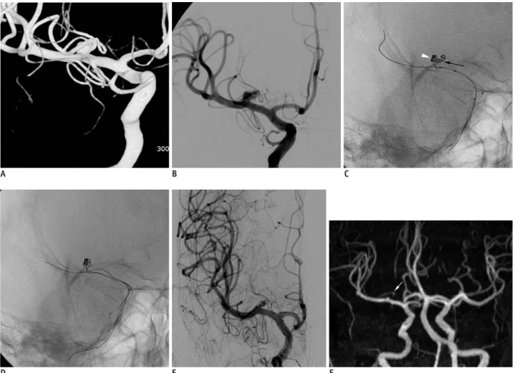
Fig. 1. Case 4. 55-year-old patient with unruptured aneurysm at right middle cerebral artery.
A, B. 3D volume rendering (A) and working projection images (B) show small aneurysm with incorporated branch from which lenticulostriate arteries arises. C. While incorporated branch is protected by 3D 2/4 coil mass (black arrow) introduced through catheter facing its origin, initial coil frame (white arrowhead) is made by another coil through another microcatheter. D. After retrieval of protection coil, initial coil frame is stable. E.
Post-treatment angiography shows near complete embolization of aneurysm sac with well-preserved incorporated branch. F. Six-month follow-up angiography reveals complete occlusion of aneurysm sac and preserved incorporated branch (white arrow). 3D = 3-dimensional
a guglielmi detachable coil 360 2/4 coil, which allowed ICA aneurysms were treated with balloon-assisted coiling
A
C
B
D
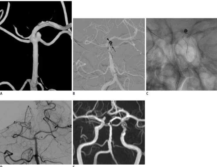
Fig. 2. Case 6. 57-year-old patient presenting with bilateral distal internal carotid artery aneurysms and right posterior inferior cerebellar artery aneurysm.
A. Right vertebral angiogram in working projection shows small aneurysm with left posterior inferior cerebellar artery incorporated into aneurysm neck. B. Initial coil basket is successfully made with coil-protection technique using helical 2/4 coil. C. Final control angiogram shows complete embolization with well-preserved posterior inferior cerebellar artery. D. Six-month follow-up angiogram reveals complete occlusion of aneurysm sac (asterisk) with well-preserved right posterior inferior cerebellar artery (arrow).
(not shown), and the left PICA aneurysm was treated with the coil-protected technique in the same session after failing the balloon-assisted technique. Seven-month follow-up angiography showed complete occlusion of all 3 aneurysms.
Case 10 (Fig. 3)
A 53-year-old patient was referred for the treatment of a left distal ICA and basilar artery-SCA aneurysm. The left distal ICA aneurysm was treated with stent-assisted coiling (not shown). The SCA aneurysm was then embolized with the coil-protected technique in the same session. Six- month follow-up angiography revealed complete occlusion of both aneurysms.
DISCUSSION
Endovascular coil embolization of intracranial aneurysms has made remarkable progress over the last decade and has been accepted as an alternative to surgical clipping showing lower morbidity and mortality rates (3). Owing to recent advances and developments of new devices and techniques for coil embolization, very small aneurysms or wide-neck aneurysms are no longer the contraindications to coiling (4-9). However, aneurysms with an incorporated branch are still regarded as relative contraindications to coil embolization because protecting the small branch arising from the neck or sac of aneurysms remains technically challenging (1, 2). Recently, Kim et al. (1) A
D
B
E
C
Fig. 3. Case 10. 53-year-old patient presenting with unruptured left distal internal carotid artery (ICA) aneurysm and left superior cerebellar aneurysm.
A. Three-dimensioal reconstruction image reveals small aneurysm with left superior cerebellar artery incorporated into aneurysm neck. B. Initial coil frame (arrowhead) is made with coil-protection (arrow) technique using helical 2/4 coil. C. After retrieval of protection coil, coil frame is stable. D. Final control angiogram reveals complete occlusion of aneurysm with preserved left superior cerebellar artery. Black asterisk indicates coil embolized left distal ICA aneurysm. E. Seven-month follow-up MR angiogram shows complete occlusion of aneurysm sac (white asterisk) and well-preserved left superior cerebellar artery.
these techniques may also be limited in treating aneurysms with an incorporated branch. Therefore, we developed a coil-protected, coil embolization technique and evaluated its efficacy and safety as an alternative to balloon- remodeling, 2-catheter, and catheter-protected techniques in the treatment of branch-incorporated aneurysms. To our knowledge, similar techniques have been described only in 2 studies in the previously reported literature (1, 4).
The coil-protected embolization technique is particularly useful in treating aneurysms that have an incorporated branch arising with an acutely inverse angle to the parent artery axis. In such cases, navigation of a microcatheter to the incorporated branch is extremely difficult and risky. In contrast, in the current case studies, whenever the tip of the microcatheter was appropriately shaped and faced the origin of the incorporated branch, the coil loop or mass could be smoothly formed or introduced into the origin of the branch. In turn, this coil loop or mass interferes with moving of the framing coil loop toward the incorporated branch. After a stable coil frame had been formed without compromise of the incorporated branch, the protection coil was carefully retrieved to confirm the stability of the framing coil. In some cases, the protection coil and framing coil were deployed alternatively rather than sequentially to increase the effect of interference with each other, resulting in the prevention of both movement of the framing coil loops toward the incorporated branch and that of the protection coil loops toward the aneurysm dome.
This technique has several advantages over the balloon or stent-assisted technique. First, the devices required for the coil embolization technique are the same as those used in conventional coil embolization. No additional femoral puncture is required, and the use of one 6 Fr guiding catheter is conventional. Second, this coil embolization technique can achieve coil stability without compromising the side branch. Various factors can influence coil stability,
mentioned above. We preferred using a smaller diameter coil for branch protection when possible. We were able to use this coil as a filling coil after making the stable coil framing basket in most of our cases. Another advantage of this technique is that it can be readily changed to a 2-catheter technique whenever needed. Once the stability of the intra-aneurysmal coil frame and preservation of the incorporated branch are confirmed, embolization of the residual aneurysmal sac can be completed by repeating the procedure or by changing to the 2-catheter technique.
Finally, the advantage of this novel coil embolization technique is that pre- or postprocedural antiplatelet aggregation therapy is not necessarily required. Therefore, following surgical procedures in patients with ruptured aneurysms, external ventricular drainage or decompressive craniotomy can be performed after endovascular treatment.
There may be potential complications associated with coil-protected embolization including thrombo-embolism or coil stretching during the procedure. However, in this study, we did not encounter any thrombo-embolisms during the procedure. This is likely related to technical considerations of the procedure. That is, the protection coil loops were not wedged into the artery to preserve antegrade blood flow through the incorporated branch and it was flushed with heparinized saline during the entire procedure. Another potential problem associated with coil manipulation is coil interlocking. However, it should be noted that that we have not yet experienced such problems. However, such problems in coil manipulation may exist and should be kept in mind.
The overall incidence of thromboembolic complications with balloon- and stent-assisted techniques ranges from 4% to 14% and from 0% to 21%, respectively (13- 18). Considering that the aneurysms in this study were of a branch-incorporated type, an increased risk of thromboembolic complications was expected. A previous study also suggested that protection of the incorporated
branch is one of the most important factors that determine the clinical outcome of the disease (2). This thromboembolic complication may be prevented by the proper use of antiplatelet agents. In this case series, 2 of 5 patients who did not receive post-treatment antiplatelet medication suffered a delayed small infarction in the relevant region supplied by the incorporated branch. In contrast, the remaining 7 patients who received post- treatment antiplatelet medication for 2-4 weeks did not experience such delayed thromboembolic complications. The delayed infarctions in the 2 patients might have been due to a delayed distal embolization of a non-detected small thrombus close to the origin of the incorporated branch.
Thus, post-treatment antiplatelet medication may be helpful in preventing such delayed embolization.
There are some limitations to the present case series. Due to the retrospective nature of this study, selection bias, which may have affected the results, cannot be overlooked.
However, all data on aneurysms with endovascular treatment had been recorded prospectively into a database; therefore, selection bias was likely minimized. Our case series showed that the coil-protected embolization technique is likely a feasible and safe option for the treatment of branch- incorporated aneurysms. This technique would be a useful alternative to traditional remodeling or protective techniques in cases with anatomical contraindications to balloon- or stent-assisted techniques, such as acute angulated parent arteries which are risky to navigate with protective devices or small side-branching arteries that are not able to be preserved by a balloon, stent or catheter.
Conclusion
In this small case series, coil-protected embolization appears to be a feasible and safe alternative option for the treatment of branch-incorporated aneurysms that are not untreatable with other techniques.
REFERENCES
1. Kim BM, Park SI, Kim DJ, Kim DI, Suh SH, Kwon TH, et al. Endovascular coil embolization of aneurysms with a branch incorporated into the sac. AJNR Am J Neuroradiol 2010;31:145-151
2. Lubicz B, Lefranc F, Levivier M, Dewitte O, Pirotte B, Brotchi J, et al. Endovascular treatment of intracranial aneurysms with a branch arising from the sac. AJNR Am J Neuroradiol 2006;27:142-147
3. Qureshi AI, Janardhan V, Hanel RA, Lanzino G. Comparison
of endovascular and surgical treatments for intracranial aneurysms: an evidence-based review. Lancet Neurol 2007;6:816-825
4. Kwon OK, Kim SH, Kwon BJ, Kang HS, Kim JH, Oh CW, et al.
Endovascular treatment of wide-necked aneurysms by using two microcatheters: techniques and outcomes in 25 patients.
AJNR Am J Neuroradiol 2005;26:894-900
5. Pierot L, Cognard C, Anxionnat R, Ricolfi F; CLARITY Investigators. Remodeling technique for endovascular treatment of ruptured intracranial aneurysms had a higher rate of adequate postoperative occlusion than did conventional coil embolization with comparable safety.
Radiology 2011;258:546-553
6. Biondi A, Janardhan V, Katz JM, Salvaggio K, Riina HA, Gobin YP. Neuroform stent-assisted coil embolization of wide-neck intracranial aneurysms: strategies in stent deployment and midterm follow-up. Neurosurgery 2007;61:460-468; discussion 468-469
7. Kelly ME, Turner R, Gonugunta V, Woo HH, Rasmussen PA, Masaryk TJ, et al. Stent reconstruction of wide-necked aneurysms across the circle of Willis. Neurosurgery 2007;61(5 Suppl 2):249-254; discussion 254-255
8. Kim BM, Kim DI, Chung EC, Kim SY, Shin YS, Park SI, et al.
Endovascular coil embolization for anterior choroidal artery aneurysms. Neuroradiology 2008;50:251-257
9. Chae KS, Jeon P, Kim KH, Kim ST, Kim HJ, Byun HS.
Endovascular coil embolization of very small intracranial aneurysms. Korean J Radiol 2010;11:536-541
10. Lubicz B, François O, Levivier M, Brotchi J, Balériaux D. Preliminary experience with the enterprise stent for endovascular treatment of complex intracranial aneurysms:
potential advantages and limiting characteristics.
Neurosurgery 2008;62:1063-1069; discussion 1069-1070 11. Ihn YK, Kim DI, Kim BS, Lee JM. Utility of catheter-assisted
Guglielmi detachable coiling in the treatment of wide-necked aneurysms. Acta Neurochir (Wien) 2006;148:1045-1052;
discussion 1052
12. Lee JY, Seo JH, Cho YD, Kang HS, Han MH. Endovascular treatment of wide-neck intracranial aneurysms using a microcatheter protective technique: results and outcomes in 75 aneurysms. AJNR Am J Neuroradiol 2011;32:917-922 13. Layton KF, Cloft HJ, Gray LA, Lewis DA, Kallmes DF. Balloon-
assisted coiling of intracranial aneurysms: evaluation of local thrombus formation and symptomatic thromboembolic complications. AJNR Am J Neuroradiol 2007;28:1172-1175 14. Pierot L, Spelle L, Leclerc X, Cognard C, Bonafé A, Moret J.
Endovascular treatment of unruptured intracranial aneurysms:
comparison of safety of remodeling technique and standard treatment with coils. Radiology 2009;251:846-855
15. Baldi S, Mounayer C, Piotin M, Spelle L, Moret J. Balloon- assisted coil placement in wide-neck bifurcation aneurysms by use of a new, compliant balloon microcatheter. AJNR Am J Neuroradiol 2003;24:1222-1225
16. Benitez RP, Silva MT, Klem J, Veznedaroglu E, Rosenwasser RH. Endovascular occlusion of wide-necked aneurysms with a