Transfemoral arterial catheterization is the most com- mon access for diagnostic and interventional proce- dures. Potential complications of arterial catheterization are hematomas, arteriovenous fistulas (AVF), pseudoa- neurysms, arterial occlusions, and peripheral emboliza- tion. The reported incidence of all vascular complica- tions ranged from 1% to 9%, and the incidence of AVF varied from 0.006% to 0.86% (1, 2). An iatrogenic AVF is the result of a communication between artery and vein, following the percutaneous puncture of diagnostic or interventional procedures.
Manual compression of AVFs by ultrasound has been advocated as an alternative treatment, but may fail in 30% (3). Several alternative treatments for AVFs have been reported in previous studies (4-6). Percutaneous transcatheter embolization with a coil is a minimally in- vasive alternative to surgery for the lesion, when exter- nal manual compression has been unsuccessful.
Here we report our experience with two iatrogenic femoral AVFs, which were successfully treated with coil embolization.
Case Presentations
Case 1
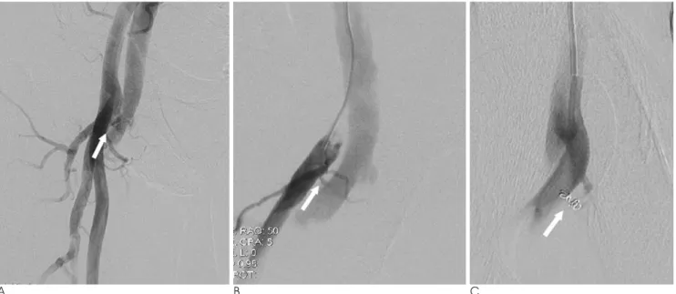
A 65-year-old man was admitted to manage an acute myocardial infarction. A coronary angiography was per- formed using the right femoral approach and confirmed a single vessel with disease, which was treated with a percutaneous transluminal coronary angioplasty (PT- CA). One day after the sheath removal for catheteriza- tion, a continuous bruit and an intense thrill were no- ticed at the right groin. A color Doppler ultrasound (CDU) examination was performed and showed an AVF between the right deep femoral artery (DFA) and the common femoral vein (CFV). Challenges to close the AVF by ultrasound-guided compression failed. Then, an angiography was performed using the left common femoral artery approach and showed an AVF between right DFA and CFV (Fig. 1A, B). The fistula was catheterized with a 3Fr microferret catheter (Cook, Bjaeverskov, Denmark) and two 3mm tornado platinum coils�(Cook, Bloomington, IN, U.S.A.) were placed in
J Korean Soc Radiol 2009;61:101-104
─ 101 ─
Iatrogenic Femoral Arteriovenous Fistulas: Endovascular Treatment with Coil Embolization in Two Patients1
Eun Jung Ahn, M.D., Jin Soo Choi, M.D., Sung Mun Lee, M.D., Young Whan Kim, M.D.
1Department of Radiology, Dongsan Medical Center, Keimyung University School of Medicine
Received March 10, 2008 ; Accepted May 11, 2009
Address reprint requests to : Jin Soo Choi, M.D., Department of Diagnostic Radiology, Dongsan Medical Center, Keimyung University College of Medicine, 194, Dongsan-dong, Jung-gu, Daegu 700-712, Korea.
Tel. 82-53-250-7767 Fax. 82-53-250-7766 E-mail: [email protected]
An iatrogenic femoral arteriovenous fistula (AVF) is an uncommon but well-recog- nized complication that has followed diagnostic or interventional procedures. We re- port two cases of postcatheterization femoral AVF that were successfully treated with embolization using microcoils through a coaxial microcatheter. The fistulous tract of these patients could not be obliterated by ultrasound-guided compression.
Index words :Femoral artery Arteriovenous fistula Embolization, therapeutic
the fistulous track. A follow-up angiography after em- bolization confirmed the obliteration of the AVF (Fig.
1C).
Case 2
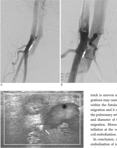
A 74-year-old man admitted due to angina symptoms and underwent a PTCA using the right femoral ap- proach. On the next day, a continuous bruit and a palpa- ble thrill was detected at the right groin. A CDU exami- nation showed an AVF between the right DFA and CFV. Both manual and ultrasound-guided compression failed to close the AVF. Then, an angiography was per- formed through the contralateral common femoral artery, which demonstrated an AVF between the right DFA and CFV (Fig. 2A, B). A coaxial microferret catheter was placed into the fistula and 5mm tornado platinum coils�were delivered into the fistulous track.
The first coil migrated to venous system and was em- bolized in the left upper segmental pulmonary artery without clinical symptoms. One coil was correctly placed in the fistulous tract. Closure of the AVF was demonstrated immediately after the embolization an- giography and CDU examination (Fig. 2C).
Discussion
An iatrogenic femoral AVF is caused by the transfixa- tion of an artery and a vein by the needle. Puncture of both vessels often occurs when performed below the
level of the femoral head, where the artery and vein are in antero-posterior relation. Other causal factors include catheterization or complex interventional procedures when employing large catheters or introducer sheaths, antegrade catheterization, obesity, difficult or repeat ar- terial puncture, simultaneous puncture of both artery and vein, anticoagulant or thrombolytic therapy and heavily calcified artery, medial calcinosis, arterial hy- pertension, and hemodialysis or diabetic patients (4).
Marsan et al. reported the relatively high incidence of AVFs following cardiac angiographies and mentioned that a very significant factor in the formation of AVFs is the distal location chosen for the cutaneous entry point and vascular puncture (7). The femoral crease that is used as a landmark by many angiographers for common femoral artery is unreliable. To avoid distal puncture and reduce the possibility of AVF formation, another study suggests that the femoral head or inguinal liga- ment should be used as a landmark (7).
The development of femoral artery AVFs represents a continuing problem after a vascular diagnostic and in- terventional procedure. For most patients, watchful waiting and ultrasound-guided compression have been an effective method of treating such complications (3, 8).
However, in patients requiring a continuous anticoagu- lant regimen, in those with large AVFs or in patients suf- fering from painful groin hematomas, the compression method is less successful (5, 9). Although surgical repair of AVFs is safe and definitive, the troublesome compli-
Eun Jung Ahn, et al: Iatrogenic Femoral Arteriovenous Fistulas
─ 102 ─
A B C
Fig. 1. A 65-year-old man presented with bruit at the right groin.
A, B. An AVF is originating from the right DFA with short segment fistula (arrow).
C. Angiography obtained immediately after coil embolization shows complete closure of the fistula (arrow).
cations include wound infection, bleeding, neuralgia, septicemia, limb swelling, and scar formation that may make future groin access difficult (6). Another alterna- tive treatment of AVFs with implantation of stent-grafts, represents an approach currently under clinical investi- gation (5). However, potential issues such as stent defor- mity and kinking, the loss of branch vessels after place- ment, stenosis and occlusion, or the misplacement of stents prevents a final evaluation of this therapeutic al- ternative thus far (9, 10). Percutaneous coil embolization of AVFs is another therapeutic option of iatrogenic femoral AVFs (4). This method is relatively easy and cost effectiveness compared to surgical repair and im- plantation of a stent graft. The important aspect for this method is the selection of a fistula and the exact place- ment of the coil within the fistula neck. The fistulous
track is uneven and anchors coils of 3-5 mm. Coil mi- grations may cause a potential problem due to high flow within the fistula. We have experienced a case of coil migration and it resulted in an asymptomatic embed at the pulmonary artery. Coils must match up to the length and diameter of the fistulous tract to prevent of distal migration. Moreover, manual compression or balloon inflation at the venous site may be helpful during the coil embolization.
In conclusion, our study suggests that a transcatheter embolization of iatrogenic femoral AVFs with a micro- coil may be a safe and effective treatment, which is easy to perform in experienced hands and relatively inexpen- sive.
References
1. Kelm M, Perings SM, Jax T, Lauer T, Schoebel FC, Heintzen MP, et al. Incidence and clinical outcome of iatrogenic femoral arteri- ovenous fistulas: implications for risk stratification and treatment.
J Am Coll Cardiol 2002;40:291-297
2. Muller DW, Shamir KJ, Ellis SG, Topol EJ. Peripheral vascular complications after conventional and complex percutaneous coro- nary intervention procedures. Am J Cardiol 1992;69:63-68 3. Fellmeth BD, Roberts ALC, Bookstein JJ, Frelschiag JA, Forsythe
JR, Buckner NK, et al. Postangiographic femoral artery injuries:
nonsurgical repair with US-guided compression. Radiology 1991;178:671-675
4. Lemaire JM, Dondelinger RF. Percutaneous coil embolization of iatrogenic femoral arteriovenous fistula or pseudoaneurysm. Eur J Radiol 1994;18:96-100
5. Thalhammer C, Kirchherr AS, Uhlich F, Waigand J, Gross CM.
Postcatheterization pseudoaneurysms and arteriovenous fistulas:
repair with percutaneous implantation of endovascular covered J Korean Soc Radiol 2009;61:101-104
─ 103 ─ C
A B
Fig. 2. A 74-year-old man presented a continuous bruit and a palpable thrill at the right groin.
A, B. A high flow AVF is originating from the right DFA with short seg- ment fistula (arrow).
C. Follow-up color Doppler ultra- sound shows complete closure of the fistula (arrow) after embolization with coil.
stents. Radiology 2000;214:127-131
6. Onal B, Ilgit ET, Akpek S, Coskun B. Postcatheterization femoral arteriovenous fistula: endovascular treatment with N-butyl-cyano- acrylate embolization. Cardiovasc Inervent Radiol 2006;29:276-278 7. Marsan RE, McDonald V, Ramamurthy S. Iatrogenic femoral arte-
riovenous fistula. Cardiovasc Intervent Radiol 1990;13:314-316 8. Schaub F, Theiss W, Heinz M, Zagel M, Schomig A. New aspects
in ultrasound-guided compression repair of postcatheterization
femoral artery injuries. Circulation 1994;90:1861-1865
9. Waigand J, Uhlich F, Gross CM, Thalhammer C, Dietz R.
Percutaneous treatment of pseudoaneurysms and arteriovenous fistulas after invasive vascular procedures. Catheter Cardiovasc Interv 1999;47:157-164
10. Sahin S, Cinar B, Bilgin SN, Celik L, Eren EE. Surgical repair of a post-traumatic arteriovenous fisfula complicated by stent-graft misplacement. Cardiovasc Intervent Radiol 2005;28:87-89
Eun Jung Ahn, et al: Iatrogenic Femoral Arteriovenous Fistulas
─ 104 ─
대한영상의학회지 2009;61:101-104
의인성 넓적다리 동정맥루: 코일색전술 2예1
1계명대학교 의과대학 영상의학과학교실 안은정∙최진수∙이성문∙김영환
혈관 조영술을 위한 천자부위로 넓적다리 동맥을 가장 많이 사용하며 시술 후에 발생한 의인성 넓적다리 동정맥루 는 드물지만 합병증 중 하나이다. 저자들은 인터벤션 시술 후 발생한 넓적다리 동정맥루에 코일 색전술을 이용하여 치료한 2예를 보고하고자 한다. 이 동정맥루는 초음파 유도 하 압박에 반응이 없었다.