Spontaneous Occluded Anterior Communicating Artery Aneurysm during Coil Embolization Treated with One Coil Insertion into Remaining Stump
Se Hun Chang, Seung Hwan Lee, Hee Sup Shin, Jun Seok Koh
Stroke and Neurological Disorders Centre, Kyung Hee University Hospital at Gangdong, College of Medicine, Kyung Hee University, Seoul, Korea
Spontaneous thrombosis of a ruptured aneurysm during coil embolization is a rare event, and some reports on recanalization of a spontaneous oc- cluded ruptured aneurysm have been published. We report on a case of a 54-year-old male who presented with a subarachnoid hemorrhage due to rupture of a small aneurysm of the anterior communicating artery (ACoA). Cerebral angiography confirmed the presence of the ACoA aneurysm, but, during coil embolization, the aneurysm was near com- pletely occluded with a remaining small neck. A small coil was inserted into the remaining stump of the neck to prevent recanalization, and the angiographic result at 1 year after coil embolization showed complete ob- literation of the aneurysm.
J Cerebrovasc Endovasc Neurosurg.
2015 September;17(3):246-251 Received : 23 June 2015
Revised : 14 August 2015 Accepted : 2 September 2015 Correspondence to Seung Hwan Lee Department of Neurosurgery, Kyung Hee University Hospital at Gangdong, 892 Dongnam-ro, Sangil-dong, Gangdong-gu, Seoul 05278, Korea
Tel : 82-2-440-6286 Fax : 82-2-440-8404 E-mail : [email protected]
ORCID : http://orcid.org/0000-0001-8043-632X
This is an Open Access article distributed under the terms of the Creative Commons Attribution Non- Commercial License (http://creativecommons.org/li- censes/by-nc/3.0) which permits unrestricted non- commercial use, distribution, and reproduction in any medium, provided the original work is properly cited.
Keywords Intracranial aneurysm, Subarachnoid hemorrhage, Thrombosis, Recanalization
INTRODUCTION
Although spontaneous thrombosis of large and giant aneurysms is not uncommon, this situation in a small ruptured cerebral aneurysm is a rare event with in- cidence of 1-2%. Its possible pathophysiology with contributing factors has been well discussed in the literature.1-3)8)12)15)18) In addition, subsequent recanaliza- tion within the next few weeks has also been described.
We report on a case of acute thrombosis of the aneur- ysm, which occurred immediately after coil extraction, and suggest the next way to prevent recanalization, which can lead to re-rupture of the aneurysm.
CASE REPORT
History
A 54-year-old male experienced a sudden onset of headache which had developed 2 days ago. After the initial diagnosis of subarachnoid hemorrhage (SAH) using brain computed tomography (CT) in another hospital, he was transferred to the emergency room where his Glasgow Coma Scale score was E4V5M5.
Despite neck stiffness, there was no focal neurological sign. His medical history was non-contributory and laboratory blood tests found no abnormalities.
Radiologic findings
A brain CT scan taken on admission showed focal SAH concentrated in the anterior portion of the basal cistern (Fig. 1). Digital subtraction angiography (DSA) confirmed the presence of the anterior communicating
SE HUN CHANG ET AL
Fig. 1. Axial brain CT scan taken on admission shows a sub- arachnoid hemorrhage on the anterior portion of the basal cistern.
CT = computed tomography.
A B
Fig. 2. Reconstruction of 3D rotational angiogram shows the dumbbell-shaped aneurysm with a very narrow neck (A). Measurement in each length of the aneurysm is 5.9 x 2.7 x 1.7 mm (width x height x neck diameter) (B).
artery (ACoA) aneurysm and 3D rotational angiog- raphy (3DRA) showed that the aneurysm had a very narrow neck, which measured 5.9 × 2.7 × 1.7 (width
× height × neck diameter) (Fig. 2). Based on the dis-
tribution of the SAH, we considered that the ACoA aneurysm had bled. As morphologic features of the aneurysm were adequate for coil embolization, we de- cided to perform an emergency endovascular coil em- bolization on the day of admission.
Endovascular treatment
The embolization procedure was performed with the patients under general anesthesia. DSA was per- formed again to assure the aneurysm. After confirm- ing that the result on DSA was identical to the pre- vious one, a microcatheter was carefully guided over a microguidewire into the aneurysm. As the tip of the catheter was kept at the neck of the aneurysm, the first coil (Target® Detachable coil 3 mm × 8 cm;
Stryker Neurovascular, Fremont, CA, USA) placement was attempted very slowly. At the last moment of de- ploying the first coil, the tip of the microcatheter was seen to move back from the aneurysm and the projec- tion of the coil loop outside the aneurysm was ob- served (Fig. 3A). Consequently, the first coil was gen- tly withdrawn from the aneurysm. However, a sub- sequent angiogram showed near obliteration of the aneurysm without evidence of vasospasm. Only a ti-
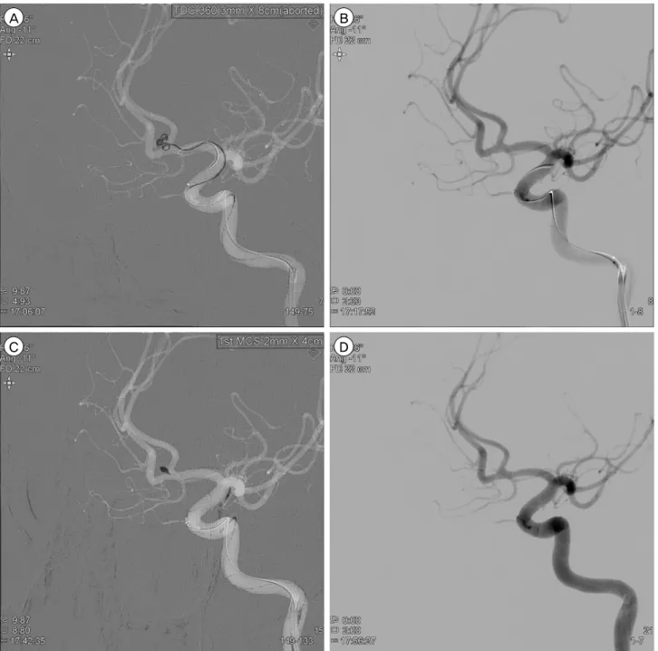
A B
C D
Fig. 3. Neurointerventional images during coil embolization. At the moment of deploying the first coil, the tip of the microcatheter is seen to move back from the aneurysm and the projection of the coil loop outside the aneurysm is observed (A). After extraction of the coil, subsequent angiogram shows near obliteration of the aneurysm (B). Insertion of a smaller coil into the remaining stump of the neck is performed (C) and post-embolization angiogram shows complete occlusion of the aneurysm (D).
ny stump at the neck was noted (Fig. 3B). Because acute thrombosis facilitated by an exposed coil within the aneurysm was assumed, and, sooner or later, re- canalization could occur, we decided to insert a smaller coil into the remaining stump of the neck.
With the coil (HyperSoft® 3D complex Coil 2 mm ×
4 cm; MicroVention, Tustin, CA, USA), embolization was performed safely and post-embolization DSA showed complete occlusion of the aneurysm (Fig. 3C, D). There was no occurrence of procedure-related complication during coil embolization and the patient recovered without neurologic deficit after the procedure.
SE HUN CHANG ET AL
A B
Fig. 4. Follow-up cerebral angiogram the next day showing persistent occlusion of the aneurysm (A, B).
Follow-up
Follow-up DSA the next day showed persistent oc- clusion of the aneurysm (Fig. 4). The patient was dis- charged without neurologic deficit of the modified Rankin scale score 0. Magnetic resonance (MR) an- giography at 3-month follow-up showed no evidence of residual neck or recurrence of recanalization. On 1 year follow-up DSA, despite change in the config- uration of the coil, the aneurysm remained to be com- pletely obliterated (Fig. 5).
DISCUSSION
The reported incidence of acute thrombosis of the aneurysm after SAH is 1-2%,7)9)17) and several possible mechanisms have been reported in the literature. In large or giant aneurysms, in particular, the incidence of spontaneous total thrombosis of the aneurysm has shown a marked increase, ranging from 13% to 20%,10) and the volume-to-orifice ratio of the aneurysm has been suggested as the most reliable mechanism of sponta- neous thrombosis.1) However, in small aneurysms, other factors including anti-fibrinolytic agent, non-ionic con-
trast media, systemic hypotension, increased blood co- agulability, increased platelet aggregation, vasospasm, and hemodynamics in the parent artery have also been suggested as an influential mechanism.3)6)8)15)18)20)
In our patient, we supposed several factors that pro- moted thrombosis of the aneurysm. The most im- portant and potent factor was a narrow neck of the aneurysm of 1.7 mm with a large volume-to-orifice ratio. Extremely narrow neck of the aneurysm was re- garded as sufficient potential to induce thrombosis under special circumstances like the following. First, the endovascular procedure - including microcatheter placement on the neck of the aneurysm and aborted first coil - could interrupt the intrasaccular blood flow and trigger aneurysm thrombosis. Second, inadequate systemic heparinization might induce spontaneous thrombosis. According to the protocol of our institute, we generally induce systemic heparinization after suc- cessful placement of the first basket coil in treatment of a ruptured cerebral aneurysm. In this patient, sys- temic heparin was not administered during the proce- dure, and the endovascular procedure was performed under inadequate systemic heparinization. Third, in-
A B
Fig. 5. One-year follow-up cerebral angiogram (A, B). Despite change in the configuration of the coil, the aneurysm remains to be completely obliterated.
duced hypotension during the procedure could be a potential factor in spontaneous thrombosis. From this experience, if the neck of a ruptured aneurysm was very narrow, a rapid and adequate endovascular pro- cedure, sufficient heparinization and exclusion of ex- cessive hypotension during the procedure should be considered in order to avoid spontaneous thrombosis of the ruptured aneurysm.
Lee et al.13) pointed out that the mechanism of re- canalization might be the result of liquefaction of the thrombus and subsequent intrathrombotic dissection by blood flow. Another opinion regarding recanalization following thrombosis of the aneurysm suggests that when the thrombosis was induced by endovascular treatment, recanalization of the thrombosed aneurysm might occur spontaneously.14) Although the pathophysiology of spon- taneous recanalization has not been fully elucidated, recanalization ensuing from complete thrombosis of aneur- ysms has been reported in many studies.4)5)11)12)16)19)21)22)
In these studies, recanalization of acute thrombosis of the aneurysm was observed in the days and weeks af- ter thrombosis. Therefore, follow-up angiogram and
careful observation during the follow-up period are required for fear of recanalization resulting in rebleed- ing of the aneurysm. In our patient, coil embolization was performed using a small coil in the tiny stump of the remaining neck. We believed that this procedure might prevent spontaneous recanalization rather than inducing recanalization because the inserted coil would generate an additional thrombus as much as to block the narrow neck, thus resulting in obstruction of blood flow. Fortunately, a satisfactory result was obtained with complete occlusion of the aneurysm after 1-year follow-up.
CONCLUSION
Thrombosis in a ruptured small aneurysm may be generated during coil insertion and the extraction process when the neck of the aneurysm is very narrow. Because recanalization may occur within the next few weeks, we believe that coil insertion on the remaining neck would provide a measure to prevent spontaneous recanalization.
SE HUN CHANG ET AL
Disclosure
The authors report no conflict of interest concerning the materials or methods used in this study or the findings specified in this paper.
REFERENCES
1. Black SP, German WJ. Observations on the relationship between the volume and the size of the orifice of ex- perimental aneurysms. J Neurosurg. 1960 Nov;17:984-90.
2. Bohmfalk GL, Story JL. Intermittent appearance of a ruptured cerebral aneurysm on sequential angiograms.
Case report. J Neurosurg. 1980 Feb;52(2):263-5.
3. Brownlee RD, Tranmer BI, Sevick RJ, Karmy G, Curry BJ. Spontaneous thrombosis of an unruptured anterior communicating artery aneurysm. An unusual cause of ischemic stroke. Stroke. 1995 Oct;26(10):1945-9.
4. Chohan MO, Westhout FD, Taylor CL. Delayed rebleed- ing of a spontaneously thrombosed aneurysm after sub- arachnoid hemorrhage. Surg Neurol Int. 2014 Mar;5:42.
5. Choi YS, Kim DW, Jang SJ, Kang SD. Recanalization of completely thrombosed non-giant saccular aneurysm mimicking as de novo aneurysm. J Korean Neurosurg Soc. 2010 Oct;48(4):354-6.
6. Cohen JE, Itshayek E, Gomori JM, Grigoriadis S, Raphaeli G, Spektor S, et al. Spontaneous thrombosis of cerebral aneurysms presenting with ischemic stroke. J Neurol Sci.
2007 Mar;254(1-2):95-8.
7. Edner G, Forster DM, Steiner L, Bergvall U. Spontaneous healing of intracranial aneurysms after subarachnoid hemorrhage. Case report. J Neurosurg. 1978 Mar;48(3):450-4.
8. Fareed J, Walenga JM, Saravia GE, Moncada RM.
Thrombogenic potential of nonionic contrast media?
Radiology. 1990 Feb;174(2):321-5.
9. Fodstad H, Liliequist B. Spontaneous thrombosis of rup- tured intracranial aneurysms during treatment with tra- nexamic acid (AMCA). Report of three cases. Acta Neurochir (Wien). 1979;49(3-4):129-44.
10. Golding R, Peatfield RC, Shawdon HH, Rice Edwards JM. Computer tomographic features of giant intracranial aneurysms. Clin Radiol. 1980 Jan;31(1):41-8.
11. Jayakumar PN, Ravishankar S, Balasubramaya KS, Chavan R, Goyal G. Disappearing saccular intracranial aneurysms:
do they really disappear? Interv Neuroradiol. 2007 Sep;13(3):247-54.
12. Kim HJ, Kim JH, Kim DR, Kang HI. Thrombosis and recanalization of small saccular cerebral aneurysm : two case reports and a suggestion for possible mechanism. Kim HJ, Kim JH, Kim DR, Kang HI. 2014 May;55(5):280-3.
13. Lee KC, Joo JY, Lee KS, Shin YS. Recanalization of completely thrombosed giant aneurysm: case report.
Surg Neurol. 1999 Jan;51(1):94-8.
14. Mericle RA, Wakhloo AK, Lopes DK, Lanzino G, Guterman LR, Hopkins LN. Delayed aneurysm regrowth and recanalization after Guglielmi detachable coil treatment. Case report. J Neurosurg. 1998 Jul;89(1):142-5.
15. Nakajima Y, Yoshimine T, Mori H, Nakamuta K, Fujimura I, Sakashita K, et al. Spontaneous disappearance and re- appearance of a ruptured cerebral aneurysm: one case found in a group of 33 consecutive patients with subarachnoid hemorrhage who underwent repeat angiography. Neurol Res. 2000 Sep;22(6):583-7.
16. Nakau H, Nagatani H, Nakau R, Ametani T. Acute dis- appearance of ruptured aneurysm located near the origin of the superior cerebellar artery - case report. Neurol Med Chir (Tokyo). 2007 Oct;47(10):468-70.
17. Spallone A, Peresedov VV, Kandel EI. Spontaneous cure of ruptured intracranial arterial aneurysms. Surg Neurol.
1981 Nov;16(5):367-70.
18. Spetzler RF, Winestock D, Newton HT, Boldrey EB.
Disappearance and reappearance of cerebral aneurysm in serial arteriograms. Case report. J Neurosurg. 1974 Oct;41(4):
508-10.
19. Su TM, Hsu SW, Chen WF, Lee TC, Cheng CH. Acute thrombosis and recanalization of a ruptured anterior communicating artery aneurysm. J Clin Neurosci. 2009 Aug;16(8):1077-9.
20. Szajner M, Jargiello T, Trojanowski T, Szczerbo-Trojanowska M. Spontaneous Thrombosis of the Pseudoaneurysm of Right SCA after an Attempt at Embolisation. A Case Report. Interv Neuroradiol. 2002 Jun;8(2):205-8.
21. Valle EP, Tamargo RJ, Gailloud P. Thrombosis and sub- sequent recanalization of a ruptured intracranial aneur- ysm in 2 children, demonstrating the value of repeating catheter angiography after an initial negative study. J Neurosurg Pediatr. 2010 Apr;5(4):346-9.
22. Wei D, Jingru Z, Cungang F, Yake X, Dongliang W, Zhengmao W, et al. Complete spontaneous thrombosis and recanalization of a ruptured posterior cerebral ar- tery aneurysm. Turk Neurosurg. 2014;24(3):406-10.