Delayed Ischemic Stroke after Flow Diversion of Large Posterior Communicating Artery Aneurysm
Si On Kim1, Yeon Gu Chung1, Yu Sam Won1, Myung Ho Rho2
1Department of Neurosurgery, Kangbuk Samsung Hospital, Sungkyunkwan University, Seoul, Korea
2Department of Radiology, Kangbuk Samsung Hospital, Sungkyunkwan University, Seoul, Korea
For securing large, giant, and wide-neck aneurysms, conventional coil em- bolization has substantial limitations, such as incomplete occlusion, recan- alization, and a high recurrence rate. To overcome these limitations, a novel paradigm was suggested and, as a result, flow-diverting device was developed. The flow-diverting device is an innovative and effective techni- que to allow securing of large, giant, and wide-neck aneurysms. In nu- merous studies, the flow-diverting device has shown better outcomes than coil embolization. However, the flow-diverting device has also some risks, including rupture of aneurysm, intracerebral hemorrhage, and ische- mic stroke. In addition, with more experience, unexpected complications are also reported.5)7) In the present case, we experienced a delayed ische- mic stroke at 27 days after endovascular treatment. The patient had mul- tiple aneurysms and, among them, we treated a large posterior communi- cating artery aneurysm using PipelineTM Embolization Device. The patient was tolerable for 25 days, but then suddenly presented intermittent right hemiparesis. In the initial diffusion magnetic resonance imaging (MRI), there was no acute lesion; however, in the follow-up MRI, an acute is- chemic stroke was found in the territory of anterior choroidal artery which was covered by Pipeline Embolization Device. We suspect that neo-intimal overgrowth or a tiny thrombus have led to this delayed complication. Through our case, we learned that the neurosurgeon should be aware of the possibility of delayed ischemic stroke after flow diversion, as well as, long-term close observation and follow-up angiog- raphy are necessary even in the event of no acute complications.
J Cerebrovasc Endovasc Neurosurg.
2016 March;18(1):19-26 Received : 21 July 2015 Revised : 21 December 2015 Accepted : 9 March 2016 Correspondence to Myung Ho Rho Department of Radiology, Kangbuk Samsung Hospital, Sungkyunkwan University School of Medicine, 29 Saemunan-ro, Jongro-gu, Seoul 03181, Korea
Tel : 82-2-2001-2330 Fax : 82-2-2001-2157 E-mail : [email protected]
ORCID : http://orcid.org/0000-0002-0556-6658 Poster presentation at Society of Korean Endovascular Neurosurgeons
This is an Open Access article distributed under the terms of the Creative Commons Attribution Non- Commercial License (http://creativecommons.org/li- censes/by-nc/3.0) which permits unrestricted non- commercial use, distribution, and reproduction in any medium, provided the original work is properly cited.
Keywords Flow diversion, Pipeline embolization device, Anterior choroidal artery oc- clusion, Ischemic stroke, Complication
INTRODUCTION
The endovascular treatment of intracranial aneurysms has become an alternative technique of surgical clip- ping for 25 years.9)18) However, coil embolization still has limitations, especially, in wide-neck, large, and giant aneurysm cases.2)5)9)13) To overcome these limi- tations, the flow-diverting device was developed and
introduced for the treatment of irresoluble aneurysms.10) After the efficacy and safety of the flow-diverting de- vice has been demonstrated in several studies, it has generally been accepted as a new solution for large or wide-neck aneurysms. Although the flow-diverting de- vice has low recanalization rate and low recurrence rate, it also has severe and unexpected complications, such as ischemic stroke, spontaneous rupture of aneurysm,
Fig. 1. 3D reconstruction of the left ICA angiography. A large PCoA aneurysm is located between PCoA and AChA (neck: 11 mm, length: 18 mm, width: 9 mm, postero-inferior projection).
Proximal PCoA originates from the aneurysm neck, but prox- imal AChA is slightly separated from the aneurysm. ACoA and left MCA aneurysms are also visible (Black star: Large PCoA aneurysm, White arrow: AChA, Arrow head: PCoA). Lt = left;
ICA = internal carotid artery; PCoA = posterior communicating artery; AChA = anterior choroidal artery; ACoA = anterior com- municating artery; MCA = middle cerebral artery.
intracerebral hemorrhage, and stent stenosis.1)2)8)11)17)19)
Among these complications of the flow-diverting de- vice, ischemic stroke due to thromboembolism and perforator occlusion is well-described and occurs most frequently. In previous reports, ischemic strokes were reported to occur in 2.5-13% of patients and most of the instances occurred during the endovascular treatment.1)3)11)17) Ischemic stroke may result from in- sufficient antiplatelet therapy, stent wall thrombus formation and occlusion, parent artery occlusion or distal thromboembolic events. It should also be noted that this complication was more frequent in posterior circulation and giant aneurysm cases.1)17)
In the present case, we observed a delayed ischemic stroke in the territory of the anterior choroidal artery which was covered by the flow-diverting device for the treatment of a large posterior communicating ar- tery aneurysm.
CASE REPORT
A 56 year-old male with histories of cerebral palsy, craniotomy for head trauma, and acute myocardial in- farction, presented at our hospital for incidentally de- tected aneurysms. Because of cerebral palsy, he al- ready had right hemiparesis (motor grade IV, modi- fied Rankin Scale (mRS) score 2), but other neurologic examinations were intact. In his brain MRA, there were four aneurysms on the right and left middle cer- ebral artery (MCA) bifurcation, anterior communicat- ing artery (ACoA), and left posterior communicating artery (PCoA). The bilateral MCA bifurcation and ACoA aneurysms were smaller than 5 mm, but the left PCoA aneurysm was measured 19 mm.
Due to the history of myocardial infarction, the pa- tient already had administered aspirin and clopidog- rel for one year. Prior to the endovascular treatment, we performed a drug resistance test for aspirin and clopigrel. Aspirin resistance test was 387 ARU (Aspirin Reaction Unit, normal range: < 550, aspirin resistance:
≥ 550) and clopidogrel resistance test was 120 PRU (P2Y12 Reaction Unit, normal range: < 240 PRU, clo-
pidogrel resistance: ≥ 240 PRU). These results con- firmed that the patient does not had resistance to as- pirin and clopidogrel.
We conducted a diagnostic transfemoral cerebral an- giography (TFCA) under local anesthesia. In TFCA, the large PCoA aneurysm was located between the left PCoA and the left anterior choroidal artery (AChA), and had a small daughter sac in the dome of the aneurysm. The left AChA was separated from the large aneurysm, but the proximal PCoA originated from the neck of the large aneurysm. Since the large aneurysm was wide-neck and involved the proximal PCoA, we decided to reschedule the operation for de- ploying the flow-diverting device at the large aneur- ysm and conventional coiling at the other aneurysms (Fig. 1).
A B
C D
Fig. 2. (A, B) Post-operative right anterior oblique and lateral view. (C, D) 3D reconstruction of PED and coils. (C, D) PED is well-at- tached to the distal ICA wall (Black star: Large PCoA aneurysm, Arrow head: Deployed PED, White arrow: coil embolization of the left ACoA, MCA aneurysms, Black arrow: cranial fixator of a previous craniotomy, Black star: Large PCoA aneurysm). PED = PipelineTM Embolization Device; ICA = internal carotid artery; PCoA = posterior communicating artery; ACoA = anterior communicating artery;
MCA = middle cerebral artery.
Under general anesthesia, the operation was per- formed under intravenous heparin infusion with the goal of maintaining an activated clotting time at 2 times of the normal value. After the puncture of the right common femoral artery, 7-French Flexor shuttle® guiding sheath (Cook Medical, Bloomington, IL, USA) was placed in the left internal carotid artery (ICA)
and the left ACoA and MCA bifurcation aneurysms were managed first using micro-catheters and coils.
After the successful coiling of the left ACoA, MCA bi- furcation aneurysms, the MarksmanTM Micro Catheter (ev3/Covidien, Irvine, CA, USA) was placed on the left proximal MCA and PipelineTM Embolization Device (PED; ev3/Covidien, Irvine, CA, USA) was de-
A B
Fig. 3. (A) Post-operative angiographyin the arterial phase. After the deployment of PED, all of the branch vessels, including AChA and PCoA, are well-maintained. (B) Post-operative angiography in the venous phase. The flow stagnation at PCoA aneurysm sac is observed (White arrow: AChA, Arrow head: PCoA). PED = PipelineTM Embolization Device; AChA = anterior choroidal artery; PCoA = posterior communicating artery.
ployed from the proximal MCA to the horizontal seg- ment of the cavernous ICA, with covering the AChA and PCoA (Fig. 2). Throughout the course of deploy- ment, the combination of forward pressure and re- traction technique was used to maximally attach PED to the ICA wall. The deployment of PED was success- fully performed and, in the post-operative angiog- raphy, whole branch vessels were not interrupted by PED including PCoA, AChA (Fig. 3). After emboliza- tion of the left side aneurysms, the femoral access site was closed with Perclose ProGlide (Abbott Vascular, Santa Clara, CA, USA) and, two days after the endo- vascular treatment, the patient was discharged with dual antiplatelet therapy, clopidogrel 75 mg, and as- pirin 100mg daily, without neurologic deficit.
Dual antiplatelet therapy was continuously main- tained and complications of the flow-diverting device were not observed during 25 days. However, 25 days after the endovascular treatment, the patient suddenly presented intermittent right hemiparesis. Although his symptom was not prominent in neurologic examina- tion and he already had right hemiparesis due to pre-
vious cerebral palsy, we readmitted him for checking the diffusion magnetic resonance imaging (MRI). In the initial diffusion MRI which was checked at 25 days after the endovascular treatment, an ischemic stroke was not found, so we recommended TFCA for further evaluation. However, he refused more tests and management, because he had phobia about the additional brain exam. We comprehended his phobia because he already had histories of cerebral palsy and craniotomy for head trauma. Because the initial MRI was fine, we maintained dual antiplatelet therapy without additional medication and closely observed his symptoms in the hospital (Fig. 4). However, as time passed, the patient complained of more frequent and worsening hemiparesis, so we rechecked dif- fusion MRI and MRA. In the second diffusion MRI which was checked at 27 days after the endovascular treatment, ischemic stroke in the territory of the left AChA was observed (Fig. 5) and his symptom, right hemiparesis, was aggravated to motor grade II and mRS score4. We recommended a follow-up TFCA to confirm the occlusion of AChA, but the patient re-
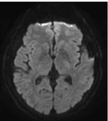
Fig. 5. Diffusion MRI scan at 27 days after the endovascular treatment. The patient's hemiparesis was aggravated, so we re- checked diffusion MRI again. In follow-up MRI, acute ischemic stroke was found in the territory of the left AChA. MRI = mag- netic resonance imaging; AChA = anterior choroidal artery.
Fig. 4. Diffusion MRI scan at 25 days after the endovascular treatment. The patient presented intermittent right hemiparesis, but there was no acute lesion in the territory of left AChA.
MRI = magnetic resonance imaging; AChA = anterior choroidal artery.
fused again. We maintained dual antiplatelet therapy without additional medication, and transferred him to the department of rehabilitation for physical training of right hemiparesis. He discharged with the dual an- tiplatelet medication at 83 days after the endovascular treatment. At the point of discharge, right hemiparesis was improved to motor grade III and mRS score 3.
DISCUSSION
After introducing of the flow-diverting device in 2011, it has become an innovative technique to allow for a more effective and safety endovascular treat- ment of large, wide-neck, and complex aneurysms.
Furthermore, its outcomes also show an excellent oc- clusion rate, a low recanalization rate, as well as ac- ceptable morbidity and mortality as compared to coil embolization. The flow-diverting device has emerged as an answer to previously irresoluble aneurysms;
however, it is not free from some complications. In re-
cent studies, the complications of flow-diverting de- vice, such as branch vessel and/or perforator occlu- sion, rupture of aneurysm, intracerebral hemorrhage (ICH), stent stenosis and even death were reported (Table 1).1-4)8)11)13)14)16)17)19)
Numerous single and multicenter studies demon- strated overall rates of adverse complications (Table 1); among these studies, intra-operative and post-op- erative rupture of aneurysm is a serious concern of flow-diverting device. Although, rupture of aneurysm was reported only 0.6-4%, however, once it occurs, it leads to serious sequelae. In Kallmesstudy,11) the over- all rupture rate was only 0.6%; however, the rupture rate was increased with the aneurysm size (giant 4.5%, large 0.6%, small aneurysm 0%). Most of this complication arose in the procedure of wire, mi- cro-catheter manipulation, deployment of device, and balloon inflation for remodeling the device. Therefore, a careful placement of micro-catheter and a gentle manipulation of the device are crucial points for pro-
Author, The year of
publication
Patient / Aneurysms
Ruptured : Unruptured aneurysms
Anterior : Posterior circulation
Intra-operative
complication Aneurysm
rupture ICH Ischemic
stroke Morbidity Mortality
Complete and near complete occlusion
rate Brinjikji et al.,
2013 1451 / 1654 4% 3% 6% 5% 4%
Kallmes et al.,
2015 793 / 906 76 : 717 838 : 59 0% 0.6% 2.4% 4.7% 7.4% 3.8%
McDonald et al.,
2015 279 / 1.4% 1.4% 5.0% 7.1% 0.7%
Saatci et al.,
2012 191 / 251 76 : 717 237 : 14 2.1% 0.5% 1.0% 6.8% 14.1% 0.5% 91.2%
O'Kelly et al.,
2013 97 / 97 0 : 97 79 : 18 3.2% 4.3% 10.3% 4.4% 6.3% 84.2%
Chalouhi et al.,
2013 40 / 40 0 : 40 36 : 4 0% 0% 5.0% 2.5% 7.5% 2.5% 84.0%
Iosif et al.,
2015 38 / 49 1 : 37 48 : 1 7.8% 0% 0% 13.2% 13.2% 0% 81.6%
Nossek et al.,
2015 27 / 28 2 : 25 26 : 2 0% 0% 0% 0% 0% 0% 100%
Monteith et al.,
2014 24 / 24 0 : 24 17 : 7 4.2% 0% 4.2% 13% 16.7% 4.2% 83.3%
Chiu et al.
2015 98 / 119 0 : 98 98 : 21 0% 0.8% 0% 4.2% 8.4% 0.8% 93.2%
Chitale et al.,
2012 36 / 42 41 : 1 2.8% 11.1% 5.6% 13.9% 2.8%
ICH = Intracerebral hemorrhage
Table 1. Complication rates of the flow-diverting device
tecting rupture of aneurysm.
Flow diversion-induced ICH is a more specific com- plication and leads to a significant morbidity and mortality. The incidence of ICH was 1-5% of the pa- tients in several retrospective reports and the exact mechanism is unclear (Table 1). However, dual anti- platelet therapy, hemorrhagic conversion of ischemic stroke, intra-operative hypertension, and altered pres- sure dynamics are suspected to be causes of ICH.2)17) As all of the above are related to blood pressure, blood pressure should be strictly controlled to prevent hemorrhagic complications.
Ischemic stroke has been reported to be a major complication accounting for 2.5-13.2% of all cases and a vast majority of strokes occurred within 30 days af- ter the endovascular treatment.10) Owing to the throm- bogenic nature of the metal component, low porosity of the flow-diverting device, and selective thrombosis in aneurysm sac, ischemic stroke is an expected complication.1)6)13)20) In most studies, the patency of
branch vessels and perforators was maintained; how- ever, in overlapping multiple devices, posterior circu- lation, large and giant aneurysm cases, occurrence risk of ischemic stroke increased more.2)7)11)12)15)16)18)
The possible mechanism of ischemic stroke is as fol- lows: intimal hyperplasia with thrombus propagation, insufficient antiplatelet activity, improper deployment, compromise of covered branch, and in-stent stenosis.16)19)
In order to prevent this complication, the dual anti- platelet therapy is required in both preoperative and postoperative settings. Similarly to other endovascular stents, the dual antiplatelet therapy is administered prior to deployment of flow-diverting devices. In most studies,9)14)16)18)19) premedication was performed prior to the operation (aspirin 81-325 mg plus clopi- dogrel 75 mg for more than five days, or loading as- pirin 325-500 mg plus clopidogrel 300-600 mg). After the operation, it is recommended to continue the dual antiplatelet therapy for 3 - 6 months; then, clopidogrel may be stopped depending on follow-up MRA or
TFCA and clinical results.4)14)16)19) There are some con- troversies about the appropriate period of clopidogrel administration. For preventing delayed ischemic strokes and complete occlusion of giant aneurysms, the pa- tients may require clopidogrel administration longer.
But if the patients have hemorrhagic risks, clopidogrel may be stopped early.4)7)16)18)21) Therefore, for prevent- ing these risks of both hemorrhagic and ischemic complications, as well as for appropriate termination of clopidogrel administration, it is necessary to peri- odically follow up angiography and to closely observe clinical symptoms.19)
In our case, we experienced a delayed ischemic stroke at 27days after PED deployment. During the operation, no complications including thromboem- bolic event occurred and PED was appropriately de- ployed at the desired location. In post-operative an- giography, the patency of AChA was well-maintained and the flow stagnation in aneurysm sac was arisen immediately after PED deployment. Despite the suc- cessful operation, we encountered an unexpected complication and the exact mechanism leading to is- chemic stroke in our patients remains unclear. We suspect that the convex curvature of PED at AChA and PCoA induces a delayed occlusion of AChA. The convexity of PED made pores slightly spread and thereby tiny thrombus in the aneurysm sac could cross to the AChA through the enlarged pores.
Another possibility is a gradual occlusion of AChA in the process of endothelial remodeling. Overgrowth of neo-intima around the aneurysm neck could be the cause of AChA occlusion.21) Intra-operatively, acute thromboembolic events can be detected easily and managed with glycoprotein IIb/IIIa platelet inhibitors;
however, post-operatively, it is difficult to predict and reduce the complications. Therefore, in order to pre- vent thromboembolic events, it is important to ob- serve neurologic changes both throughout and after the operation and to continue the dual antiplatelet therapy. The reported case suggest that, in order to prevent the occurrence of a delayed ischemic stroke, it is necessary to administer the dual antiplatelet ther-
apy for over 3 months, as well as to check the fol- low-up angiography, and to closely observe the patient.
CONCLUSION
The flow-diverting device is an innovative technique allowing for a more effective and safe endovascular treatment of previously untreatable aneurysms; the outcomes of PED are also considerably better than the coil embolization. However, there are still complica- tions, such as rupture of aneurysm, ischemic stroke, and flow diversion-induced ICH. In our case, despite the successful deployment of flow-diverting device, we observed a delayed ischemic stroke as an un- expected complication. Therefore, the neurosurgeon should be aware of the possibility of delayed compli- cations, even in cases when no acute complications during the operation are observed. Furthermore, in order to prevent these delayed complications of the flow-diverting device, it is crucial to efficiently ad- minister the dual antiplatelet therapy, as well as to check follow-up angiography and closely observe clin- ical symptoms.
Disclosure
The authors report no conflict of interest concerning the materials or methods used in this study or the findings specified in this paper.
REFERENCES
1. Brinjikji W, Murad MH, Lanzino G, Cloft HJ, Kallmes DF. Endovascular treatment of intracranial aneurysms with flow diverters: a meta-analysis. Stroke. 2013 Feb;44(2):442-7.
2. Chalouhi N, Tjoumakaris S, Starke RM, Gonzalez LF, Randazzo C, Hasan D, et al. Comparison of flow di- version and coiling in large unruptured intracranial saccular aneurysms. Stroke. 2013 Aug;44(8):2150-4.
3. Chitale R, Gonzalez LF, Randazzo C, Dumont AS, Tjoumakaris S, Rosenwasser R, et al. Single center expe- rience with pipeline stent: feasibility, technique, and complications. Neurosurgery. 2012 Sep;71(3):679-91.
4. Chiu AH, Cheung AK, Wenderoth JD, De Villiers L, Rice H, Phatouros CC, et al. Long-term follow-up re- sults following elective treatment of unruptured intra- cranial aneurysms with the Pipeline embolization device.
AJNR Am J Neuroradiol. 2015 Sep;36(9):1728-34.
5. Ding D, Starke RM, Liu KC. Microsurgical strategies fol- lowing failed endovascular treatment with the pipeline embolization device: case of a giant posterior cerebral artery aneurysm. J Cerebrovasc Endovasc Neurosurg.
2014 Mar;16(1):26-31.
6. Eller JL, Dumont TM, Sorkin GC, Mokin M, Levy EI, Snyder KV, et al. The Pipeline embolization device for treatment of intracranial aneurysms. Expert Rev Med Devices. 2014 Mar;11(2):137-50.
7. Fiorella D, Hsu D, Woo HH, Tarr RW, Nelson PK.
Very late thrombosis of a pipeline embolization device construct: case report. Neurosurgery. 2010 Sep;67(3 Suppl Operative):onsE313-4; discussion onsE314.
8. Iosif C, Camilleri Y, Saleme S, Caire F, Yardin C, Ponomarjova S, et al. Diffusion-weighted imaging-detected ischemic lesions associated with flow-diverting stents in intracranial aneurysms: safety, potential mechanisms, clinical outcome, and concerns. J Neurosurg. 2015 Mar;122(3):627-36.
9. Jeon HJ, Kim DJ, Kim BM, Lee JW. Pipeline emboliza- tion device for giant internal carotid artery aneurysms:
9-month follow-up results of two cases. J Cerebrovasc Endovasc Neurosurg. 2014 Jun;16(2):112-8.
10. Kallmes DF, Ding YH, Dai D, Kadirvel R, Lewis DA, Cloft HJ. A new endoluminal, flow-disrupting device for treatment of saccular aneurysms. Stroke. 2007 Aug;38(8):
2346-52.
11. Kallmes DF, Hanel R, Lopes D, Boccardi E, Bonafé A, Cekirge S, et al. International retrospective study of the pipeline embolization device: a multicenter aneurysm treat- ment study. AJNR Am J Neuroradiol. 2015 Jan;36(1):108-15.
12. Lall RR, Crobeddu E, Lanzino G, Cloft HJ, Kallmes DF.
Acute branch occlusion after Pipeline embolization of in- tracranial aneurysms. J Clin Neurosci. 2014 Apr;21(4):668-72.
13. McDonald RJ, McDonald JS, Kallmes DF, Lanzino G, Cloft HJ. Periprocedural safety of Pipeline therapy for unruptured cerebral aneurysms: Analysis of 279 Patients
in a multihospital database. Interv Neuroradiol. 2015 Feb;21(1):6-10.
14. Monteith SJ, Tsimpas A, Dumont AS, Tjoumakaris S, Gonzalez LF, Rosenwasser RH, et al. Endovascular treat- ment of fusiform cerebral aneurysms with the Pipeline Embolization Device. J Neurosurg. 2014 Apr;120(4):945-54.
15. Nelson PK, Lylyk P, Szikora I, Wetzel SG, Wanke I, Fiorella D. The pipeline embolization device for the in- tracranial treatment of aneurysms trial. AJNR Am J Neuroradiol. 2011 Jan;32(1):34-40.
16. Nossek E, Chalif DJ, Chakraborty S, Lombardo K, Black KS, Setton A. Concurrent use of the Pipeline Embolization Device and coils for intracranial aneurysms: technique, safety, and efficacy. J Neurosurg. 2015 Apr;122(4):904-11.
17. O'Kelly CJ, Spears J, Chow M, Wong J, Boulton M, Weill A, et al. Canadian experience with the pipeline embolization device for repair of unruptured intracranial aneurysms. AJNR Am J Neuroradiol. 2013 Feb;34(2):381-7.
18. Piano M, Valvassori L, Quilici L, Pero G, Boccardi E.
Midterm and long-term follow-up of cerebral aneurysms treated with flow diverter devices: a single-center experience.
J Neurosurg. 2013 Feb;118(2):408-16.
19. Saatci I, Yavuz K, Ozer C, Geyik S, Cekirge HS.
Treatment of intracranial aneurysms using the pipeline flow-diverter embolization device: a single-center experi- ence with long-term follow-up results. AJNR Am J Neuroradiol. 2012 Sep;33(8):1436-46.
20. Tan LA, Keigher KM, Munich SA, Moftakhar R, Lopes DK.
Thromboembolic complications with Pipeline Embolization Device placement: impact of procedure time, number of stents and pre-procedure P2Y12 reaction unit (PRU) value. J Neurointerv Surg. 2015 Mar;7(3):217-21.
21. Yeung TW, Lai V, Lau HY, Poon WL, Tan CB, Wong YC. Long-term outcome of endovascular reconstruction with the Pipeline embolization device in the manage- ment of unruptured dissecting aneurysms of the intra- cranial vertebral artery. J Neurosurg. 2012 Apr;116(4):882-7.