214 Korean J Radiol 3(3), September 2002
MR Imaging and Histopathologic Findings of A Case of Cerebral Ganglioneurocytoma
We report a case of ganglioneurocytoma manifesting as a complex partial seizure in a young adult male. MR images depicted a well-marginated cystic mass with a heterogeneous solid portion abutting the dura in the parietal lobe.
The solid portion showed minimal heterogeneous enhancement, and pressure erosion of the overlying calvarium had occurred. Following gross total resection, the clinical outcome was satisfactory, with no further seizures, and during the five-year follow-up period, the tumor did not recur.
anglioneurocytoma is a very rare variant of neuronal tumor and is char- acterized by differentiation toward neurocytes and ganglion cells. The histopathological characteristics of ganglioneurocytoma match those of central neurocytoma, except that the former shows ganglioid differentiation, frequent- ly forms a cystic lesion, and arises extraventricularly (1). We describe a case of gan- glioneurocytoma involving the left parietal lobe and discuss the definition and histoge- nesis of this rare tumor.
CASE REPORT
A 23-year-old man whose birth and postnatal development were uneventful pre- sented with a longstanding history of complex partial seizures, first experienced at the age of two and subsequently occurring once or twice yearly. He was alert and of nor- mal intelligence, and physical examination revealed no neurologic abnormalities.
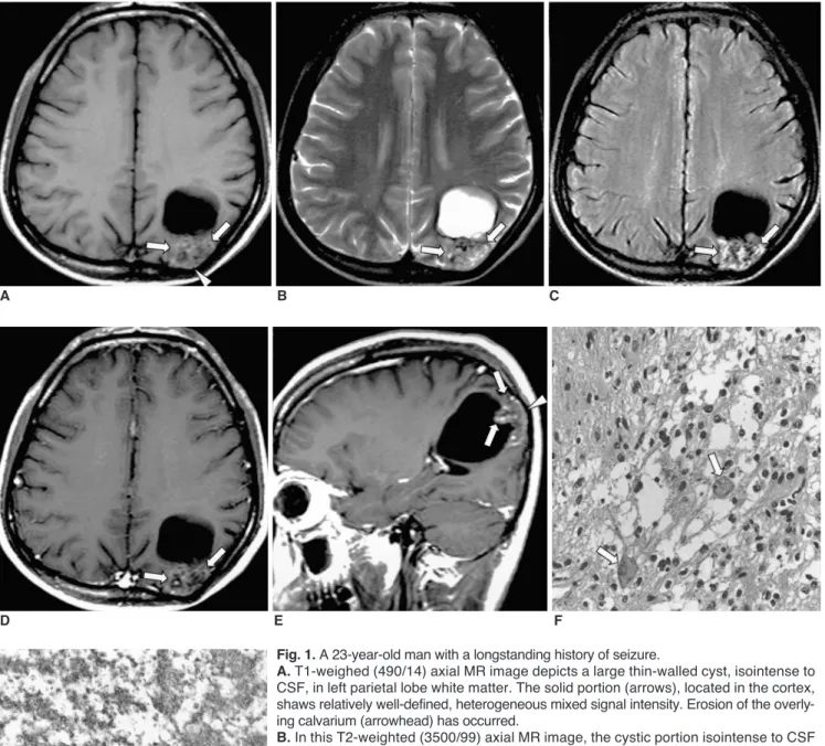
MR images indicated that in the left parietal lobe, a large, thin-walled cyst with a sol- id portion was present. The signals of the cystic portion were hypointense / hyperin- tense at T1-/ T2-weighted imaging, respectively, similar to those of cerebrospinal fluid (CSF) (Figs. 1A, B). The solid portion demonstrated heterogeneous mixed signal inten- sity and was located along the cortex, with erosion of the overlying calvarium. At FLAIR (fluid attenuated inversion recovery) imaging (Fig. 1C), the cystic portion also showed low signal intensity, similar to that of CSF, and heterogeneous high signal in- tensity of the solid portion was observed, with a lobulating contour. Gd-enhanced T1- weighted imaging revealed minimal enhancement of the solid portion (Figs. 1D, E), and there was no evidence of leptomeningeal or intraventricular seeding. The preoper- ative diagnosis was low-grade glioma, such as oligodendroglioma.
Gross total resection of the mass was performed using stereotactic instruments and intraoperative ultrasonography, and the tumor, together with a minimal amount of surrounding brain tissue, was removed. The solid portion was brownish in color and of relatively firm consistency. The patient’s post-operative course was uneventful.
Microscopically, the tumor was composed of small round cells with clear cytoplasm, Ji Hoon Shin, MD1
Ho Kyu Lee, MD1 Jung-Kyo Lee, MD2 Shin Kwang Khang, MD3 Choong Gon Choi, MD1 Dae Chul Suh, MD1
Index terms : Brain, neoplasms Brain, neoplasms, MR
Korean J Radiol 2002;3:214-217 Received May 8, 2002; accepted after revision March 18, 2002.
Departments of 1Radiology, 2Neuro- surgery, and 3Pathology, Asan Medical Center, University of Ulsan College of Medicine
Address reprint requests to :
Ho Kyu Lee, M.D., Department of Radiology, Asan Medical Center, 388-1 Poongnap-dong, Songpa-gu, Seoul 138- 736, South Korea.
Telephone: (822) 3010-4325 Fax: (822) 476-4719 E-mail: [email protected]
G
which were lobulated by a well-developed vascular net- work mimicking the histology of central neurocytoma or oligodendroglioma. Ganglioid differentiation was noted throughout the tumor and was represented by scattered in- dividual ganglion cells, or groups of these, among small round cells (Fig. 1F). The tumor cells were embedded in a
neurophil-like fibrillary background which was strongly immunoreactive for synaptophysin (Fig. 1G), and GFAP (glial fibrillary acidic protein) immunostaining disclosed that among them, numerous, large reactive astrocytes with stellate processes were present. All these histopathologic findings indicated the presence of a ganglioneurocytoma.
MR Imaging and Histopathologic Findings of A Case of Cerebral Ganglioneurocytoma
Korean J Radiol 3(3), September 2002 215
A B C
Fig. 1. A 23-year-old man with a longstanding history of seizure.
A. T1-weighed (490/14) axial MR image depicts a large thin-walled cyst, isointense to CSF, in left parietal lobe white matter. The solid portion (arrows), located in the cortex, shaws relatively well-defined, heterogeneous mixed signal intensity. Erosion of the overly- ing calvarium (arrowhead) has occurred.
B. In this T2-weighted (3500/99) axial MR image, the cystic portion isointense to CSF shows high signal intensity, while the solid portion shows heterogeneous high signal in- tensity (arrows).
C. FLAIR (9999/119) image more clearly demonstrates the heterogeneous high signal in- tensity of the solid portion (arrows).
D, E. Contrast-enhanced T1-weighed (490/14) axial (D) and sagittal (E) MR images show minimal enhancement of the solid portion (arrows).
F. Photomicrograph (H & E staining, 200) depicts ganglionic cells (arrows) among the small round cells.
G. Photomicrograph (synaptophysin staining, 200) reveals tumor cells embedded in a neurophil-like fibrillary background which is strongly immunoreactive for synaptophysin.
G
D E F
Surgery was performed five years ago, and the seizures have subsequently shown complete remission. MR images obtained one year ago revealed no residual tumor, and at that time, no tumor recurrence was noted.
DISCUSSION
Neuronal tumors of the central nervous system are known to show wide morphologic variability, and hence their histologic diagnosis is based on the degree of differen- tiation of their neuronal elements as well as the relative proportion of neuronal to glial elements within a single tu- mor (2, 3). Because of the difficulties in diagnosis due to the variability encountered at pathologic examination, this group of tumors has, however, been poorly understood, and there is still considerable controversy regarding their exact classification and nomenclature. A central neurocy- toma is characterized by its intraventricular location, a small uniform neoplastic cell population with features of neuronal differentiation, and the absence of any ganglionic neuronal cells (4, 5). In recent years, however, several cases of central neurocytomas with an extraventricular location and ganglioid differentiation have been reported (1, 5 8).
Hence, this tumor is composed at least of two distinctive cell types: large ganglionic and small round cells, both showing the morphologic characteristics of neuronal cells.
Cases of ganglioneurocytoma were first reported by Nishio et al. (5), in 1988, and von Deimling et al. (8), in 1990, but few radiologic descriptions of these tumors are available.
Patients who present with the symptoms of ganglioneu- rocytoma are mostly children or adults aged less than thir- ty. As in our case, the symptoms in the reported cases were nonspecific and varied according to the location of the tumor. They included seizure, increased intracranial pressure, neurologic deficit, and headache (1, 5 7). In our patient, the presenting symptom was longstanding partial complex seizure, and in view of his long history and the pressure erosion of the calvarium revealed by MR images, the tumor was presumed to be slow-growing and benign.
The MR imaging findings for this tumor are described in only two case reports. Funato et al. (1) stated that Gd-en- hanced T1-weighted MR imaging revealed a large cyst with an enhanced mural nodule, though did not mention T2- weighted MR imaging. The case reported by Chan et al. (6) involved a patient in whom a heterogeneous hyperintense mass was revealed by T2-weighted MR imaging, and ex- tensive, heterogeneous enhancement by Gd-enhanced T1- weighed MR imaging; the moderate mass effect mimicked a malignant tumor. The MR imaging findings in our case in- cluded those of both previous reports: the tumor had a large cystic area as well as a heterogeneous solid compo-
nent. The degree of enhancement, however, was less than previously reported, and it thus appears that this varies in both ganglioneurocytoma and other neuronal tumors. The reported CT findings, namely a large cystic mass with an enhancing solid nodule and occasional calcification, did not differ from those of other neuronal tumors (1, 5 7).
Although our patient did not undergo CT scanning, we in- ferred from MR imaging findings that the CT findings would be similar to those of previous reports.
Differentiation between this tumor and low-grade gliomas or ganglion cell tumors is difficult or even impossi- ble. Low-grade gliomas including oligodendroglioma can show similar imaging findings to those of this present case, though in a glioma a large cystic component is uncommon.
Ganglion cell tumors such as ganglioglioma or gangliocy- toma have been described as cystic masses with a variously enhancing solid portion, the so-called mural nodule, and occasional calcification (9, 10). In addition, it is important to distinguish ganglioneurocytomas from neoplasms con- taining neuroblastic elements: the latter have a less favor- able prognosis, and ganglioneurocytoma can sometimes mimic a malignant neoplasm (6).
Complete surgical excision is the treatment of choice, though post-operative radiotherapy is a possible adjunctive treatment (5, 7). Since, in our patient, there was no evi- dence of recurrence after four years, nor after seven years in a patient described in a previous report (5), this tumor is thought to have a favorable prognosis.
Acknowledgement: We are very grateful to Bonnie Hami, M.A., Department of Radiology, University Hospitals of Cleveland (U.S.A.) for her editorial assistance.
References
1. Funato H, Inoshita N, Okeda R, Yamamoto S, Aoyagi M. Cystic ganglioneurocytoma outside the ventricular region. Acta Neuropathol 1997;94:95-98
2. Nishio S, Takeshita I, Fukui M. Primary cerebral ganglioneuro- cytoma in an adult. Cancer 1990;66:358-362
3. Shimada H. Transmission and scanning electron microscopic studies on the tumors of neuroblastoma group. Acta Pathol Jpn 1982;32:415-426
4. Chang KH, Han MH, Kim DG, et al. MR appearance of central neurocytoma. Acta Radiol 1993;34:520-526
5. Nishio S, Tashima T, Takeshita I, Fukui M. Intraventricular neu- rocytoma: clinicopathological features of six cases. J Neurosurg 1988;68:665-670
6. Chan A, McAbee G, Queenan J, Manning A. Ganglioneurocy- toma mimicking a malignant tumor: case report with a literature review of the MRI appearance of neurocytomas and gangli- ogliomas. J Neuroimaging 2001;11:47-50
7. Biernat W, Zakrzewski K, Liberski PP. Twelve-year-old boy with recent onset seizures. Brain Pathol 2000;10:313-314, 319 8. von Deimling A, Janzer R, Kleihues P, Wiestler OD. Patterns of
differentiation in central neurocytoma: an immunohistochemical study of eleven biopsies. Acta Neuropathol 1990;79:473-479 Shin et al.
216 Korean J Radiol 3(3), September 2002
MR Imaging and Histopathologic Findings of A Case of Cerebral Ganglioneurocytoma
Korean J Radiol 3(3), September 2002 217
9. Castillo M, Davis PC, Takei Y, Hoffman JC Jr. Intracranial gan- glioglioma: MR, CT, and clinical findings in 18 patients. Am J Neuroradiol 1990;11:109-114
10. Kim HS, Lee HK, Jeong AK, Shin JH, Choi CG, Khang SK.
Supratentorial gangliocytoma mimicking extra-axial tumor: a re- port of two cases. Korean J Radiol 2001;2:108-112