Copyright © 2015 Korean Dementia Association 135 An 83-year-old male was admitted to the emergency de-
partment with two days of cognitive impairment. On initial presentation, his neurologic examination showed him to be
alert but disoriented with a mini-mental status examination score of zero. An initial CT and diffusion-weighted image of the brain showed no abnormalities. A chest X-ray showed a
Limbic Encephalitis with Hyperintense Cerebrospinal Fluid on Fluid Attenuated Inversion Recovery Images
Inha Hwang,1 In Joong Kim,2 Byung Joon Lee,3 Sang-Won Ha1
Departments of 1Neurology, 2Radiology, and 3Pulmonology, Veterans Health Service Medical Center, Seoul, Korea
Received: September 7, 2015 Revised: September 11, 2015 Accepted: September 11, 2015
Correspondence: Sang-Won Ha, MD, Department of Neurology, Veterans Health Service Medical Center, 53 Jinhwangdo-ro 61-gil, Gangdong-gu, Seoul 05368, Korea
Tel: +82-2-2225-4601, Fax: +82-2-2225-1327, E-mail: [email protected]
cc This is an Open Access article distributed under the terms of the Creative Commons Attribution Non-Commercial License (http://creativecommons.org/
licenses/by-nc/3.0) which permits unrestricted non-commercial use, distribution, and reproduction in any medium, provided the ori-ginal work is properly cited.
DND
Print ISSN 1738-1495 / On-line ISSN 2384-0757
Dement Neurocogn Disord 2015;14(3):135-136 / http://dx.doi.org/10.12779/dnd.2015.14.3.135
LETTER TO THE EDITOR
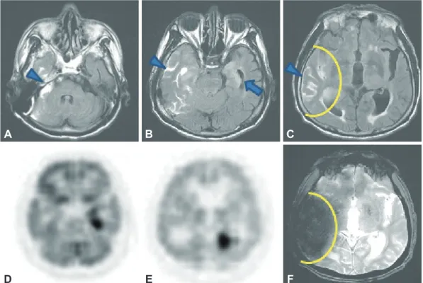
Fig. 1. Fluid-attenuated inversion recovery images (A), (B), and (C) show hyperintense cerebrospinal fluid (CSF) on right temporal area (ar- rowhead) and a high signal intensity lesion on left medial temporal area suspicious of limbic encephalitis (B) (arrow). Positron emission to- mography-computed tomography images (D) and (E) show an asymmetric increase of fluorodeoxyglucose uptake in the left medial tempo- ral area matching the high signal intensity lesion seen in the image (B, arrow). Gradient recalled echo T2-weighted image (F) shows magnetic susceptibility artifact matching hyperintense CSF, seen in fluid-attenuated inversion recovery image (C, yellow curved line).
A
D
B
E
C
F
Inha Hwang et al.
Limbic Encephalitis with Hyperintense CSF on FLAIR
136 Dement Neurocogn Disord 2015;14(3):135-136
3×3 cm mass on left upper lobe. Biopsy confirmed mass to be a squamous cell carcinoma. Examination of the cerebrospi- nal fluid (CSF) was within normal limits.
Fluid attenuated inversion recovery images (FLAIR) showed suspicious high signal intensity lesion on left medial temporal lobe and hyperintense CSF on right temporal area (Fig. 1A, B, and C). Axial T1- and T2-weighted images showed no abnor- mality on corresponding same region. Whole-body positron emission tomography (PET) showed glucose uptake on left medial temporal lobe indicating limbic encephalitis. At first glance, hyperintense CSF lesion on right temporal lobe were thought to be more remarkable than left medial temporal lobe lesions, causing confusion to diagnosis. With PET showing glucose uptake on left medial temporal lobe, we could sus- pect limibic encephalitis.
The term “hyperintense CSF” is used to describe failed sup- pression, or hyperintensity, of CSF on FLAIR of brain.1 Patho- logic conditions associated with hyperintense CSF includes
subarachnoid hemorrhage, meningitis, and acute infarction following gadolinium administration.1 Some non-pathologic conditions showing hyperintense CSF are images obtained during oxygen inhalation, CSF flow-related artifact.1 Gradient echo imaging showed magnetic susceptibility artifact match- ing hyperintense CSF seen in FLAIR image (Fig. 1). We reached an agreement that the lesion on FLAIR image is an artifact in the process of nulling CSF signal during acquiring FLAIR im- age. Knowledge of pathologic conditions of hyperintense CSF needed for correct diagnosis.
Conflicts of Interest
The authors have no financial conflicts of interest.
REFERENCE
1. Tha KK, Terae S, Kudo K, Miyasaka K. Differential diagnosis of hy- perintense cerebrospinal fluid on fluid-attenuated inversion recovery images of the brain. Part II: non-pathological conditions. Br J Radiol 2009;82:610-614.