Cardioprotective Effect of Fimasartan, a New Angiotensin Receptor Blocker, in a Porcine Model of Acute Myocardial Infarction
Cardioprotective effect of fimasartan, a new angiotensin receptor blocker (ARB), was evaluated in a porcine model of acute myocardial infarction (MI). Fifty swine were randomized to group 1 (sham, n = 10), group 2 (no angiotensin-converting enzyme inhibitor [ACEI] or ARB, n = 10), group 3 (perindopril 2 mg daily, n = 10), group 4 (valsartan 40 mg daily, n = 10), or group 5 (fimasartan 30 mg daily, n = 10). Acute MI was induced by occlusion of the left anterior descending artery for 50 min. Echocardiography, single photon emission computed tomography (SPECT), and F-18 fluorodeoxyglucose cardiac positron emission tomography (PET) were performed at baseline, 1 week, and 4 weeks. Iodine-123 meta-iodobenzylguanidine (MIBG) scan was done at 6 weeks for visualization of cardiac sympathetic activity. Left ventricular function and volumes at 4 weeks were similar between the 5 groups. No difference was observed in groups 2 to 5 in SPECT perfusion defect, matched and mismatched segments between SPECT and PET at 1 week and 4 weeks. MIBG scan showed similar uptake between the 5 groups. Pathologic analysis showed similar infarct size in groups 2 to 5. Infarct size reduction was not observed with use of fimasartan as well as other ACEI and ARB in a porcine model of acute MI.
Keywords: Angiotensin-Converting Enzyme Inhibitors; Angiotensin Receptor Blockers;
Myocardial Infarction Doo Sun Sim,1 Myung Ho Jeong,1
Ho Chun Song,2 Jahae Kim,2 Ari Chong,2 Hee Seung Bom,2 In Seok Jeong,3 Sang Gi Oh,3 Jong Min Kim,1 Dae Sung Park,1 Jung Ha Kim,1 Kyung Seob Lim,1 Min Suk Kim,1 Shi Hyun Ryu,1 Hyun Kuk Kim,1 Sung Soo Kim,1 Su Young Jang,1 Jae Yeong Cho,1 Hae Chang Jeong,1 Ki Hong Lee,1 Keun Ho Park,1 Nam Sik Yoon,1 Hyun Ju Yoon,1 Kye Hun Kim,1 Young Joon Hong,1 Hyung Wook Park,1 Ju Han Kim,1 Youngkeun Ahn,1 Jeong Gwan Cho,1 Jong Chun Park,1 and Jung Chaee Kang1
1The Heart Research Center of Chonnam National University Hospital Designated by Korea Ministry of Health, Welfare and Family Affairs, Gwangju;
2Department of Nuclear Medicine, and 3Department of Cardiothoracic Surgery, Chonnam National University Hospital, Gwangju, Korea Received: 11 June 2014
Accepted: 29 August 2014 Address for Correspondence:
Myung Ho Jeong, MD
Director of The Heart Research Center of Chonnam National University Hospital Designated by Korea Ministry of Health, Welfare and Family Affairs, 671 Jaebong-ro, Dong-gu, Gwangju 501-757, Korea
Tel: +82.62-220-6243, Fax: +82.62-228-7174 E-mail: myungho@chollian.net
Funding: This study was supported by grants of the Korean Health Technology R&D Project, Ministry of Health & Welfare (HI13C1527) and Boryung Pharmaceutical Co., Ltd, Seoul, Republic of Korea.
http://dx.doi.org/10.3346/jkms.2015.30.1.34 • J Korean Med Sci 2015; 30: 34-43
INTRODUCTION
The role of angiotensin receptor blockers (ARBs) in attenuating ischemia-reperfusion injury is not fully elucidated. It has been suggested that the myocardial protective ef- fects of ARB against ischemia-reperfusion injury is mediated by activation of angioten- sin II type 2 (AT2) receptors by angiotensin II, while the deleterious effects of AT1 re- ceptors are blocked by the ARB (1, 2), leading to release of bradykinin with downstream activation of protein kinase C, nitric oxide synthase and eicosanoid release (2-4).
Fimasartan (BR-A-657-K, KANARB®, Boryung Pharmaceutical Co., Ltd, Seoul, Re- public of Korea) is a new ARB developed for the first time in Korea. It is a selective block- er of AT1 receptor subtype and has shown rapid and potent antihypertensive effect in a number of clinical trials (5-8). Furthermore, anti-atherosclerotic effects of fimasartan were demonstrated in animal studies (9, 10). However, the efficacy of fimarsartan in acute MI is not elucidated. The goal of the present study is, therefore, to evaluate the ef- fect of fimasartan in a porcine model of acute myocardial infarction (MI).
MATERIALS AND METHODS Induction of acute MI
This study was conducted on a total of 50 swine at the animal catheterization laborato- ry of Chonnam National University Hospital in Gwangju, Korea between May 2011 and June 2013. Landrace swine (20-25 kg) were provided and observed in the animal breed- ing house of Chonnam National University Medical Institute for 3-5 days before the ex-
periment. All swine were given loading doses of aspirin (300 mg) and clopidogrel (300 mg) on the morning of the experiment, followed by aspirin 100 mg and clopidogrel 75 mg daily through- out the study period of 6 weeks. Experiment was done under anesthesia with zolazepam (2.5 mg/kg, intramuscular), tiletamine (2.5 mg/kg, intramuscular), xylazine (3 mg/kg, intramuscular), and azaperone (6 mg/kg, intramuscular). A 7F arterial sheath was placed in the left carotid artery under local anesthesia with 2% lidocaine. After infusion of 10,000 units of heparin, a 7F cor-
onary artery guiding catheter was placed within the opening of the coronary artery and baseline coronary angiogram was ob- tained under the fluoroscopic guidance by mobile C-arm (Phil- lips BV-25 Gold). Acute MI model was induced with inflation of a balloon (3.0×20 mm, Terumo Co. Tokyo, Japan) just distal to the first diagonal branch or the septal branch. Complete occlu- sion was maintained by balloon dilatation (up to 8 atmospheres) for 50 min (Fig. 1). During the experiment oxygen and normal saline were supplied continuously and the anesthesia maintain-
A
C
B
D Fig. 1. Coronary angiograms during and after induction of anterior wall myocardial infarction. (A) Baseline. (B) Occlusion of the mid-left anterior descending artery just distal to the diagonal branch (black arrowheads) with a 3.0 mm balloon (white arrowheads). (C) 50 min after occlusion. (D) Follow-up angiogram at 6 weeks.
ed with an additional administration of zolazepam (1.25 mg/
kg, intravenous injection), tiletamine (1.25 mg/kg, intravenous injection), xylazine (1.5 mg/kg, intravenous injection), and aza- perone (6 mg/kg, intramuscular injection). Continuous electro- cardiographic monitoring was performed to confirm normal ST segment at baseline and ST elevation during the ischemic period and to monitor occurrence of cardiac arrhythmia. After induction of acute MI, each swine was closely observed for 1 hr for development of ventricular tachycardia or fibrillation, after which the swine was carried back to the breeding house and monitored until recovery.
Study groups and medications
The swine were randomly divided into 5 groups: group 1 (sham operation without induction of acute MI, n = 10); group 2 (no ACEI or ARB post-MI, n = 10); group 3 (perindopril 2 mg daily post-MI, n = 10); group 4 (valsartan 40 mg daily post-MI, n = 10);
and group 5 (fimasartan 30 mg daily post-MI, n = 10). Drug do- sage was arbitrarily selected as half the initial dose in adult hu- mans with hypertension. Study medications were administered orally 6-12 hr after the experiment and maintained for 4 weeks afterwards. Given the rapid growth of the animals (10-15 kg wei- ght gain over a few weeks), the dose of each study medication was doubled after 2 weeks and maintained throughout the rest of the study period (Fig. 2).
Two-dimensional echocardiography
All swine underwent 2-dimensional transthoracic echocardio- graphic examination at baseline (before the procedure), 1 week, and 4 weeks after induction of acute MI (Fig. 2). Left ventricular ejection fraction (LVEF) and LV end-systolic volume (LVESV) and LV end-diastolic volume (LVEDV) were determined by mo- dified biplane Simpson’s rule in the 2- and 4-chamber views (11).
Considering the rapid growth of the animal, LVESV and LVEDV were normalized to the animal’s body surface area in order to more adequately present the data over time, as both volumes naturally increase with growth of the animal (12). A global dia-
stolic function was assessed using transmitral inflow parame- ters: peak early (E) and peak late (A) velocities, E-wave deceler- ation time (DT) and E/A ratio. In addition, tissue Doppler im- aging of mitral annular velocities (E´, A´) was measured. For the prediction of LV filling pressures, E/E´ was calculated (13).
Tc-99m sestamibi myocardial perfusion SPECT
All swine underwent technetium (Tc)-99m sestamibi myocar- dial perfusion single photon emission computed tomography (SPECT) at resting state 3 times at baseline (before the proce- dure) and 1 week and 4 weeks after the procedure (Fig. 2 and 3).
Resting ECG-gated Tc-99m sestamibi SPECT imaging was per- formed in concordance with standards of the American Society of Nuclear Cardiology (14). The swine were fasted overnight, Tc-99m sestamibi 111 MBq was injected intravenously at rest.
Forty minutes after the injection, the planar and SPECT images were acquired in the supine position with ECG-gated technique using eight frames for a cardiac cycle. The SPECT data was ac- quired using a dual-headed SMV DST-XLi gamma camera (GE Medical systems, Milwaukee, WI, USA) with low-energy, all purpose (LEAP) collimator, setting the energy photo-peak at 140 keV with a 20% symmetric window and a 90˚ acquisition arc. The SPECT acquisition was undertaken in 16 steps (32 pro- jections) and each step collect counts for 30 sec. Reconstruc- tion of the images was performed by filtered back projection using butterworth filter. After reconstruction by filtered back pro- jection using butterworth filter, transaxial slices along the verti- cal long axis, the horizontal long axis, and the short axis were ge- nerated.
F-18 FDG cardiac PET/CT
After Tc-99m sestamibi myocardial perfusion SPECT, F-18 fluo- rodeoxyglucose (FDG) cardiac positron emission tomography (PET)/computed tomography (CT) (FDG PET) was done (Fig. 2 and 3). Imaging was performed on a Discovery STE PET/CT sys- tem (GE medical systems, Milwaukee, WI, USA). Preparation was performed using simplified glucose/insulin loading (20 g
Fig. 2.
Fig. 2. Scheme of the study protocol. ACEI, angiotensin-converting enzyme inhibitor; ARB, angiotensin-receptor blocker; CAG, coronary angiography; 2DE, 2-dimensional echo- cardiography; MIBG scan, Iodine-123 meta-iodobenzylguanidine cardiac scintigraphy; PET, F-18 fluorodeoxyglucose cardiac positron emission tomography/computed tomogra- phy; SPECT, Tc-99m sestamibi myocardial perfusion single photon emission computed tomography; TTC, 2,3,5-triphenyl tetrazolium chloride.
of dextrose intravenously with simultaneous intravenous and subcutaneous insulin to adjust blood glucose to 5 mM/L) (15).
PET/CT acquisitions for the heart were started at 40 min after the injection of 7.4 MBq per kilogram of body weight. CT imag- es were acquired using parameters with a peak voltage of 140 keV, a tube current of 20 mA×16 sec, a rotation time of 1.0 sec, a field of view of 50 cm and a slice thickness of 3.27 mm. Immedi- ately following the CT acquisition, the PET data were acquired in the same anatomic locations in the 2D mode with acquisi- tion time of 15 min. The CT data were used for attenuation cor- rection and the images were reconstructed using a convention- al iterative algorithm (OSEM). A Xeleris workstation (GE medi- cal systems) providing multiplanar reformatted images was used for image display and analysis.
SPECT, PET data analysis
Perfusion SPECT and FDG PET which were done on the same day were evaluated simultaneously on the consensus of 2 inde- pendent nuclear physicians. LVEF was derived from the ECG- gated images from perfusion SPECT (16). Static tomographic images and polar maps were normalized to their maximum and used for visual analysis of regional perfusion/metabolism pat- terns using the American Heart Association 17-segment model.
Perfusion defect size was quantified on polar maps according to a method described earlier (17). Visual classification was done for normal, matched defect, or mismatched segments. Segments with decreased uptake on perfusion SPECT were visually de- fined as matched defect segments if FDG uptake was concor- dantly decreased on PET. Segments with decreased uptake on perfusion SPECT were defined as mismatch ed segments if FDG uptake was not concordantly decreased on PET. Perfusion de- fect assessment on SPECT was performed using an automated
quantification of total perfusion defect that was calculated as the percentage of the total surface area of the LV below the pre- defined uniform average deviation threshold weighted on a pixel-by-pixel basis by the severity of perfusion on a scale from 0 to 4 using Quantitative Perfusion SPECT (QPS) software (Ce- dars Sinai, Los Angeles, CA, USA).
I-123 MIBG cardiac scintigraphy
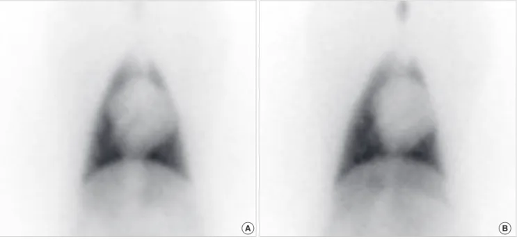
Two weeks after cessation of medication, Iodine-123 (I-123) meta-iodobenzylguanidine (MIBG) scintigraphy was done by method described previously (18). I-123 MIBG 111 MBq was injected intravenously. After 15 min postinjection, imaging was acquired using a dual-headed SMV DST-XLi gamma camera (GE Medical systems) (Fig. 4). A 10-min planar image acquisi- tion of the anterior thorax was performed at 15 min, followed immediately by SPECT image acquisition. These 2 procedures were later repeated starting at 3 hr 50 min post-injection (Fig. 4).
All imaging was performed with low-energy/high resolution collimators, with the camera peaked at 159 keV with a symmet- ric 20% energy window. Planar images were acquired at 30- and 70-degree left anterior oblique views and anterior views in 128×
128 matrix. SPECT images were obtained with a minimum of 60 projections (30 sec/stop) over a 180-degree arc. All SPECT images were stored in a 64×64 matrix, with the use of camera zoom factor as appropriate to achieve a nominal pixel size of 6.4 mm (range: 6.0 to 6.8 mm). The heart/mediastinum count (H/
M) ratio was determined from the delayed anterior planar I-123 MIBG image. The heart region of interest (ROI) was drawn man- ually to include all visible ventricular activity. A square medias- tinal ROI (7 ×7 pixels) was drawn in the upper mediastinum, using the apices of the lungs as anatomic landmarks. The H/M ratio was calculated as the ratio of the counts/pixel in the 2
A B
SPECT FDG PET SPECT FDG PET
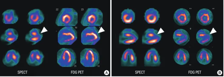
Fig. 3. SPECT and PET images at 1 week (A) and 4 weeks (B) showing anterior wall myocardial infarction. (A) At 1 week, SPECT shows moderate to severe, large perfusion de- fect in the apex and anterior wall (arrowhead). PET reveals preserved FDG metabolism in the apex and anterior wall (arrowhead), demonstrating perfusion-metabolism mismatch pattern, which reflects viable myocardium. (B) At 4 weeks, SPECT shows remaining perfusion defect in the apex and anterior wall. PET reveals reduced FDG uptake in the apex and anterior wall, demonstrating perfusion-metabolism match pattern, which reflects nonviable myocardium. PET, F-18 fluorodeoxyglucose cardiac positron emission tomogra- phy/computed tomography; SPECT, Tc-99m sestamibi myocardial perfusion single photon emission computed tomography.
ROIs. The heart/mediastinum count (H/M) ratio was determin- ed from the delayed anterior planar I-123 MIBG image. The wash- out rate (WR) was calculated using the following formula: ([H/
M]early – [H/M]late])/(H/M)early × 100 (%).
Pathological examination of infarcted myocardium Six weeks later, a follow-up coronary angiography was perform- ed according to the same protocol described above, after which the swine was sacrificed and the heart was extracted. The ex- tracted heart was rinsed, and the myocardial sections including the left and right ventricle were obtained at 1-cm intervals us-
ing a microtome knife. The myocardial sections were incubated in 2,3,5-triphenyl tetrazolium chloride (TTC) solution until por- tions of viable myocardium turned brick red (Fig. 5). The speci- mens were reviewed by a cardiac pathologist for pathological changes in the infarcted myocardium. Histomorphometric in- farct size was estimated on digital photographs of TTC staining by outlining the LV area and TTC negative infarct area. Infarct size was then expressed as % LV area (19).
Study endpoints
The primary end-point of the study was scintigraphic and patho- Fig. 4. I-123 MIBG cardiac scintigraphy at 6 weeks. Planar images in the anterior view, acquired at 15 min (A) and 3 hr 50 min (B) after I-123 MIBG injection. Heart-to-medias- tinum ratios were 1.64 and 1.51 at early and late phases with washout rate of 29%, indicating reduced cardiac sympathetic nerve activity. I-123 MIBG, Iodine-123 meta-iodo- benzylguanidine.
A B
Fig. 5. 2,3,5-triphenyl tetrazolium chloride staining at 6 weeks showing areas of infarcted myocardium in the anterior wall (arrowheads).
logic myocardial infarct size. Secondary end-points were LV func- tion and volumes on echocardiography and sympathetic nerve activity on MIBG scintigraphy.
Statistical analysis
All data were expressed as mean ± standard deviation. Analysis was performed using SPSS statistical software, version 18.0 (SPSS Inc., Chicago, IL, USA). One-way ANOVA with Bonferro- ni’s post hoc test for multiple comparisons was used to test for within-group and between-group comparisons. For all statisti- cal analyses, the null hypothesis was rejected at the 95% confi- dence level, considering a P value < 0.05 significant. To avoid type II errors (missing significant differences when the sample size is too small) we determined the required sample size for the present study using an approximation for α equal to 0.05 and power to 0.90 (20). Accordingly, 9 swine per group was the minimal sample size to detect differences of 10 mL in infarct size with a standard deviation of 5.1 based on our previous data recorded in identical experimental conditions (21). To compen- sate for possible dropouts, 10 animals were investigated in each group.
Ethics statement
All animals received humane care. All procedures were approv-
ed by the Institutional Animal Care and Use Committee (IACUC) of Chonnam National University Hospital (IACUC approval No.
CNU IACUC-H-2010-19).
RESULTS
The results of SPECT and PET scans are described in Table 1.
Heart rates were not different between the groups at baseline, 1 week, and 4 weeks. No significant difference was observed in SPECT perfusion defect extent, total perfusion defect, number of SPECT and PET defects across the groups at baseline, 1 week, and 4 weeks. The number of matched and mismatched seg- ments between SPECT and PET at 1 week and 4 weeks was not different in groups 2 to 5, suggesting similar infarct size and via- bility. On MIBG scan at 6 weeks, groups 2 to 5 showed compa- rable H/M ratios for I-123 MIBG cardiac uptake at early (15 min) and late (3 hr 50 min) phases without significant difference in washout rate, indicating similar recovery of cardiac sympathetic nerve activity after acute MI (Table 2). Two-dimensional echo- cardiographic study revealed no difference in LV function and volumes at baseline (Table 3). At 1 week, LVEF was significantly lower in group 2 to 5 ranging from 29.3% ± 7.4% to 38.6% ± 14.7%, compared to sham control group (57.2% ± 7.1%) (P < 0.001). At 4 weeks, however, LV systolic and diastolic functions improved Table 1. SPECT and PET data of the experimental animals
Parameters Sham control (n = 10) AMI only (n = 10) Perindopril (n = 10) Valsartan (n = 10) Fimasartan (n = 10) P value Baseline
Heart rate (bpm) 61.0 ± 32.0 51.0 ± 11.4 49.5 ± 5.4 47.0 ± 12.8 65.8 ± 29.4 0.535
SPECT perfusion defect extent (%) 18.1 ± 5.2 18.0 ± 3.5 21.0 ± 2.9 19.8 ± 4.2 17.8 ± 5.7 0.653
SPECT total perfusion defect (%) 13.2 ± 3.4 13.0 ± 2.0 14.6 ± 2.0 13.8 ± 3.2 12.5 ± 3.5 0.742
Number of segments SPECT defect PET defect Match Mismatch
0 1.3 ± 1.2
0 1.3 ± 1.2
0 0.5 ± 1.2
0 0.5 ± 1.2
0 0.5 ± 1.2
0 0.5 ± 1.2
0 1.0 ± 1.2
0 1.0 ± 1.2
0 1.0 ± 1.2
0 1.0 ± 1.2
0.730 0.730 1 week
Heart rate (bpm) 61.3 ± 27.3 45.3 ± 7.9 44.3 ± 10.1 62.2 ± 25.5 53.5 ± 11.2 0.513
SPECT perfusion defect extent (%) 16.4 ± 3.2 17.9 ± 3.1 18.2 ± 4.1 18.1 ± 2.0 20.0 ± 1.8 0.332
SPECT total perfusion defect (%) 12.1 ± 2.3 13.3 ± 2.0 13.3 ± 2.4 13.6 ± 1.1 15.2 ± 2.0 0.143
Number of segments SPECT defect PET defect Match Mismatch
0 1.9 ± 1.1
0 1.9 ± 1.1
0 1.3 ± 2.0
0 1.3 ± 2.0
0 6.5 ± 6.8
0 4.5 ± 6.7
0 3.0 ± 5.4
0 3.0 ± 5.4
0.2 2.0 ± 1.4
0.2 1.8 ± 1.6
0.356 0.181 0.356 0.625 4 weeks
Heart rate (bpm) 57.3 ± 22.7 54.9 ± 15.1 71.0 ± 12.6 51.5 ± 2.1 54.6 ± 20.1 0.440
SPECT perfusion defect extent (%) 15.7 ± 5.8 14.9 ± 4.2 20.0 ± 2.4 15.6 ± 4.5 20.8 ± 8.7 0.208
SPECT total perfusion defect (%) 11.7 ± 4.1 11.3 ± 2.6 14.7 ± 1.5 11.6 ± 2.9 16.2 ± 6.4 0.108
Number of segments SPECT defect PET defect Match Mismatch
0 2.9 ± 3.3
0 2.9 ± 3.3
0 0.6 ± 1.1
0 0.6 ± 1.1
0 3.7 ± 3.4
0 3.7 ± 3.4
0 2.1 ± 1.3
0 2.1 ± 1.3
0.5 ± 0.8 2.8 ± 0.8 0.5 ± 0.8 2.3 ± 0.5
0.069 0.166 0.069 0.179 AMI, acute myocardial infarction; bpm, beats per minute; PET, F-18 fluorodeoxyglucose cardiac positron emission tomography/computed tomography; SPECT, Tc-99m sestamibi myocardial perfusion single photon emission computed tomography.
Fig. 6. Infarct size by pathology between groups 2-5. Bonferroni’s post hoc test be- tween AMI control (group 2) and AMI with use of perindopril, valsartan, and fimasar- tan (groups 3-5). AMI, acute myocardial infarction.
%LV area
25
20
15
10
5
0 AMI only (n = 10)
Perindopril (n = 10)
Valsartan (n = 10)
Fimasartan (n = 10) P = 0.454 P = 0.630
P = 0.872
Table 2. Results of I-123 MIBG cardiac scintigraphy at 6 weeks
Parameters Sham control (n = 10) AMI only (n = 10) Perindopril (n = 10) Valsartan (n = 10) Fimasartan (n = 10) P value
H/M ratio (15 min) 21.8 ± 10.7 21.1 ± 12.3 25.5 ± 11.7 26.3 ± 8.6 24.5 ± 7.5 0.854
H/M ratio (3 hr 50 min) 19.0 ± 12.2 19.0 ± 8.9 22.0 ± 6.6 20.9 ± 5.8 22.2 ± 5.7 0.916
Washout (%) 10.0 ± 33.9 2.1 ± 25.5 -1.1 ± 50.4 12.4 ± 41.1 8.0 ± 10.7 0.953
AMI, acute myocardial infarction; H/M ratio, heart/mediastinum count ratio; I-123 MIBG, Iodine-123 meta-iodobenzylguanidine.
Table 3. Two-dimensional echocardiography results
Parameters Sham control (n = 10) AMI only (n = 10) Perindopril (n = 10) Valsartan (n = 10) Fimasartan (n = 10) P value Baseline
LVEF (%)
LVEDV index (mL/m2) LVESV index (mL/m2) E/A
E/E´
58.0 ± 4.2 52.1 ± 16.2 21.9 ± 7.4
1.0 ± 0.4 13.7 ± 4.9
59.2 ± 5.7 59.2 ± 23.3 24.2 ± 9.9
1.0 ± 0.5 13.0 ± 5.8
55.8 ± 5.6 63.3 ± 31.5 27.2 ± 11.3 0.9 ± 0.2 11.4 ± 3.8
61.3 ± 6.5 68.6 ± 26.3 27.6 ± 14.5 1.1 ± 0.3 12.3 ± 2.7
61.2 ± 6.9 60.6 ± 10.8 23.7 ± 6.1
1.1 ± 0.3 11.7 ± 3.8
0.432 0.747 0.825 0.902 0.865 1 week
LVEF (%)
LVEDV index (mL/m2) LVESV index (mL/m2) E/A
E/E´
57.2 ± 7.1 52.4 ± 13.3 22.9 ± 8.5
0.9 ± 0.3 13.4 ± 4.1
32.9 ± 7.0 66.1 ± 20.3 43.8 ± 11.8 0.6 ± 0.2 12.9 ± 3.4
29.3 ± 7.4 62.4 ± 12.3 44.2 ± 10.6 0.8 ± 0.3 11.4 ± 3.1
37.0 ± 5.6 66.2 ± 17.7 42.3 ± 14.5 1.0 ± 0.5 12.3 ± 4.5
38.6 ± 14.7 69.3 ± 27.6 45.0 ± 24.0 0.8 ± 0.1 11.9 ± 2.7
< 0.001 0.530 0.051 0.201 0.878 4 weeks
LVEF (%)
LVEDV index (mL/m2) LVESV index (mL/m2) E/A
E/E´
51.2 ± 9.9 39.7 ± 13.0 19.7 ± 9.5
1.0 ± 0.2 10.5 ± 2.8
56.4 ± 19.6 54.9 ± 18.8 26.4 ± 18.7 0.9 ± 0.1 11.1 ± 4.2
56.9 ± 6.9 58.6 ± 28.1 26.3 ± 14.3 1.2 ± 0.3 9.9 ± 2.7
57.5 ± 15.2 53.0 ± 17.0 24.3 ± 15.4 1.2 ± 0.4 10.7 ± 2.9
57.7 ± 19.3 55.3 ± 17.7 23.7 ± 14.3 1.2 ± 0.4 10.3 ± 2.6
0.926 0.445 0.917 0.304 0.969 AMI, acute myocardial infarction; LVEDV, left ventricular end-diastolic volume; LVEF, left ventricular ejection fraction; LVESV, left ventricular end-systolic volume.
Table 4. Pathologic results
Parameters Sham control (n = 10) AMI only (n = 10) Perindopril (n = 10) Valsartan (n = 10) Fimasartan (n = 10) P value*
LV area (mm2) 0 2,052 ± 3,131 3,163 ± 3,545 2,060 ± 3,569 1,931 ± 3,046 0.387
Infarct area (mm2) 0 150 ± 184 297 ± 322 111 ± 167 97 ± 111 0.291
Infarct size (%LV area) 0 7.3 ± 9.2 9.4 ± 10.5 5.4 ± 9.2 5.0 ± 7.8 0.393
*Comparison was.
to normal limits without between-group differences (Table 3).
Pa thologic analysis showed no significant difference in infarct
size in groups 2 to 5 ranging from 5.0% ± 7.8% to 9.4% ± 10.5%
of LV area (P = 0.393) (Table 4, Fig. 6).
DISCUSSION
In the present study, use of fimasartan, a new ARB, after acute MI did not reduce infarct size in swine, compared to control groups. Myocardial viability, cardiac sympathetic nerve activity, LV function by SPECT, PET, MIBG scan and echocardiography as well as infarct size by SPECT, PET, and pathologic studies were not different between ACEI, ARB, and control groups.
Fimasartan (BR-A-657-K, KANARB®) is a new ARB developed for the first time in Korea. It is a selective blocker of AT1 recep- tor subtype and has shown rapid and potent antihypertensive effect in clinical trials (5-8). Fimasartan was recently approved by Korea Food and Drug Administration for its safe and potent blood pressure lowering effect in patients with hypertension.
However, there is a paucity of data on the efficacy of fimarsartan in atherosclerosis including acute MI. Lee et al. (9) reported that fimasartan attenuated atherosclerosis progression and reduced
macrophage accumulation in the rabbit aortic plaques. Han et al. (10) observed that fimasartan pretreatment reduced isch- emia/reperfusion injury and infarct size in rat hearts, indicating that fimasartan may prevent mitochondrial dysfunction and apoptosis accompanied by ischemia/reperfusion injury.
There have been inconsistent results concerning the effects of angiotensin inhibition on infarct size in animal models of acute MI (1-3, 17, 22-27). In canine models, captopril adminis- tered after coronary occlusion reduced infarct size (17). In rat models of acute MI, losartan and ramiprilat protected the heart against ischemia/reperfusion injury independently of their he- modynamic effects but in a time-dependent manner (25). In contrast, several studies failed to demonstrate reduced infarct size with ramiprilat administered before, during, or after induc- tion of myocardial ischemia in canine and mice hearts (26-28).
The failure to reduce infarct size in the present study utilizing porcine models may in part be explained from several perspec- tives. Firstly, angiotensin inhibition may exert time-dependent cardioprotective effects. Zhu et al. observed that pretreatment with losartan for 4 and 10 weeks, compared to pretreatment for 1 day or 1 week, before induction of acute MI reduced infarct size in a rat model of ischemia-reperfusion, suggesting time-depen- dent cardiovascular protective effects of losartan (29, 30). In the present study, ACEI and ARB were administered orally several hours after acute MI when the swine recovered from the anes- thesia. This may have attenuated the effect of the study drugs in terms of protection from ischemia-reperfusion injury. Second- ly, the doses of ACEI and ARB were arbitrarily chosen as half the initial dose in humans for essential hypertension and were doubled at 2 weeks considering the rapid growth of the swine.
Due to lack of non-invasive methods available for serial blood pressure measurement in swine, we were unable to find the op- timal tolerable doses that would maximize the beneficial effects of ACEI and ARB post-MI. On the contrary, the cardioprotective effect assessed by infarct size in the present study may have been confounded by the potent antihypertensive effect of the study drugs leading to increased infarct size compared to that of the AMI only group. However, since blood pressure measurements were not available in this study, it is difficult to provide evidence to support the impact of hemodynamic change on infarct size.
Thirdly, most of the swine with persistent severe LV dysfunction after MI failed to survive and were dropped out early in the study period (overall mortality 45.6%) and the majority of the swine which completed the study showed preserved LV function. This again may have diluted cardioprotective effect of ACEI and ARB, which would play a major role in ischemic heart failure post- MI, leading to only modest infarct size ranging from 5%-9% of LV. Still, successful creation of a porcine acute MI model with severe LV dysfunction is a technical challenge with high peri- procedural mortality. Finally, cardioprotective effect failed to be shown possibly due to the shorter duration of the study. The
effect of blocking the renin-angiotensin-aldosterone system may take a longer period to be significant as shown in The On- going Telmisartan Alone and in Combination with Ramipril Global Endpoint Trial (ONTARGET) (31, 32). In addition, the favorable effect of this class of drugs in heart failure and renal protection supports that a longer duration may be needed in order to show its benefit. Nevertheless, the rapidly growing swine and the failure of the animal to survive the acute events as a re- sult of severe heart failure reflects the difficulty of conducting this study. It remains to be further investigated whether cardio- vascular protective benefits of ACEI and ARB including fima- sartan are dependent on the time and duration of angiotensin inhibition and the drug doses before, during, or after induction of myocardial ischemia. In conclusion, the present study did not demonstrate infarct size reduction with use of a new ARB, fimasartan, as well as other ACEI and ARB in a porcine model of acute MI. The time- and dose-dependent cardiovascular pro- tective effects of angiotensin inhibition needs to be determined.
DISCLOSURE
The authors have no conflicts of interest to disclose.
AUTHOR CONTRIBUTION
Conceived and designed the experiments: DS Sim, MH Jeong, JG Cho, JC Park, JC Kang. Performed the experiments: DS Sim, MH Jeong, HC Song, J Kim, A Chong, IS Jeong, SG Oh, JM Kim, DS Park, JH Kim, KS Lim, MS Kim, SH Ryu, HK Kim, SS Kim, SY Jang, JY Cho, HC Jeong, KH Lee, KH Park, NS Yoon, HJ Yoon, KH Kim, YJ Hong, HW Park, JH Kim, Y Ahn. Analyzed the data: DS Sim, HC Song, J Kim, A Chong, JH Kim. Contributed reagents/
materials/analysis tools: HC Song, J Kim, A Chong, HS Bom. Wrote the first draft of the manuscript: DS Sim, MH Jeong. Wrote the paper: DS Sim, MH Jeong. Manuscript approval: all authors.
ORCID
Doo Sun Sim http://orcid.org/0000-0003-4162-7902 Myung Ho Jeong http://orcid.org/0000-0003-4173-1494 Ho Chun Song http://orcid.org/0000-0002-6407-8160 Jahae Kim http://orcid.org/0000-0003-4077-7722 Ari Chong http://orcid.org/0000-0001-9327-9810 In Seok Jeong http://orcid.org/0000-0002-2249-0667 Sang Gi Oh http://orcid.org/0000-0001-9394-4980 Dae Sung Park http://orcid.org/0000-0002-9948-7546 Jung Ha Kim http://orcid.org/0000-0003-3334-3432 Kyung Seob Lim http://orcid.org/0000-0002-9117-887X Shi Hyun Ryu http://orcid.org/0000-0001-8376-5946 Hyun Kuk Kim http://orcid.org/0000-0002-4554-041X Sung Soo Kim http://orcid.org/0000-0002-5190-227X
Su Young Jang http://orcid.org/0000-0001-5843-0802 Jae Yeong Cho http://orcid.org/0000-0002-9393-2821 Hae Chang Jeong http://orcid.org/0000-0002-1273-3864 Ki Hong Lee http://orcid.org/0000-0002-9938-3464 Nam Sik Yoon http://orcid.org/0000-0001-9112-150X Young Joon Hong http://orcid.org/0000-0003-0192-8161 Hyung Wook Park http://orcid.org/0000-0002-9630-0467 Jong Chun Park http://orcid.org/0000-0002-3168-2054
REFERENCES
1. Jalowy A, Schulz R, Dörge H, Behrends M, Heusch G. Infarct size reduc- tion by AT1-receptor blockade through a signal cascade of AT2-receptor activation, bradykinin and prostaglandins in pigs. J Am Coll Cardiol 1998;
32: 1787-96.
2. Jugdutt BI, Menon V. AT2 receptor and apoptosis during AT1 receptor blockade in reperfused myocardial infarction in the rat. Mol Cell Biochem 2004; 262: 203-14.
3. Jugdutt BI, Balghith M. Enhanced regional AT(2)-receptor and PKC (ep- silon) expression during cardioprotection induced by AT(1)-receptor block- ade after reperfused myocardial infarction. J Renin Angiotensin Aldoste- rone Syst 2001; 2: 134-40.
4. Sato M, Engelman RM, Otani H, Maulik N, Rousou JA, Flack JE 3rd, De- aton DW, Das DK. Myocardial protection by preconditioning of heart with losartan, an angiotensin II type 1-receptor blocker: implication of bradykinin-dependent and bradykinin-independent mechanisms. Cir- culation 2000; 102: Iii346-51.
5. Lee SE, Kim YJ, Lee HY, Yang HM, Park CG, Kim JJ, Kim SK, Rhee MY, Oh BH; Investigators. Efficacy and tolerability of fimasartan, a new an- giotensin receptor blocker, compared with losartan (50/100 mg): a 12- week, phase III, multicenter, prospective, randomized, double-blind, par- allel-group, dose escalation clinical trial with an optional 12-week ex- tension phase in adult Korean patients with mild-to-moderate hyperten- sion. Clin Ther 2012; 34: 552-68, 68. e1-9.
6. Lee H, Yang HM, Lee HY, Kim JJ, Choi DJ, Seung KB, Jeon ES, Ha JW, Rim SJ, Park JB, et al. Efficacy and tolerability of once-daily oral fima- sartan 20 to 240 mg/d in Korean patients with hypertension: findings from Two Phase II, randomized, double-blind, placebo-controlled stud- ies. Clin Ther 2012; 34: 1273-89.
7. Park JB, Sung KC, Kang SM, Cho EJ. Safety and efficacy of fimasartan in patients with arterial hypertension (Safe-KanArb study): an open-label observational study. Am J Cardiovasc Drugs 2013; 13: 47-56.
8. Lee H, Kim KS, Chae SC, Jeong MH, Kim DS, Oh BH. Ambulatory blood pressure response to once-daily fimasartan: an 8-week, multicenter, ran- domized, double-blind, active-comparator, parallel-group study in Ko- rean patients with mild to moderate essential hypertension. Clin Ther 2013; 35: 1337-49.
9. Lee JY, Lee CW, Kim WJ, Ahn JM, Park DW, Kang SJ, Lee SW, Kim YH, Son WC, Jung S, et al. Antiatherosclerotic effects of the novel angiotensin receptor antagonist Fimasartan on plaque progression and stability in a rabbit model: a double-blind placebo-controlled trial. J Cardiovasc Phar- macol 2013; 62: 229-36.
10. Han J, Park SJ, Thu VT, Lee SR, Long le T, Kim HK, Kim N, Park SW, Jeon ES, Kim EJ, et al. Effects of the novel angiotensin II receptor type I antag-
onist, fimasartan on myocardial ischemia/reperfusion injury. Int J Car- diol 2013; 168: 2851-9.
11. Schiller NB, Shah PM, Crawford M, DeMaria A, Devereux R, Feigen- baum H, Gutgesell H, Reichek N, Sahn D, Schnittger I, et al. Recommen- dations for quantitation of the left ventricle by two-dimensional echo- cardiography. American Society of Echocardiography Committee on Standards, Subcommittee on Quantitation of Two-Dimensional Echo- cardiograms. J Am Soc Echocardiogr 1989; 2: 358-67.
12. Hawk CT, Leary SL, Morris TH; American College of Laboratory Ani- mal Medicine; European College of Laboratory Animal Medicine. For- mulary for laboratory animals. 3rd ed. Ames, Iowa: Blackwell Pub., 2005.
13. Wang J, Khoury DS, Thohan V, Torre-Amione G, Nagueh SF. Global dia- stolic strain rate for the assessment of left ventricular relaxation and fill- ing pressures. Circulation 2007; 115: 1376-83.
14. American Society of Nuclear Cardiology. Imaging Guidelines Overview:
ASNC Imaging Guidelines for Nuclear Cardiology Procedures. Available at http://www.asnc.org/content_1884.cfm [accessed on 13 November 2007].
15. Lautamäki R, Schuleri KH, Sasano T, Javadi MS, Youssef A, Merrill J, Ne- kolla SG, Abraham MR, Lardo AC, Bengel FM. Integration of infarct size, tissue perfusion, and metabolism by hybrid cardiac positron emis- sion tomography/computed tomography: evaluation in a porcine model of myocardial infarction. Circ Cardiovasc Imaging 2009; 2: 299-305.
16. Liu YH, Sinusas AJ, Khaimov D, Gebuza BI, Wackers FJ. New hybrid count- and geometry-based method for quantification of left ventricular volumes and ejection fraction from ECG-gated SPECT: methodology and valida- tion. J Nucl Cardiol 2005; 12: 55-65.
17. Nuyts J, Mortelmans L, Suetens P, Oosterlinck A, de Rou M. Model-based quantification of myocardial perfusion images from SPECT. J Nucl Med 1989; 30: 1992-2001.
18. Jacobson AF, Lombard J, Banerjee G, Camici PG. 123I-mIBG scintigra- phy to predict risk for adverse cardiac outcomes in heart failure patients:
design of two prospective multicenter international trials. J Nucl Cardiol 2009; 16: 113-21.
19. Fonge H, Vunckx K, Wang H, Feng Y, Mortelmans L, Nuyts J, Bormans G, Verbruggen A, Ni Y. Non-invasive detection and quantification of acute myocardial infarction in rabbits using mono-[123I]iodohypericin micro- SPECT. Eur Heart J 2008; 29: 260-9.
20. Dawson B, Trapp RG. Basic & clinical biostatistics. 4th ed. New York:
Lange Medical Books-McGraw-Hill, Medical Pub. Division, 2004.
21. Sim DS, Jeong MH, Kim YH, Choi S, Lim KS, Kim JH, Cho KH, Kim MC, Kim HK, Kim SS, et al. Effects of sildenafil in combination with angio- tensin-converting enzyme inhibitor on limiting infarct expansion in a porcine model of acute myocardial infarction. Int J Cardiol 2011; 146:
459-60.
22. Weidenbach R, Schulz R, Gres P, Behrends M, Post H, Heusch G. En- hanced reduction of myocardial infarct size by combined ACE inhibition and AT(1)-receptor antagonism. Br J Pharmacol 2000; 131: 138-44.
23. Messadi-Laribi E, Griol-Charhbili V, Gaies E, Vincent MP, Heudes D, Meneton P, Alhenc-Gelas F, Richer C. Cardioprotection and kallikrein- kinin system in acute myocardial ischaemia in mice. Clin Exp Pharma- col Physiol 2008; 35: 489-93.
24. Messadi-Laribi E, Griol-Charhbili V, Pizard A, Vincent MP, Heudes D, Meneton P, Alhenc-Gelas F, Richer C. Tissue kallikrein is involved in the cardioprotective effect of AT1-receptor blockade in acute myocardial isch-
emia. J Pharmacol Exp Ther 2007; 323: 210-6.
25. Safari F, Hajizadeh S, Shekarforoush S, Bayat G, Foadoddini M, Khosh- baten A. Influence of ramiprilat and losartan on ischemia reperfusion injury in rat hearts. J Renin Angiotensin Aldosterone Syst 2012; 13: 29-35.
26. Black SC, Driscoll EM, Lucchesi BR. Effect of ramiprilat or captopril on myocardial infarct size: assessment in canine models of ischemia alone and ischemia with reperfusion. Pharmacology 1998; 57: 35-46.
27. Lange SA, Wolf B, Schober K, Wunderlich C, Marquetant R, Weinbrenner C, Strasser RH. Chronic angiotensin II receptor blockade induces cardio- protection during ischemia by increased PKC-epsilon expression in the mouse heart. J Cardiovasc Pharmacol 2007; 49: 46-55.
28. Xu J, Carretero OA, Shesely EG, Rhaleb NE, Yang JJ, Bader M, Yang XP.
The kinin B1 receptor contributes to the cardioprotective effect of angio- tensin-converting enzyme inhibitors and angiotensin receptor blockers in mice. Exp Physiol 2009; 94: 322-9.
29. Zhu B, Sun Y, Sievers RE, Browne AE, Lee RJ, Chatterjee K, Parmley WW.
Effects of different durations of pretreatment with losartan on myocardi- al infarct size, endothelial function, and vascular endothelial growth factor. J Renin Angiotensin Aldosterone Syst 2001; 2: 129-33.
30. Zhu B, Sun Y, Sievers RE, Browne AE, Pulukurthy S, Sudhir K, Lee RJ, Chou TM, Chatterjee K, Parmley WW. Comparative effects of pretreat- ment with captopril and losartan on cardiovascular protection in a rat model of ischemia-reperfusion. J Am Coll Cardiol 2000; 35: 787-95.
31. ONTARGET Investigators, Yusuf S, Teo KK, Pogue J, Dyal L, Copland I, Schumacher H, Dagenais G, Sleight P, Anderson C. Telmisartan, ramipril, or both in patients at high risk for vascular events. N Engl J Med 2008;
358: 1547-59.
32. Mann JF, Schmieder RE, McQueen M, Dyal L, Schumacher H, Pogue J, Wang X, Maggioni A, Budaj A, Chaithiraphan S, et al. ONTARGET in- vestigators. Renal outcomes with telmisartan, ramipril, or both, in peo- ple at high vascular risk (the ONTARGET study): a multicentre, rando- mised, double-blind, controlled trial. Lancet 2008; 372: 547-53.