Case Report The Korean Journal of Sports Medicine 2012;30(2):148-151 http://dx.doi.org/10.5763/kjsm.2012.30.2.148
148 대한스포츠의학회지
관절경하 반월연골판 부분절제술 후 발생한 총장골정맥의 심부정맥 혈전증의 증례보고
국민건강보험공단 일산병원 정형외과
1, 연세대학교 의과대학 정형외과학교실
2유주형
1
ㆍ장기준2
ㆍ이윤태1
ㆍ박상훈1
ㆍ심동우2
A Case of Deep Vein Thrombosis in Common Iliac Vein after Arthroscopic Partial Meniscectomy
Ju-Hyung Yoo, MD 1 , Ki-Joon Jang, MD 2 , Yun-Tae Lee, MD 1 , Sang-Hoon Park, MD 1 , Dong-Woo Shim, MD 2
1
Department of Orthopedic Surgery, National Health Insurance Corporation Ilsan Hospital, Goyang,
2
Department of Orthopedic Surgery, Yonsei University College of Medicine, Seoul, Korea
Knee arthroscopy is generally considered a very safe operation with very high success rates. Few reported cases of complications arising from only arthroscopic partial meniscectomy include deep vein thrombosis and pulmonary embolism. In this study, we present the case of a 44-year-old female patient with complications of deep vein thrombosis arising after undergoing an arthroscopic partial meniscectomy. Eight days post-operation, the patient presented with pain and swelling of the lower limb and inguinal area of the same side as the operation and was diagnosed by computed tomography scan with deep vein thrombosis. Apart from obesity, the patient presented with no other risk factors for deep vein thrombosis. The patient was given heparin treatment and discharged once her symptoms were relieved.
Key Words: Arthroscopy, Venous thrombosis, Common iliac vein
Received: July 30, 2012 Revised: September 7, 2012 Accepted: September 18, 2012
Correspondence: Sang-Hoon Park
Department of Orthopedic Surgery, National Health Insurance Corporation Ilsan Hospital, 100 Ilsan-ro, Ilsandong-gu, Goyang 410-719, Korea
Tel: +82-31-900-0540, Fax: +82-31-900-0343 E-mail: orthomania@gmail.com
CC
This is an Open Access article distributed under the terms of the Creative Commons Attribution Non-Commercial License (http://creativecommons.org/
licenses/by-nc/3.0) which permits unrestricted non-commercial use, distribution, and reproduction in any medium, provided the original work is properly cited.
서 론
관절경수술의 전반적인 합병증의 발생률은 최고 8.2%로
보고되며 1) , 이 중 반월연골판 부분절제술 후 발생한 심부정맥 혈전증(deep vein thrombosis) 및 폐색전증은 그 보고가 매우 드물다 . 정맥 혈전증은 특히 원위 대퇴부에서 발생이 흔하며 총장골정맥 등 근위부에서는 드문 것으로 알려져 있다. 본 증례에서는 비만 이외에 특별한 위험인자가 없는 44세 여자환 자에서, 성공적인 반월연골판 부분절제술 후 총장골정맥 (common iliac vein)의 심부정맥 혈전증이 발생한 1예에 대해 보고하고자 한다.
증 례
44세 여자 환자에서 이학적 검사와 자기 공명영상 검사를
통해 확인한 좌측 슬관절의 내측 반월연골판 파열에 대해
유주형 외. 관절경하 반월연골판 부분절제술 후 발생한 총장골정맥의 심부정맥 혈전증의 증례보고
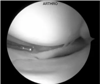
제30권 제2호 2012 149 Fig. 1. Arthroscopic finding showed complex tear (includ-
ing radial and horizontal tear) in posterior horn of medial meniscus.
Fig. 2. Abdomen-pelvic computed tomography showed an intra-luminal filling defect (white arrow) in the common iliac vein (A), and in the external, internal iliac vein (B).
수술적 치료를 계획하였다. 수술 전 신장 151.9 cm, 체중 68.7 kg, 체질량 지수(body mass index, BMI)는 29.86 kg/m 2 이었다.
다른 특이 기왕력은 없었다.
척추 마취하에 시행한 관절경수술에서 내측 반월연골판 후각 부위에 방사성 파열과 수평 파열을 동반한 복합 파열을 확인하고 부분 절제술을 시행하였다(Fig. 1). 총 수술 시간은 25분이 소요 되었으며 수술 중 특이 소견은 관찰되지 않았다.
수술 전 또는 이후 혈전색전증 발생에 대한 특별한 예방 조치는 취하지 않았으며, 환자는 수술 당일부터 보행을 허용하 였다 . 수술 후 1일째 특이 소견은 없었으며, 수술 전과 비교하여 통증의 감소에 대해 만족하였고, 보행의 제한이 없었다. 수술 후 2일째 정상 퇴원하였다.
이후 수술 후 8일째부터 특이 수상력 없이 발생한 좌측 서혜부 및 하지의 전반적인 부종 및 통증을 주소로 술 후 10일째 응급실 내원하였다. 이학적 검사상 좌측 하지 대퇴부, 슬관절 및 경골부에 걸쳐 부종이 관찰되었고, 국한된 부위의 압통이나, 열감, 피부의 홍반 등은 관찰되지 않았으며, 정상적 으로 동맥혈의 촉진이 되었다 . 당시 환자의 신체 활력 지수는 정상이었으며, 시행한 혈액 검사 결과 white blood cell, 12,380 (segemented neutrophil 85.8%); hemoglobin, 11.5; hematocrit, 34.5; platelet count 281 k, erythrocyte sedimentation rate 47, C-reactive protein 2.41, prothrombin time 10.3 s, prothrombin time (international normalized ratio) 0.97, activated partial thromboplastin time 22.2 s로 특이 소견은 관찰되지 않았다.
복부-골반 조영증강 전산화단층촬영(abdomen-pelvic contrast computed tomography) 소견상 좌측 총장골정맥, 외장골정맥 (external iliac vein), 내장골정맥(internal iliac vein)의 정맥염 소견과 함께 심부정맥 혈전증이 발견되었다 (Fig. 2). 즉시 헤파 린나트륨(heparin sodium) 25,000 U 정맥 주사하였으며, 와파린 (Warfarin) 5 mg 경구 투여하였다. 입원 7일간 헤파린나트륨 25,000 U 정맥 주사 지속하였으며 prothrombin time (PT) international normalized ratio (INR) 2.49 보여 헤파린나트륨의 투여를 중지하였다. 입원 5일간 와파린 5 mg 경구 투여 후 PT의 INR 2.34 보여 6일째부터 와파린 3 mg으로 감량하였다.
이후 검사결과상 PT의 INR이 2–3 사이로 유지되었으며, 이학
적 검사상으로 좌측 하지의 부종은 감소하였고, 통증은 완화
되었다 . 환자는 와파린 3 mg 경구투여 유지한 채 입원 9일째
퇴원하였다.
JH Yoo, et al. A Case of Deep Vein Thrombosis in Common Iliac Vein after Arthroscopic Partial Meniscectomy
150 대한스포츠의학회지
고 찰
전체적으로 슬관절경수술의 합병증 발병률은 0.8–8.2%인 것으로 보고되었다 1,2) . 이런 관절경수술의 합병증으로는 관절 면 마모, 인대 손상, 혈색전증, 폐색전증, 감염, 혈관 손상, 혈종, 구획증후군, 활액막루(synovial fistulas), 골괴사 등이 있 으며, 이 중 슬관절경수술 후 심부정맥 혈전증의 발생률은 0.1–17.9% 인 것으로 보고된다 2,3) . Small 4) 은 반월상 연골 수술 에 대한 합병증을 보고 하였는데, 심부정맥 혈전증 등을 모두 포함한 총 합병증 발생률이 내측 반월연골판의 경우 1.78%, 외측 반월연골판의 경우 1.48%이었다. 실제로 관절경수술 중 특히 반월연골판에 해당하는 수술 과정 후 발생한 정맥 혈전증의 발생은 그 빈도가 매우 적으며, 공식적 보고 역시 드물다.
인공 관절 치환술에서 예방적 조치가 없을 때 정맥 혈전증의 발병률은 약 40–70%에 달한다고 보고되고 있다 5) . 심부 정맥에 서 형성된 혈전이 폐 순환계로 유입되면서 색전증을 발생시키 고, 이는 치명적 결과를 초래할 수 있다. 이러한 혈전의 70%
이상은 하지의 심부 정맥에서 유래한다 6) . 일반적으로 저분자 량 헤파린(low molecular weight heparin, LMWH)이 혈전의 발생을 예방하는데 효과적이라 받아들여지고 있으며, 주로 인공관절 치환술을 받은 환자들에게 널리 사용되고 있다 6) . 하지만 이러한 항응고제의 사용은 수술 부위의 major bleeding 을 유발할 수 있고, 출혈 소인을 높여 타 부위 출혈 질환을 발생시킬 수 있는 문제가 있다 7) .
저분자량 헤파린을 받거나 받지 않은 슬관절경수술환자들 사이에서 심부정맥 혈전증율을 판단하기 위한 연구가 진행되 었다. Wirth 등 8) 은 저분자량 헤파린을 투여받지 않은 군에서 심부정맥 혈전증 발생률을 4.1%였으나 저분자량 헤파린을 투여받은 군에서는 단 0.85%이었음을 발견했는데, 이는 예방 적 저분자량 헤파린(prophylactic LMWH) 사용의 필요성을 시사한다.
관절경수술에 있어 항혈전 예방적 치료의 필요성은 여전히 논의 대상으로 남아있는데 , 그 이유는 예방적 저분자량 헤파린 사용에도 불구하고 혈전색전성 합병증(thromboembolic com- plications)이 발생할 수 있고 예방적 사용 또한 그에 동반되는 합병증이 있기 때문이다. 특히, 슬관절경수술에서 혈전색전성 합병증 발생률이 낮다는 이유로 아시아, 태평양 지역에서는 관절경수술을 받는 환자들에게 일반적으로 예방 조치가 취해 지지 않고 있는 실정이다 5) .
심부정맥 혈전증 발생의 후천적 위험 인자는 수술, 외상, 고령, 흡연, 비만, 경구 피임약 복용, 호르몬 치료, 혈전증 기왕 력, 장기간의 침상 안정 등이고, 선천적 위험 인자는 혈액 응고 인자의 이상 즉, homocysteine, Anti-thrombin III 결핍, protein C 결핍, protein S 결핍 등으로 선천적 위험인자를 가지 고 있는 사람은 경한 외상 혹은 수술에 의해서도 혈전증이 생길 수 있다고 보고되었다 6,9) . 지혈대의 사용 시간이 60분을 넘는 경우와 혈전증 기왕력이 있거나 위험인자가 두 개 이상 존재할 때 혈전증의 발생 빈도가 현저히 증가한다고 하였으며, 전신마취에 비해 척추마취하의 수술 시 발생빈도가 낮다는 보고가 있다 2,9) .
본 사례에서 지혈대(tourniquet)는 심각한 위험 인자로 알려 져 있는 60분보다 짧은 단 25분간 사용되었다. 더불어 환자는 혈전증 기왕력이 없으며, 시행한 혈액 검사상 선천적 위험인자 도 발견되지 않았고, 척추마취하에 수술을 진행하였다. 따라 서 본 환자에서는 BMI 만이 29.86 kg/m 2 으로, 비만만이 유일한 위험인자였다.
본 증례에서와 같이 비만 이외에 위험요소가 없음에도 불구 하고 슬관절경수술 , 특히 비교적 수술 시간이 짧은 반월연골판 부분절제술만을 시행하였음에도 근위부의 심부정맥 혈전증 이 발생하였음을 상기해야 하겠다. 이러한 가능성을 항상 염두 에 두고 수술 전 환자 교육 및 수술 후 조기 보행 , 압박 스타킹 및 간헐적 압박 펌프의 사용, 그리고 적극적인 예방법 사용 등의 노력이 필요할 것으로 생각된다.
참 고 문 헌