280 https://e-kcj.org
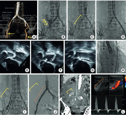
A 64-year-old woman with end-stage renal disease was diagnosed with symptomatic severe aortic stenosis. Because of high risk for operation, she underwent transcatheter aortic valve replacement (TAVR). Computed tomography (CT) showed acceptable calibers of the right iliofemoral arteries, but severe calcification was revealed (Figure 1A). After passing the delivery system of a 29-mm Evolut R (Medtronic, Minneapolis, MN, USA) by right transfemoral access, acute hemodynamic deterioration occurred. Angiography revealed massive contrast media extravasation at the right iliac artery (Figure 1B, Supplementary Video 1). Because of emergency, it was impossible to identify the true injured length of the vessel and therefore, an occlusive balloon was promptly installed by contralateral access and bleeding ceased after implantation of a SEAL bifurcated stent graft extension 10×60 mm (S&G, Biotech Inc., Seoul, Korea) at the ruptured site (Figure 1C). To proceed with the TAVR, the delivery system was advanced by left femoral access (Figure 1D). TAVR was successfully performed with absolute reliance on transesophageal echocardiography after a few trials of repositioning (Figure 1E-H, Supplementary Videos 2, 3, and 4). However, just after valve implantation, abrupt hemodynamic collapse recurred, and the stent graft was found in her abdomen (Figure 1I). After then, two stent grafts (a limb stent of Endurant II 13×13×82 mm (Medtronic) and a SEAL bifurcated stent graft extension 10×100 mm) with larger size were implanted at the right iliac artery, and hemodynamic status improved (Figure 1J, Supplementary Video 5). CT revealed migration of the stent graft inside a massive retroperitoneal hematoma (Figure 1K). Her peripheral arterial ultrasound revealed good flow through the stent-grafts (Figure 1L). Fortunately, after completely recovered, she has been doing well without any symptoms or events for 1 year follow up.
SUPPLEMENTARY MATERIALS
Supplementary Video 1
Peripheral angiography showing iliac artery rupture.
Click here to view
Supplementary Video 2
Transesophageal echocardiography showing too deep positioning of aortic valve prosthesis.
Click here to view Korean Circ J. 2019 Mar;49(3):280-281
https://doi.org/10.4070/kcj.2018.0266 pISSN 1738-5520·eISSN 1738-5555
Received: Aug 13, 2018 Revised: Nov 12, 2018 Accepted: Jan 16, 2019 Correspondence to Chi Young Shim, MD
Division of Cardiology, Severance Cardiovascular Hospital, Yonsei University College of Medicine, 50, Yonsei-ro, Seodaemun-gu, Seoul 03722, Korea.
E-mail: [email protected]
Copyright © 2019. The Korean Society of Cardiology
This is an Open Access article distributed under the terms of the Creative Commons Attribution Non-Commercial License (https://
creativecommons.org/licenses/by-nc/4.0) which permits unrestricted noncommercial use, distribution, and reproduction in any medium, provided the original work is properly cited.
ORCID iDs Jah Yeon Choi
https://orcid.org/0000-0002-6793-4137 Chi Young Shim
https://orcid.org/0000-0002-6136-0136 Geu-Ru Hong
https://orcid.org/0000-0003-4981-3304 Chul-Min Ahn
https://orcid.org/0000-0002-7071-4370 Young-Guk Ko
https://orcid.org/0000-0001-7748-5788 Myeong-Ki Hong
https://orcid.org/0000-0002-2090-2031 Conflict of Interest
The authors have no financial conflicts of interest.
Jah Yeon Choi , MD, Chi Young Shim , MD, Geu-Ru Hong , MD, Chul-Min Ahn , MD, Young-Guk Ko , MD, and Myeong-Ki Hong , MD
Division of Cardiology, Severance Cardiovascular Hospital, Yonsei University College of Medicine, Seoul, Korea
Iliac Artery Rupture and
Retroperitoneal Migration of a Stent Graft during Transcatheter Aortic Valve Replacement
Images in
Cardiovascular Medicine
Supplementary Video 3
Transesophageal echocardiography showing too shallow positioning of aortic valve prosthesis.
Click here to view
Supplementary Video 4
Transesophageal echocardiography showing good positioning of aortic valve prosthesis.
Click here to view
Supplementary Video 5
Peripheral angiography showing successful implantation of two stent grafts.
Click here to view
281 https://e-kcj.org https://doi.org/10.4070/kcj.2018.0266
Iliac Artery Rupture during Transcatheter Aortic Valve Replacement
Author Contributions
Conceptualization: Choi JY, Shim CY, Hong GR.
Data curation: Shim CY. Investigation: Shim CY.
Supervision: Shim CY, Ahn CM, Ko YG, Hong MK. Visualization: Shim CY. Writing - original draft: Choi JY, Shim CY. Writing - review &
editing: Shim CY, Hong GR, Ahn CM, Ko YG, Hong MK.
A B C D
E F G H
K
I J L
Figure 1. Computed tomographic, fluoroscopic, and echocardiographic images. (A) Severe calcification from the iliac arteries to the descending aorta on computed tomography (CT). (B) Massive contrast media extravasation at the right iliac artery on abdominal aortic angiography (arrow). (C) Implantation of a 10×60 mm stent graft at the right iliac artery (solid line). (D) Advance of transcatheter aortic valve replacement (TAVR) system through the left iliac artery with balloon supporting at right common artery (E) Too deep positioning of aortic valve prosthesis on transesophageal echocardiography. (F) Too shallow positioning of aortic valve prosthesis on transesophageal echocardiography. (G) Good positioning of aortic valve prosthesis on transesophageal echocardiography. (H) Good positioning of aortic valve prosthesis on fluoroscopy. (I) Migration of the stent graft into her abdominal space (solid line). (J) No contrast media extravasation after implantation of 2 stent grafts (dotted line). (K) The migrated stent graft inside a massive retroperitoneal hematoma on CT (solid line). (L) Good flow through the stent-grafts on Doppler ultrasound (dotted line).