http://dx.doi.org/10.5624/isd.2016.46.3.159
Introduction
Increases in life expectancy worldwide have led to an increased demand for oral rehabilitation surgical pro
cedures, such as implant therapies and bone grafts. Al
though dental implant placement in the mandibular inter
foraminal region is regarded as a relatively safe proce
dure, patients may experience sensory disturbances after surgery.1
Some anatomical structures should be carefully observ
ed when considering surgery in the anterior mandible, such as the anterior loop of the mental nerve and the man
dibular incisive canal, to avoid the risk of nerve injury and hemorrhage.1
The anterior loop is defined as an anterior/mesial exten
sion of the mental nerve in the interforaminal region of the mandible, with a path that creates a loop before turn
ing posterosuperiorly to exit the bone through the mental foramen.29
The mandibular incisive canal is a mesial extension of the mandibular canal, containing the incisive nerve and vessels, which irrigate and innervate the lower anterior teeth.10 The intraosseous pathway of the incisive canal can be a determining factor in the success of surgical pro
cedures in the interforaminal region.11
Panoramic radiographs underestimate extensions of the anterior loop and mandibular incisive canal
Ana Caroline Ramos de Brito1,*, Yuri Nejaim1, Deborah Queiroz de Freitas1, Christiano de Oliveira Santos2
1Department of Oral Diagnosis, Division of Oral Radiology, Piracicaba Dental School, University of Campinas, São Paulo, Brazil
2Department of Stomatology, Public Oral Health and Forensic Dentistry, School of Dentistry of Ribeirão Preto, University of São Paulo, São Paulo, Brazil
AbstrAct
Purpose: The purpose of this study was to detect the anterior loop of the mental nerve and the mandibular incisive canal in panoramic radiographs(PAN) and conebeam computed tomography(CBCT) images, as well as to determine the anterior/mesial extension of these structures in panoramic and crosssectional reconstructions using PAN and CBCT images.
Materials and Methods: Images(both PAN and CBCT) from 90 patients were evaluated by 2 independent observers. Detection of the anterior loop and the incisive canal were compared between PAN and CBCT. The anterior/mesial extension of these structures was compared between PAN and both crosssectional and panoramic CBCT reconstructions.
results: In CBCT, the anterior loop and the incisive canal were observed in 7.7% and 24.4% of the hemimandibles, respectively. In PAN, the anterior loop and the incisive canal were detected in 15% and 5.5% of cases, respectively.
PAN presented more difficulties in the visualization of structures. The anterior/mesial extensions ranged from 0.0 mm to 19.0mm on CBCT. PAN underestimated the measurements by approximately 2.0mm.
conclusion: CBCT appears to be a more reliable imaging modality than PAN for preoperative workups of the anterior mandible. Individual variations in the anterior/mesial extensions of the anterior loop of the mental nerve and the mandibular incisive canal mean that is not prudent to rely on a general safe zone for implant placement or bone surgery in the interforaminal region.(Imaging Sci Dent 2016; 46: 159-65)
Key words: Radiography, Panoramic; ConeBeam Computed Tomography; Mandible; Anatomic Variation
Copyright ⓒ 2016 by Korean Academy of Oral and Maxillofacial Radiology
This is an Open Access article distributed under the terms of the Creative Commons Attribution NonCommercial License(http://creativecommons.org/licenses/bync/3.0) which permits unrestricted noncommercial use, distribution, and reproduction in any medium, provided the original work is properly cited.
Imaging Science in Dentistry·pISSN 22337822 eISSN 22337830 Received November 26, 2015; Revised April 8, 2016; Accepted May 6, 2016
*Correspondence to : Dr. Ana Caroline Ramos de Brito
Department of Oral Diagnosis, Division of Oral Radiology, Piracicaba Dental School, University of Campinas. Av. Limeira, 901. Areião. Zip 13414903, Piracicaba, São Paulo, Brazil
Tel) 551921065327, Fax) 551934210144, Email) [email protected]
A large number of surgical procedures are still conduct
ed using only conventional radiographs. However, visual
ization of neurovascular bony canals in the mandible us
ing 2dimensional(2D) radiographs may be difficult. Al
ternatively, crosssectional techniques such as conebeam computed tomography(CBCT) may provide 3dimen
sional images, resulting in a more accurate preoperative examination because such techniques allow the fuller as
sessment of the relevant structures.9 Several studies have evaluated the prevalence of the anterior loop and inci
sive canal, either in direct evaluations of cadavers or in anatomical samples.46,10,12,13 Twodimensional imaging techniques such as panoramic(PAN) and intraoral radio
graphs,1014 have also been used to evaluate these variations, as well as CT scans.12,1421 However, results regarding the prevalence of these structures are still contradictory, and conflicting results have also been obtained in compari
sons of different imaging methods in detecting anatomical variations. The prevalence of the anterior loop has been reported to range from 28% to 71%.27,9,22,23 The preva
lence of the incisive canal has been found to range from 15% in PAN images to as high as 93% on CT scans.11
This crosssectional observational study aimed to assess the detection of the anterior loop and incisive canal by PAN and CBCT, and to measure and compare the anteri
or/mesial extension of these anatomical structures from the mental foramen in PAN and CBCT reconstructions.
Materials and Methods
The Research Ethics Committee of Piracicaba Dental School, State University of Campinas, approved this work without restrictions(Protocol 102/2012).
The sample consisted of PAN and CBCT images of 180 hemimandibles from 90 patients(56 males and 34 females;
age range, 1576 years; mean age, 26.3 years). The images had been previously taken for clinical purposes(removal of third molars and orthognathic surgeries), and retrieved from a database of the institutional imaging center. The following inclusion criteria were used: the presence of at least 8 lower teeth from the first right premolar to first left premolar, PAN and CBCT images taken within an in
terval of <6 months, and the absence of local or systemic osseous pathologies in the anterior mandible. PAN images with positioning errors were excluded.
PAN images were acquired with a digital charge coupled device sensor(OP100D; Instrumentarium Corp., Imag ing Division, Tuusula, Finland), with an acquisition protocol of 66kVp, 12mA, and an exposure time of 17.6s. CBCT images were acquired with a Classic iCAT apparatus (Imaging Sciences International Inc., Hatfield, PA, USA), with an acquisition protocol of 120kVp, 7mA, an expo
sure time of 40s, 0.25mm voxels, and field of view of 16×8cm(removal of third molars) or 16×13cm(orthog
nathic surgery).
All images were evaluated on a highresolution 19inch monitor(LG FLATRON W1942SB, LG Electronics Inc., Seoul, Korea), at 32bit, 60Hz, and 1680×1050 resolu
tion. Brightness, contrast, and zoom were manipulated freely for the best visualization of the structures of inter
est.
The following parameters were assessed on the PAN and CBCT images for each hemimandible: presence of the anterior loop of the mental nerve; presence of the mandibular incisive canal; and mesial extension of the an
terior loop and/or incisive canal.
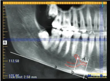
Fig. 1. A panoramic radiograph shows the anterior loop and incisive canal(right side) and the anterior loop(left side). 1. The lower man
dibular cortex as the plane of refer
ence. 2. The line perpendicular to the line passing through the mesial border of the mental foramen. 3. The line perpendicular to the line pass
ing through the most mesial point of the anterior loop of the mental nerve with the mandibular incisive canal. 4. The distance between lines 2 and 3, corresponding to the mesial length of the extent of the anterior loop or incisive canal.
Visualization of the structures(anterior loop and inci
sive canal) was scored as present, absent, or difficult to interpret. To ensure that the findings were clinically rel
evant, visualization of these structures was considered positive only when the bony canal was at least 1mm in diameter.
PAN images were analyzed on Radiocef Studio 2 soft
ware(Radio Memory, Belo Horizonte, Brazil). In order to obtain more reliable linear measurements, the magnifica
tion was estimated by taking a PAN image of 3 dry man
dibles with an orthodontic wire(9mm long and 0.7mm thick) placed horizontally on the alveolar crest, adjacent to the mental foramen. This was performed on the same panoramic machine from which data were collected. The magnification was then estimated based on the average of 3 repeated measurements in each image. The mean hori
zontal magnification was 8.3%.
CBCT images were analyzed on iCAT Vision software (Imaging Sciences International Inc., Hatfield, PA, USA).
Panoramic reconstructions(CBCTp) and crosssections (CBCTcs) were analyzed. The thickness used in CBCTcs was 0.25mm. The thickness of CBCTp could be adjusted freely to better visualize the structures.
The extension of the anterior loop and incisive canal consisted of the distance between the mental foramen and the most mesial part of these structures. For PAN(Fig.
1) and CBCTp(Fig. 2), the mandibular lower cortex was used as the guiding plane. In CBCTcs(Fig. 3), measure
ments were obtained by counting the number of slices mesial to the mental foramen in which the structures were identifiable, based on the occlusal plane of the patient.
Initially, two experienced(5 years of experience) oral radiologists who had previously undergone calibration training evaluated all images individually in a quiet, dimly lit room. All measurements were performed 3 times; the mean was considered to be the definitive measurement.
In cases of disagreement between observers, the images were reassessed and a consensus was reached. One month after the first assessments, onethird of the sample(30 PANs and 30 CBCTs) was reassessed to evaluate intraob
server consistency.
Fig. 2. Visualization of the anterior loop and incisive canal(red dotted line) on the panoramic reconstructions. a: The lower man
dibular cortex as the plane of reference. b: The line perpendicular to the line passing through the mesial border of the mental fo
ramen. c: The line perpendicular to the line passing through the most mesial point of the anterior loop of the mental nerve and the mandibular incisive canal. d: The distance between lines b and c, corresponding to the mesial length of the extent of the anterior loop or incisive canal.
Fig. 3. Crosssectional images(perpendicular to the occlusal plane) demonstrate the presence of the anterior loop(red arrows) and the inci
sive canal(green arrows).
The statistical analysis was performed using IBM SPSS Statistics for Windows version 22.0(IBM Corp., Armonk, NY, USA) at the 5% significance level. Intraobserver con
sistency for structure visualization was determined using the kappa coefficient. Intraobserver consistency for mea
surements was assessed using the intraclass correlation coefficient(ICC). The McNemarBowker test was used to compare the accuracy of each imaging technique(PAN and CBCT) for visualization of the structures. Analysis of variance(ANOVA) with the posthoc Tukey test was used to compare the measurements obtained using the 3 imag
ing methods(PAN, CBCTp, and CBCTcs). The t test was used to compare differences between the left and right sides and between genders.
results
Intraobserver agreement for structure visualization was substantial for CBCT, ranging from 0.715(incisive canal) to 0.802(anterior loop), and almost perfect for PAN, rang
ing from 0.849(incisive canal) to 0.878(anterior loop), according to the interpretation of kappa presented by Landis and Koch.24 Intraobserver agreement(ICC) was excellent for both of the CBCT measures(0.971, pan
oramic reconstruction; and 0.931, crosssectional recon
struction) and for the PAN measurements(0.961).
The responses of the evaluators for PAN images regard
ing the visualization of anatomical structures, in com
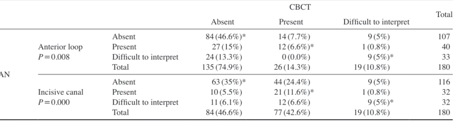
parison to the results obtained for the reference standard (CBCT), are shown in Table 1. The McNemarBowker test indicated that the PAN images disagreed with the ref
erence standard for both anatomical structures(P<0.05).
The incisive canal was detected more frequently in CBCT images. The anterior loop was detected more frequently in the PAN images. PAN was the method that presented the highest frequency of cases scored as difficult to inter
pret.
The mesial extensions of the anterior loop and inci
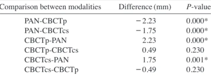
sive canal in the 3 imaging methods(PAN, CBCTp, and CBCTcs) are shown in Table 2. ANOVA using the post hoc Tukey test demonstrated that the measurements were significantly shorter in PAN images than in CBCT images (mean, 2.0mm shorter; P<0.05)(Table 3). The difference between the two types of CBCT reconstructions was sub
millimetric and had no statistical significance(P>0.05).
No significant difference was found in the visualization of the anterior loop of the mental nerve or the mandibular incisive canal, or according to genders or the side of the
Table 1. Contingency table comparing the responses obtained using panoramic radiography(PAN) and conebeam computed tomography (CBCT) for the visualization of the anterior loop and incisive canal.
CBCT Total
Absent Present Difficult to interpret
PAN
Anterior loop P=0.008
Absent Present
Difficult to interpret Total
84(46.6%)*
27(15%) 24(13.3%) 135(74.9%)
14(7.7%) 12(6.6%)*
0(0.0%) 26(14.3%)
9(5%) 1(0.8%) 9(5%)*
19(10.8%)
10740 18033
Incisive canal P=0.000
Absent Present
Difficult to interpret Total
63(35%)*
10(5.5%) 11(6.1%) 84(46.6%)
44(24.4%) 21(11.6%)*
12(6.6%) 77(42.6%)
9(5%) 1(0.8%) 9(5%)*
19(10.8%)
11632 18032
*Indicates agreement between the radiographic methods evaluated. Pvalues<.05 were considered to indicate statistical significance(McNemarBowker test).
Table 2. Minimum and maximum values, means, medians, and standard deviations, in millimeters, of the mesial length of the structures from the mental foramen in panoramic radiography(PAN), conebeam computed tomography panoramic reconstruction(CBCTp), and conebeam computed tomography crosssection(CBCTcs).
Minimum1 Minimum2 Maximum Mean(SD)1 Mean(SD)2 Median1 Median2
PANCBCTp CBCTcs
0.000.00 0.00
1.852.10 1.00
19.09 18.22 19.00
2.28(3.46) 4.51(4.48) 4.02(4.01)
6.14(2.93) 7.89(2.88) 6.13(3.41)
0.004.93 3.50
5.427.61 5.50
1All cases, including those where the anterior loop or incisive canal was not present.
2Considering only cases where the anterior loop and/or the incisive canal was present.
mandible. However, incisive canal detection was slightly higher for the right side in CBCT images(56%).
discussion
Visualization of anterior loop and incisive canal The presence and extension of the anterior loop have been reported to vary widely in the radiology literature. In the present study, the anterior loop was visualized in 7.7%
of the CBCT images and 15% of the PAN images. Previ
ous anatomical studies in cadavers and anatomical speci
mens reported the prevalence of the anterior loop to range from 10% to 62.7%.25,8,25,26 Kieser et al.’s study27 did not show the measurable anterior loop that would have any significant impact on treatment planning for implants in the anterior mandible. Benninger et al.8 considered the anterior loop to be an anomaly, rather than an anatomical finding. Rosenquist28 visualized the anterior loop direct
ly in inferior alveolar nerve transposition operations, re
porting that the loop was absent in 74.1% of the sample and, when present, it measured <1mm. However, even though the anterior loop was found to have a low preva
lence in our study, implants should not be installed close to the mental foramen without a careful evaluation.
The importance of the location of the incisive canal should be highlighted. Direct contact of an implant with this structure can lead to migration of the soft tissue around the metallic device, preventing osseointegration, and, upon reaching its neurovascular content, may cause sensory disturbances and bleeding in the region.28 The prevalence of the mandibular incisive canal ranged from 20% to 100% in previous anatomical studies.10,13 Inci
sive canal detection in CT scans ranged from 71.9% to 100%,14,1621 whereas in PAN and intraoral radiographs, it ranged from 11% to 56%, depending on the degree of canal corticalization.10,11,13,14 In our study, incisive ca
nal visualization ranged from 5.5% in PAN to 24.4% in CBCT. CBCT provides accurate linear measurements,
so this imaging modality was considered to be reference standard in this study. Discrepancies observed between CBCT and PAN confirm the limitations of 2D imaging in the assessment of the anterior loop and incisive canal, as demonstrated by the higher difficulty in detecting these structures and the underestimation of the length of the an
terior loop and incisive canal. These results mean that the use of only PAN should be discouraged for surgical plan
ning in the anterior mandible.
In the present study, although the anterior loop and inci
sive canal were considered isolated structures in terms of determining their presence, they were not considered sep
arately when performing the measurements for their an
terior/mesial extensions. Detecting the anterior loop and the incisive canal is a difficult task. The sample size(180 hemimandibles) may also have contributed to the low incidence of these two structures. However, the ICC for these structures was satisfactory, ranging from 0.931 to 0.971. Thus, although the difficulty in distinguishing be
tween the anterior loop and incisive canal may have influ
enced the ability of the observer to detect these structures, measurements of their anterior/mesial extension from the mental foramen, which did not require such a distinction, were more reliable and reproducible.
Significant differences were found between PAN and CBCT images regarding the detection of both the anteri
or loop and incisive canal. Moreover, significantly more PAN images were difficult to interpret. Previous studies have shown that the interpretation of 2D images has limitations and often results in false negatives and false positives, as well as misestimating anterior loop exten
sion.2,3,22,26 The presence of the anterior loop was overes
timated by as much as 40% in PAN images in a previous study, in which false positives were determined by dissec
tion of the radiographed anatomical specimens.2 Image overlapping and the degree of corticalization of the bony canals are factors that may affect the visualization of structures such as the anterior loop and incisive canal.9,11
Statistically significant differences in laterality or gen
der were not identified with regards to the presence and length of anterior loop. However, some authors have found such structures more frequently in men,5,6,29 and suggested that visibility of the anterior loop decreases with age.7,19 For the anterior loop, no significant differ
ences regarding its measurements were found using either imaging modality. However, there was a slightly higher (56%) detection rate of this variant on the right side on CBCT exams.
Table 3. Comparison among measures for the mesial length from the mental foramen in the 3 imaging modalities.
Comparison between modalities Difference(mm) Pvalue PANCBCTp
PANCBCTcs CBCTpPAN CBCTpCBCTcs CBCTcsPAN CBCTcsCBCTp
-2.23 -1.75 2.230.49 1.75 -0.49
0.000*
0.000*
0.000*
0.230 0.001*
0.230 lway ANOVA with post hoc comparisons using the Tukey test. *P<0.05
Mesial distances from the mental foramen and clinical implications
Mesial extensions from the mental foramen have been measured thoroughly to determine a safe zone for implant placement in the interforaminal region.2,3,5,6,9,22,29,30 How
ever, an important factor to consider is the reference used to conduct the measurements. Depending on the reference plane adopted, the distances from the mental foramen are different. Uchida et al.5,6 used only the lower border of the mandible as the reference, while de OliveiraSantos et al.9 and CoutoFilho et al.26 used two references: the low
er border of the mandible(measurements performed in CBCTp) and the occlusal plane, which could be more re
presentative of the field of vision of the surgeon during the implant placement procedure(for CBCTcs).
In this study, when cases of anterior loop and incisive canal absence were included, the mean mesial distances from the mental foramen in CBCTp and CBCTcs were similar(4.51mm and 4.02mm, respectively). Excluding cases where the anterior loop and incisive canal were not present, the mean measures were 7.89mm(CBCTp) and 6.13mm(CBCTcs). Comparing the imaging methods based on the accuracy of measurements, the difference between measurements in the two reconstruction planes was submillimetrical. We found no statistically significant difference between the imaging methods, suggesting that both methods are comparable. It is always prudent to as
sess all preoperative CBCT reconstructions in the anterior mandible.
PAN images underestimated the distances by a mean of 2.0mm, which was found to be statistically significant.
PAN images underestimated the presence and extent of these structures, suggesting that this 2D imaging modal
ity does not offer reliable information about the location of the neurovascular structures of the anterior mandible.
Some studies have suggested a safe zone for implant placement in the anterior mandible.3,30 In the present study, the overall mean mesial distance from the mental fora
men, regardless of the presence and absence of the anteri
or loop or the incisive canal ranged from 2.28mm in PAN to 4.51mm in CBCT. Considering only cases where the anterior loop and incisive canal were present, the mean distance was approximately 8mm. Our results were indic
ative of considerable anatomical variability, as distances from the mental foramen reached up to 20mm(2.0cm).
CoutoFilho et al.,26 evaluating only the anterior loop, ob
tained mesial measures of the mental foramen similar to those obtained this study. This variability could be related to the incisive canal, a structure not evaluated in the pre
vious study by CoutoFilho et al.26 Due to these discrep
ancies, it is recommended that all surgical cases involving the anterior mandible be assessed individually rather than relying on averages.
Considering the previously known limitations of 2D imaging, as well as the discrepancies observed between PAN and CBCT in visualization and measurements of the extension of the anterior loop of the mental nerve and the mandibular incisive canal, CBCT is the best choice as an imaging method for preoperative planning for procedures involving the anterior mandible. Moreover, the consider
able individual variation for measurements obtained from both imaging methods(PAN and CBCT) shows that it is not prudent to rely on a general safe zone for implant placement or bone surgery in the interforaminal region.
references
1. Liang X, Lambrichts I, Corpas L, Politis C, Vrielinck L, Ma GW, et al. Neurovascular disturbance associated with implant placement in the anterior mandibular and its surgical implica
tions: literature review including report of a case. Chin J Dent Res 2008; 11: 5664.
2. Mardinger O, Chaushu G, Arensburg B, Taicher S, Kaffe I.
Anterior loop of the mental canal: an anatomicalradiologic study. Implant Dent 2000; 9: 1205.
3. Kuzmanovic DV, Payne AG, Kieser JA, Dias GJ. Anterior loop of the mental nerve: a morphological and radiographic study. Clin Oral Implants Res 2003; 14: 46471.
4. Hu KS, Yun HS, Hur MS, Kwon HJ, Abe S, Kim HJ. Branch
ing patterns and intraosseous course of the mental nerve. J Oral Maxillofac Surg 2007; 65: 228894.
5. Uchida Y, Yamashita Y, Goto M, Hanihara T. Measurement of anterior loop length for the mandibular canal and diameter of the mandibular incisive canal to avoid nerve damage when installing endosseous implants in the interforaminal region. J Oral Maxillofac Surg 2007; 65: 17729.
6. Uchida Y, Noguchi N, Goto M, Yamashita Y, Hanihara T, Takamori H, et al. Measurement of anterior loop length for the mandibular canal and diameter of the mandibular incisive canal to avoid nerve damage when installing endosseous im
plants in the interforaminal region: a second attempt introduc
ing cone beam computed tomography. J Oral Maxillofac Surg 2009; 67: 74450.
7. Ngeow WC, Dionysius DD, Ishak H, Nambiar P. A radio
graphic study on the visualization of the anterior loop in den
tate subjects of different age groups. J Oral Sci 2009; 51: 231
8. Benninger B, Miller D, Maharathi A, Carter W. Dental im7.
plant placement investigation: is the anterior loop of the men
tal nerve clinically relevant? J Oral Maxillofac Surg 2011; 69:
1825.
9. de OliveiraSantos C, Souza PH, de Azambuja BertiCouto S, Stinkens L, Moyaert K, RubiraBullen IR, et al. Assessment
of variations of the mandibular canal through cone beam com
puted tomography. Clin Oral Investig 2012; 16: 38793.
10. Mardinger O, Chaushu G, Arensburg B, Taicher S, Kaffe I.
Anatomic and radiologic course of the mandibular incisive canal. Surg Radiol Anat 2000; 22: 15761.
11. Jacobs R, Mraiwa N, Van Steenberghe D, Sanderink G, Qui
rynen M. Appearance of the mandibular incisive canal on pan
oramic radiographs. Surg Radiol Anat 2004; 26: 32933.
12. Mraiwa N, Jacobs R, Moerman P, Lambrichts I, van Steenber
ghe D, Quirynen M. Presence and course of the incisive canal in the human mandibular interforaminal region: twodimen
sional imaging versus anatomical observations. Surg Radiol Anat 2003; 25: 41623.
13. Jalili MR, Esmaeelinejad M, Bayat M, Aghdasi MM. Ap
pearance of anatomical structures of mandible on panoramic radiographs in Iranian population. Acta Odontol Scand 2012;
70: 3849.
14. Pires CA, Bissada NF, Becker JJ, Kanawati A, Landers MA.
Mandibular incisive canal: cone beam computed tomography.
Clin Implant Dent Relat Res 2012; 14: 6773.
15. Jacobs R, Mraiwa N, vanSteenberghe D, Gijbels F, Quiry
nen M. Appearance, location, course, and morphology of the mandibular incisive canal: an assessment on spiral CT scan.
Dentomaxillofac Radiol 2002; 31: 3227.
16. Makris N, Stamatakis H, Syriopoulos K, Tsiklakis K, van der Stelt PF. Evaluation of the visibility and the course of the mandibular incisive canal and the lingual foramen using cone
beam computed tomography. Clin Oral Implants Res 2010;
21: 76671.
17. Sokhn S, Nasseh I, Noujeim M. Using cone beam computed tomography to determine safe regions for implant placement.
Gen Dent 2011; 59: e727.
18. Parnia F, Moslehifard E, Hafezeqoran A, Mahboub F, Mo
javerKahnamoui H. Characteristics of anatomical landmarks in the mandibular interforaminal region: a conebeam com
puted tomography study. Med Oral Patol Oral Cir Bucal 2012;
17: e4205.
19. Kajan ZD, Salari A. Presence and course of the mandibular incisive canal and presence of the anterior loop in cone beam computed tomography images of an Iranian population. Oral Radiol 2012; 28: 5561.
20. Apostolakis D, Brown JE. The dimensions of the mandibular incisive canal and its spatial relationship to various anatomical landmarks of the mandible: a study using cone beam comput
ed tomography. Int J Oral Maxillofac Implants 2013; 28: 117
21. AlAni O, Nambiar P, Ha KO, Ngeow WC. Safe zone for 24.
bone harvesting from the interforaminal region of the mandi
ble. Clin Oral Implants Res 2013; 24 Suppl A100: 11521.
22. Kaya Y, Sencimen M, Sahin S, Okcu KM, Dogan N, Bahceci
tapar M. Retrospective radiographic evaluation of the anterior loop of the mental nerve: comparison between panoramic radiography and spiral computerized tomography. Int J Oral Maxillofac Implants 2008; 23: 91925.
23. Kilic C, Kamburoğlu K, Ozen T, Balcioglu HA, Kurt B, Kuto
glu T, et al. The position of the mandibular canal and histolog
ic feature of the inferior alveolar nerve. Clin Anat 2010; 23:
3442.
24. Landis JR, Koch GG. The measurement of observer agree
ment for categorical data. Biometrics 1977; 33: 15974.
25. Liang X, Jacobs R, Corpas LS, Semal P, Lambrichts I. Chro
nologic and geographic variability of neurovascular structures in the human mandible. Forensic Sci Int 2009; 190: 2432.
26. CoutoFilho CE, Moraes PH, Alonso MB, HaiterNeto F, Olate S, AlbergariaBarbora JR. Accuracy in the diagnosis of the mental nerve loop. A comarative study between panoramic radiography and cone beam computed tomography. Int J Mor
phol 2015; 33: 32732.
27. Kieser J, Kuzmanovic D, Payne A, Dennison J, Herbison P.
Patterns of emergence of the human mental nerve. Arch Oral Biol 2002; 47: 7437.
28. Rosenquist B. Is there an anterior loop of the inferior alveolar nerve? Int J Periodontics Restorative Dent 1996; 16: 405.
29. Rosa MB, SottoMaior BS, Machado Vde C, Francischone CE. Retrospective study of the anterior loop of the inferior al
veolar nerve and the incisive canal using cone beam computed tomography. Int J Oral Maxillofac Implants 2013; 28: 38892.
30. Apostolakis D, Brown JE. The anterior loop of the inferior alveolar nerve: prevalence, measurement of its length and a recommendation for interforaminal implant installation based on cone beam CT imaging. Clin Oral Implants Res 2012; 23:
102230.