This is an open-access article distributed under the terms of the Creative Commons Attribution Non-Commercial License (http://creativecommons.org/
licenses/by-nc/4.0/), which permits unrestricted non-commercial use, distribution, and reproduction in any medium, provided the original work is properly cited.
CC
Oral rehabilitation of Papillon–Lefèvre syndrome patients by dental implants: a systematic review
Fazele Atarbashi-Moghadam1, Saede Atarbashi-Moghadam2, Setare Kazemifard3, Soran Sijanivandi3, Mahshid Namdari4,5
1Department of Periodontics, Dental School of Shahid Beheshti University of Medical Sciences,
2Department of Oral and Maxillofacial Pathology, School of Dentistry, Shahid Beheshti University of Medical Sciences,
3Dental Research Center, Research Institute of Dental Sciences, Shahid Beheshti University of Medical Sciences,
4Community Oral Health Department, School of Dentistry, Shahid Beheshti University of Medical Sciences,
5Department of Biostatistics, School of Allied Medical Sciences, Shahid Beheshti University of Medical Sciences, Tehran, Iran
Abstract(J Korean Assoc Oral Maxillofac Surg 2020;46:220-227)
Objectives: Papillon–Lefèvre syndrome (PLS) is a rare autosomal recessive disorder. These patients lose their teeth at a young age and are in need of prosthetic rehabilitation. The aim of this systematic review was to assess the success of dental implant placement in these patients.
Materials and Methods: An electronic search was performed in PubMed Central, Scopus, and Web of Science using the keyword “Papillon–Lefèvre syndrome” AND “dental implant” OR “prosthodontics”. Articles reporting implant placement in patients with PLS until July 2019 were included.
Results: Assessment of the included 11 articles reporting 15 cases showed 136 implant placements in these patients. Implant failure occurred in 3 patients (20 implants). The peri-implantitis and failure rate was higher in the maxilla. Meta-analysis showed the probability of failure to be 7% (95%
confidence interval [CI] 0%-31%) for maxillary implants and 2% (95% CI 0%-9%) for mandibular implants. The follow-up time ranged between 1 and 20 years. Healing after bone graft and implant placement in these patients was uneventful.
Conclusion: Dental implants may be a viable treatment option for PLS patients. Implantation can help preserve alveolar bone if the patients’ immuno- logical and growing conditions are well-considered and proper oral hygiene and compliance with the maintenance program are continued.
Key words: Papillon–Lefèvre syndrome, Dental implants, Oral rehabilitation, Prosthodontics, Rapid progressive periodontitis
[paper submitted 2020. 3. 4 / revised 2020. 6. 5 / accepted 2020. 6. 5]
Copyright © 2020 The Korean Association of Oral and Maxillofacial Surgeons. All rights reserved.
I. Introduction
Full or partial edentulism in children and adolescents oc- curs most commonly as a result of trauma or genetic disor- ders, and these patients encounter esthetic and functional problems1. Esthetic problems can cause psychologic disorders and affect social activity1,2. Long-standing edentulism leads to a senile profile and a decreased facial height3. The functional problems experienced include difficulty in eating and speak-
ing1,2. Papillon–Lefèvre syndrome (PLS; OMIM#245000) is one of the disorders that can cause early tooth loss3,4, and those affected by PLS usually need oral rehabilitation to overcome these problems3.
PLS is a rare autosomal recessive disorder with dermal and oral manifestations4,5. Mutation in the cathepsin C gene (CTSC), which is localized on chromosome 11q14-q21, in these patients causes the loss of cathepsin C function. Ca- thepsin C plays a role in epithelial differentiation and des- quamation6. The severity of dermal manifestations, including diffuse hyperkeratosis affecting the palms of the hands and soles of the feet, varies in patients from mild psoriasiform scaly skin to overt hyperkeratosis4,5. The soles of the feet are usually affected more than the palms, and the degree of hy- perkeratosis may be affected by seasonal changes. There is evidence that the hyperkeratosis severity is associated with periodontal involvement5. PLS patients’ neutrophil functions are affected, and these dysfunctions appear to be localized to Soran Sijanivandi
Dental Research Center, Research Institute of Dental Sciences, Shahid Beheshti University of Medical Sciences, Tehran 1983969411, Iran TEL: +98-920-618-3943 FAX: +98-21-44424248
E-mail: [email protected]
ORCID: https://orcid.org/0000-0003-4663-4423
tissues that are under direct and chronic bacterial attacks like the periodontal tissue. This has been suggested to be the pos- sible reason for PLS patients’ lack of systemic infections7.
Rapid progressive destructive periodontitis around primary and permanent dentitions is the oral manifestation of PLS8. The inflammatory gingival changes and periodontitis around primary dentition appear concomitant with palmoplantar keratosis and subside after tooth loss5. Because of this, almost all PLS patients lose their teeth at a young age. Surprisingly, third molars, which erupt at older ages, are not affected3,5. PLS patients’ immune system function improves with age and their polymorphonuclear (PMN) cells’ chemotaxis and phagocytosis abilities become normal after some years of be- ing edentulous8,9.
Dental implants can be a viable treatment option for these patients in order to overcome esthetic and functional pros- thetic problems10. However, issues surrounding implantation raise success concerns in these patients; these issues include impaired patient immune systems and severe bone loss that results in the need for complex ridge augmentations10,11. The aim of this systematic review was to assess the clinical out- come and the survival rate of dental implants used for the oral rehabilitation of PLS patients.
II. Materials and Methods
The present study is a systematic review that was per- formed in accordance with the “Preferred Reporting Items for Systematic Review and Meta-Analyses Protocols” (PRISMA- P, 2015)12. The search was performed using “MeSH” terms and keywords based on the elements of the PICO question:
1. Participants (P): PLS patients
2. Intervention (I): Oral rehabilitation using dental implants 3. Comparison or control (C): Not applicable
4. Outcome measures (O): Success of dental implant 1. Information sources and search strategy
An exhaustive search of the literature available in PubMed Central, Scopus, and Web of Science’s electronic databases until July 2019 was conducted. The following keywords were used in the search strategy: “Papillon–Lefèvre syndrome”
AND “dental implant” OR “prosthodontics”. The full title and abstract of each article were screened by two independent authors (F.A. and S.K.) using predetermined inclusion and exclusion criteria.(Fig. 1) All of the references were selected from the EndNote X9 (Thomson Reuters, Philadelphia, PA, USA). In order to ascertain whether any relevant studies were neglected in the initial search, the bibliographies of the
IncludedEligibilityScreeningIdentification
Records after duplicates removed (n=26)
Records screened (n=26)
Full-text articles assessed for eligibility
(n=11)
Studies included in qualitative synthesis
(n=11)
Records excluded (n=10) Reasons:
- Not concerning implants in PLS patients - A 1996 study was excluded due to repetition
of results in the follow up study on 2004 Records identified through
electronic search (n=80)
Additional records identified through other sources
(n=1)
Studies included in quantitative synthesis (meta-analysis)
(n=11)
Full-text articles excluded (n=2)
Reasons:
No implant placements
Fig. 1. Diagram of literature search and selection criteria adapted from PRISMA (Preferred Reporting Items for System- atic Reviews and Meta-Analyses)12. (PLS: Papillon–Lefèvre syndrome) Fazele Atarbashi-Moghadam et al: Oral rehabilita- tion of Papillon–Lefèvre syndrome patients by dental implants: a systematic review. J Korean Assoc Oral Maxillofac Surg 2020
Table 1. Data extracted from selected articles StudySexAge (yr)1 Treatment modalityImplant numberFollow up (yr)2 OutcomeProbable reason Kinaia et al.20 (2017)M21Rehabilitation of maxilla & mandible using calvarium bone graft Implant placement (after 3 months) Prosthetic rehabilitation (after 3 months)
Max: 9 Man: 61Healthy Nickles et al.11 (2013) Case 1F20Dental implant placementMax: 6 Man: 44Incipient peri- implantitisPoor oral Hygiene Case 2F20Dental implant placementMax: 6 Man: 42Healthy Case 3F18Dental implant placementMax: 6 Man: 8Max: 4 Man: 20Peri-implantitis Max: 4 implants lostPoor oral Hygiene Case 4F19Dental implant placementMax: 10 Man: 10Max: 8 Man: 10Peri-implantitis Max: 9 implants lost Man: 6 implants lost
Poor oral Hygiene & compliance Rai et al.23 (2014) M26Extraction of all teeth Maxillary conventional denture Mandibular implant supported overdenture (6 months after implant placement)
Man: 21Healthy Al Farraj AlDosari21 (2013) F19Extraction of all teeth except second molars Transitional lower partial denture Dental implant insertion (after 2 months) Simultaneous bone grafting with Bio-Oss in needed sites Prosthetic rehabilitation (after 4 months) Periodic follow-up
Max: 8 Man: 61Healthy Senel et al.16 (2012) M18Saving two third molars Implant placement and prosthetic rehabilitation (after 12 months) Regular follow-up
Max: 6 Man: 63Max: 1 implant lost (early failure)Lack of osseointegration Ahmadian et al.10 (2011) F21Complete denture (interim) Mandibular set back (sagittal split ramus osteotomy) Another mandibular denture (after 1 month) Nerve repositioning for posterior mandible & simultaneous implant placement (after 9 months) Bilateral open sinus augmentation (tibia bone graft) Maxillary implant placement (after 9 months) Vestibuloplasty for increasing attached gingiva & prosthetic rehabilitation (after 6 months)
Max: 8 Man: 84Healthy Etöz et al.17 (2010)F34Implant-supported overdenture in mandible using short implants (6 mm in height and 4.1 mm in diameter at the canine regions) Prosthetic rehabilitation (after 3 months)
Man: 21Healthy
selected studies and Google Scholar were also reviewed. Any differences in the selection of the studies were resolved by discussion with a third reviewer (S.A.).
2. Eligibility criteria
Inclusion criteria were articles written in English with full text availability that reported on oral rehabilitation of PLS patients with dental implants, including, for example, case reports and case series.
Exclusion criteria were articles on other prosthetic reha- bilitation methods besides dental implants or articles without treatment outcome.
3. Data collection
Data were extracted from the included articles by two inde- pendent authors (F.A. and S.K.). A third author resolved any disagreements in the extracted data. Microsoft Excel 2016 (Microsoft, Redmond, WA, USA) was used for standardiza- tion and organization of the extracted data.
The following information was collected (if available) and is provided in Table 1:
1. Patients’ sex and age at the time of implant placement 2. Treatment modality, additional procedures, and grafting donor site or material
3. The number of inserted implants and the outcome of treatment (for example, implant failure, peri-implantitis, or healthy implant)
4. The probable reason for peri-implant diseases 5. The follow-up period after implant placement 6. General characteristics of the selected studies 4. Quality assessment of studies
The quality of each study was assessed using CARE (CAse REport) guidelines13 to estimate risk of bias. The quality of all thirteen parts was considered.
5. Statistical method
Data analysis was performed using Stata statistical soft- ware (release 15; StataCorp, College Station, TX, USA).
Descriptive data were expressed as percentages. In order to examine the statistical heterogeneity of the data, the I2 statis- tic was used. To analyze and integrate the results, random and fixed effect methods were used for maxillary and mandibular
Table 1. Continued StudySexAge (yr)1 Treatment modalityImplant numberFollow up (yr)2 OutcomeProbable reason Toygar et al.3 (2007) F18Alveolar bone augmentation (demineralized bone matrix and a titanium membrane) Membrane removal (after 8 weeks) Implant placement (after 3 months) and simultaneous repeated augmentation for bone thickening (esthetic requirement)
Max: 2NMHealthy Dhanrajani22 (2004) Case 1MNMFixed implant-retained mandibular prosthesesMan: 69Healthy Case 2MNMFixed implant-retained mandibular prosthesesMan: 69Healthy Woo et al.18 (2003) M14Implant-retained overdenture 4 months after implant placementMan: 21Healthy Ullbro et al.8 (2000) F25Dental implant placement Prosthetic rehabilitation (after 3 months)Man: 54.5Gingival hyperplasia around 1 implant Healthy
Lack of keratinized gingiva (M: male, F: female, Max: maxilla, Man: mandible, NM: not mentioned) 1 Age at the time of implant placement. 2 Dental implant follow-up. Fazele Atarbashi-Moghadam et al: Oral rehabilitation of Papillon–Lefèvre syndrome patients by dental implants: a systematic rev
iew. J Korean Assoc Oral Maxillofac Surg 2020
implants, respectively.
III. Results
The primary electronic search resulted in 80 studies. Af- ter exclusion of irrelevant studies, 11 articles with 15 cases were included in this systematic review.(Table 1) These cases included six male and nine female patients afflicted by PLS who were treated by dental implants. The mean age of pa- tients at the time of implant placement was 20.69±4.96 years, and most of implant placement was done after the age 18. A total number of 136 dental implant placements were reported for these patients. Most of the treatment plans called for full mouth rehabilitation followed by mandibular overdenture.
One case reported two single implant placements of maxil- lary central incisors, the only maxillary teeth lost. The patient was under meticulous follow-up before and during perma- nent teeth eruption. Her mandibular incisors were planned for future reconstruction3.
Peri-implantitis was reported in three patients with poor oral hygiene, and a total of 20 dental implants failed (14.70%).
Implant failures occurred in three patients, and these rates were obtained without considering the replaced implants. The reasons of implant failure were peri-implantitis (2 patients)
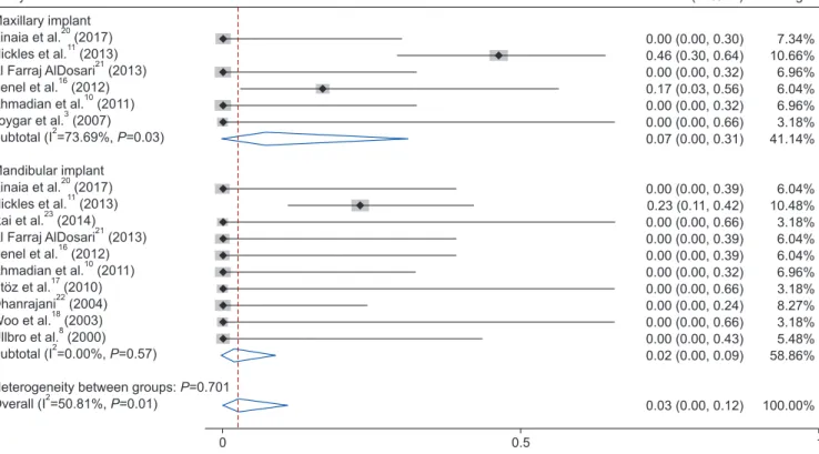
and lack of osseointegration (1 patient). Gingival hyperplasia around one implant due to lack of attached gingiva was re- ported. The failure rate was higher in maxillae. Meta-analysis of the probability of failure was 7% (95% confidence interval [CI] 0%-31%) for maxillary implants and 2% (95% CI 0%- 9%) for mandibular implants. The value of I2 was 73% and 0% for maxillary and mandibular implants, respectively. The overall result for all I2 was 50.81% with a pooled random ef- fect estimate of 3% (95% CI 0%-12%). A forest plot of the meta-analysis is provided in Fig. 2. None of the patients had healing problems after the placement of implants or bone graft. The mean and median follow-up after prosthetic reha- bilitation was 5.16±5.08 years and 4 years (Q1=1, Q2=8.75), respectively. The follow-up time ranged between 1 years and 20 years.
IV. Discussion
Periodontitis is an important manifestation of PLS and, in these patients, the conventional periodontal treatments usual- ly cannot prevent the progressive attachment loss8,14. In addi- tion to periodontal treatment, antimicrobial therapies includ- ing erythromycin, tetracycline, penicillin, and amoxicillin- metronidazole have been suggested. However, controversial
Study
Maxillary implant Kinaia et al. (2017) Nickles et al. (2013) Al Farraj AlDosari (2013) Senel et al. (2012) Ahmadian et al. (2011) Toygar et al. (2007) Subtotal (I =73.69%, =0.03)
Mandibular implant Kinaia et al. (2017) Nickles et al. (2013) Rai et al. (2014)
Etoz et al. (2010) Dhanrajani (2004) Woo et al. (2003) Ullbro et al. (2000) Subtotal (I =0.00%, =0.57)
Heterogeneity between groups: =0.701 Overall (I =50.81%, =0.01)
20 11
21 16
10 3 2
20 11 23
17 22 18
8
P
Al Farraj AlDosari (2013) Senel et al. (2012) Ahmadian et al. (2011)
21 16
10
2
2
P P P
Weight
7.34%
10.66 6.96 6.04 6.96 3.18 41.14
6.04 10.48 3.18 6.04 6.04 6.96 3.18 8.27 3.18 5.48 58.86
100.00
%
%
%
%
%
%
%
%
%
%
%
%
%
%
%
%
%
% ES (95% CI)
0.00 (0.00, 0.30) 0.46 (0.30, 0.64) 0.00 (0.00, 0.32) 0.17 (0.03, 0.56) 0.00 (0.00, 0.32) 0.00 (0.00, 0.66) 0.07 (0.00, 0.31)
0.00 (0.00, 0.39) 0.23 (0.11, 0.42) 0.00 (0.00, 0.66) 0.00 (0.00, 0.39) 0.00 (0.00, 0.39) 0.00 (0.00, 0.32) 0.00 (0.00, 0.66) 0.00 (0.00, 0.24) 0.00 (0.00, 0.66) 0.00 (0.00, 0.43) 0.02 (0.00, 0.09)
0.03 (0.00, 0.12)
0 0.5 1
Fig. 2. Probability of implant failure based on maxillary and mandibular implant. (ES: effect size, CI: confidence interval)
Fazele Atarbashi-Moghadam et al: Oral rehabilitation of Papillon–Lefèvre syndrome patients by dental implants: a systematic review. J Korean Assoc Oral Maxillofac Surg 2020
results have been reported15. Continuous attachment and tooth loss are present in some patients11,14. These patients ex- perience partial or complete edentulism during their adoles- cence8 and need oral reconstruction. Conventional complete denture or overdenture is the traditional prosthetic treatment for PLS patients, but this treatment causes esthetic and func- tional problems that lead patients to seek a more comprehen- sive treatment10. The chief patient complaints were the lack of stability and retention of their mandibular prosthesis8,16-18. Dental implants can provide the necessary support, stability, and retention for dental prostheses10 and are, therefore, help- ful to PLS patients. The benefits of implants include not only enhanced prosthesis stability and retention, but also preserva- tion of the supporting bone and prevention of further bone loss8.
Extraction of all PLS patient primary teeth so that the pa- tient experiences an edentulism period between primary and permanent dentition has been reported to be helpful9,14,19.
Although implants help patients with edentulism, lack of available bone for dental implant placement as the result of progressive periodontitis and/or continuous use of full dentures for many years is the major problem for these pa- tients10,17,18.Therefore, an implant-based treatment plan for these patients is, in many cases, restricted to overdentures unless complicated pre-surgical bone augmentations are required10. In this review, pre-surgical augmentation was reported in three patients. These included sinus augmenta- tion, inferior alveolar nerve repositioning and guided bone regeneration (GBR) using extra-oral harvesting bone (calvaria and tibia) or bone substitute material3,10,20. Simultaneous GBR was also performed during implant placement if needed21. In all cases with bone augmentation, the healing period was nor- mal and uneventful3,10,20,21.
To avoid these complicated and expensive procedures, implant placement between the mental foramina for fixed mandibular prosthesis could be the treatment of choice if sufficient bone is available in the anterior mandible8,22. Inser- tion of two implants in the anterior mandible was another treatment modality for an implant-supported removable overdenture17,18,23. Etöz et al.17 used short dental implants for mandibular overdenture support. Their patient showed severe ridge atrophy so distraction osteogenesis had a potential for bone fracture17.
Osseointegration occurred successfully except for the early failure of one implant. For areas lacking soft and hard tissue, distraction osteogenesis is suggested3.
Another consideration for PLS patients is the age at edentu-
lism. Most of the patients lost their teeth early; these patients lost most of their permanent teeth by 14 years5. However, dental implants act as ankylosed teeth and are contraindicated in teenagers and growing individuals24. Dental implants in- serted in patients under the age of 18 led to infra-occlusal po- sitioning of the maxillary dental implant. Insertion of dental implants in the anterior mandible encountered less complica- tions1. Ullbro et al.8 suggested that dental implant complica- tions in growing PLS patients was less important than bone preservation. Bohner et al.1 suggested that, whenever growing patients may benefit more from dental implants, the implan- tation can be performed cautiously; maintenance follow-ups and implant-supported prosthesis adjustments are required until growth cessation. However, in their systematic review only anterior region implant placements were considered1. Dental implant placement before the cessation of growth had been performed in patients with ectodermal dysplasia. A sys- tematic review25 demonstrated that the rate of dental implants failure in these patients was relatively low (5.3%-7.2%).
Impaired immune systems of young PLS patients is an- other consideration11. PLS patient neutrophils are deficient in the ability to establish neutrophil extracellular traps (NET), and chemotactic velocity is also reduced in PLS patient neu- trophils7. However, clinical evaluation and long-term follow- up of PLS patients have shown that the function of PMNs in PLS patients improves with age8,9. Tinanoff et al.9 reported normal PMN chemotaxis and adherence in their patient after 15 years follow-up (at age 24 years). Ullbro et al.8 tested the PMN chemotaxis and phagocytosis of their patient before implant placement (at age 25 years) which had improved to normal values. Therefore, the insertion of PLS patient dental implants at younger ages may result in the same inflamma- tory process as the one that occurs in the teeth. Based on these articles, the optimum age for implant placement in PLS patients is still unclear; and, if implant treatment is performed at an early age, immunological analysis is necessary. Most of the cases in this review received dental implant treatment after the age of 18.
This systematic review assessed the results of dental im- plant treatment in PLS patients. The longest follow-up pe- riods after implant placement reported in these studies were 20 years and 10 years11. Other extended follow-up periods included 9 years22, 4.5 years8, and 4 years10,11. In 40% of pa- tients the implant follow-up time was 2 years or less.
Peri-implantitis occurred in three patients. Implant failure (19 implants) resulted in two of these patients. Poor oral hy- giene and poor compliance with the maintenance program
were reported as a probable cause of implant failure. Another implant failure occurred due to lack of osseointegration; im- plant replacement was successful. One implant showed gin- gival hyperplasia due to lack of attached gingiva. Poor oral hygiene and lack of regular attendance at recall visits were reported as important factors in occurrence of peri-implant diseases26-28. These results emphasize that oral hygiene and compliance with follow-up programs have important roles in PLS patient implant success. The results of this study showed a higher rate of maxillary peri-implantitis and implant failure.
(Table 1, Fig. 2) The data concerning the higher prevalence of maxillary peri-implantitis was heterogeneous and this rela- tionship was not proven29.
More cases with long-term follow-up results are required for drawing definite conclusions about dental implant treat- ment modalities in PLS patients.
ORCID
Fazele Atarbashi-Moghadam, https://orcid.org/0000-0002- 3499-2250
Saede Atarbashi-Moghadam, https://orcid.org/0000-0002- 1731-2786
Setare Kazemifard, https://orcid.org/0000-0002-2892-5019 Soran Sijanivandi, https://orcid.org/0000-0003-4663-4423 Mahshid Namdari, https://orcid.org/0000-0002-7069-6977
Author’s Contributions
F.A.M. and S.A.M. conceived the ideas. S.K., F.A.M., and S.S. collected the data. M.N. analyzed the data. F.A.M. and S.S. did the writing.
Conflict of Interest
No potential conflict of interest relevant to this article was reported.
References
1. Bohner L, Hanisch M, Kleinheinz J, Jung S. Dental implants in growing patients: a systematic review. Br J Oral Maxillofac Surg 2019;57:397-406. https://doi.org/10.1016/j.bjoms.2019.04.011 2. Heuberer S, Dvorak G, Mayer C, Watzek G, Zechner W. Dental
implants are a viable alternative for compensating oligodontia in adolescents. Clin Oral Implants Res 2015;26:e22-7. https://doi.
org/10.1111/clr.12323
3. Toygar HU, Kircelli C, Firat E, Guzeldemir E. Combined therapy
in a patient with Papillon-Lefèvre syndrome: a 13-year follow- up. J Periodontol 2007;78:1819-24. https://doi.org/10.1902/
jop.2007.070004
4. Machado RA, Cuadra-Zelaya FJM, Martelli-Júnior H, Miranda RT, Casarin RCV, Corrêa MG, et al. Clinical and molecular analysis in Papillon-Lefèvre syndrome. Am J Med Genet A 2019;179:2124- 31. https://doi.org/10.1002/ajmg.a.61285
5. Hart TC, Shapira L. Papillon-Lefèvre syndrome. Periodontol 2000 1994;6:88-100. https://doi.org/10.1111/j.1600-0757.1994.tb00029.x 6. Nagy N, Vályi P, Csoma Z, Sulák A, Tripolszki K, Farkas K, et al.
CTSC and Papillon-Lefèvre syndrome: detection of recurrent mu- tations in Hungarian patients, a review of published variants and database update. Mol Genet Genomic Med 2014;2:217-28. https://
doi.org/10.1002/mgg3.61
7. Roberts H, White P, Dias I, McKaig S, Veeramachaneni R, Thak- ker N, et al. Characterization of neutrophil function in Papillon- Lefèvre syndrome. J Leukoc Biol 2016;100:433-44. https://doi.
org/10.1189/jlb.5A1015-489R
8. Ullbro C, Crossner CG, Lundgren T, Stålblad PA, Renvert S.
Osseointegrated implants in a patient with Papillon-Lefèvre syn- drome. A 4 1/2-year follow up. J Clin Periodontol 2000;27:951-4.
https://doi.org/10.1034/j.1600-051x.2000.027012951.x
9. Tinanoff N, Tempro P, Maderazo EG. Dental treatment of Papillon-Lefèvre syndrome: 15-year follow-up. J Clin Periodon- tol 1995;22:609-12. https://doi.org/10.1111/j.1600-051x.1995.
tb00813.x
10. Ahmadian L, Monzavi A, Arbabi R, Hashemi HM. Full-mouth rehabilitation of an edentulous patient with Papillon-Lefèvre syndrome using dental implants: a clinical report. J Prosthodont 2011;20:643-8. https://doi.org/10.1111/j.1532-849X.2011.00768.x 11. Nickles K, Schacher B, Ratka-Krüger P, Krebs M, Eickholz P.
Long-term results after treatment of periodontitis in patients with Papillon-Lefèvre syndrome: success and failure. J Clin Periodontol 2013;40:789-98. https://doi.org/10.1111/jcpe.12120
12. Moher D, Liberati A, Tetzlaff J, Altman DG; PRISMA Group. Pre- ferred reporting items for systematic reviews and meta-analyses:
the PRISMA statement. Ann Intern Med 2009;151:264-9, W64.
https://doi.org/10.1371/journal.pmed.1000097
13. Riley DS, Barber MS, Kienle GS, Aronson JK, von Schoen-Anger- er T, Tugwell P, et al. CARE guidelines for case reports: explana- tion and elaboration document. J Clin Epidemiol 2017;89:218-35.
https://doi.org/10.1016/j.jclinepi.2017.04.026
14. De Vree H, Steenackers K, De Boever JA. Periodontal treatment of rapid progressive periodontitis in 2 siblings with Papillon-Lefèvre syndrome: 15-year follow-up. J Clin Periodontol 2000;27:354-60.
https://doi.org/10.1034/j.1600-051x.2000.027005354.x
15. Pacheco JJ, Coelho C, Salazar F, Contreras A, Slots J, Velazco CH. Treatment of Papillon-Lefèvre syndrome periodontitis. J Clin Periodontol 2002;29:370-4. https://doi.org/10.1034/j.1600- 051x.2002.290414.x
16. Senel FC, Altintas NY, Bagis B, Cankaya M, Pampu AA, Satiroglu I, et al. A 3-year follow-up of the rehabilitation of Papillon-Lefèvre syndrome by dental implants. J Oral Maxillofac Surg 2012;70:163- 7. https://doi.org/10.1016/j.joms.2011.03.058
17. Etöz OA, Ulu M, Kesim B. Treatment of patient with Papil- lon-Lefèvre syndrome with short dental implants: a case re- port. Implant Dent 2010;19:394-9. https://doi.org/10.1097/
ID.0b013e3181ed0798
18. Woo I, Brunner DP, Yamashita DD, Le BT. Dental implants in a young patient with Papillon-Lefèvre syndrome: a case re- port. Implant Dent 2003;12:140-4. https://doi.org/10.1097/01.
id.0000041223.08656.a7
19. Wiebe CB, Häkkinen L, Putnins EE, Walsh P, Larjava HS. Suc- cessful periodontal maintenance of a case with Papillon-Lefèvre syndrome: 12-year follow-up and review of the literature. J Peri- odontol 2001;72:824-30. https://doi.org/10.1902/jop.2001.72.6.824 20. Kinaia BM, Hope K, Zuhaili A, Tulasne JF. Full-mouth rehabilita-
tion with calvarium bone grafts and dental implants for a Papillon- Lefèvre syndrome patient: case report. Int J Oral Maxillofac Im- plants 2017;32:e259-64. https://doi.org/10.11607/jomi.6282 21. Al Farraj AlDosari A. Oral rehabilitation of a case of Papillon-
Lefèvre syndrome with dental implants. Saudi Med J 2013;34:424- 22. Dhanrajani PJ. Re: "Dental implants in a young patient with 7.
Papillon-Lefèvre syndrome: a case report" (Implant Dent.
2003;12(2):140-4). Implant Dent 2004;13:280. https://doi.
org/10.1097/01.id.0000148561.03643.75
23. Rai R, Kumar A, Deshpande V. Papillon–Lefèvre syndrome – the prosthodontic management. J Pierre Fauchard Acad 2014;28:23-7.
https://doi.org/10.1016/j.jpfa.2014.03.002
24. Oesterle LJ, Cronin RJ Jr, Ranly DM. Maxillary implants and the growing patient. Int J Oral Maxillofac Implants 1993;8:377-87.
25. Chrcanovic BR. Dental implants in patients with ectodermal dys- plasia: a systematic review. J Craniomaxillofac Surg 2018;46:1211- 7. https://doi.org/10.1016/j.jcms.2018.05.038
26. Cortellini S, Favril C, De Nutte M, Teughels W, Quirynen M. Pa- tient compliance as a risk factor for the outcome of implant treat- ment. Periodontol 2000 2019;81:209-25. https://doi.org/10.1111/
prd.12293
27. Lin CY, Chen Z, Pan WL, Wang HL. The effect of supportive care in preventing peri-implant diseases and implant loss: a systematic review and meta-analysis. Clin Oral Implants Res 2019;30:714-24.
https://doi.org/10.1111/clr.13496
28. Atarbashi-Moghadam F, Atarbashi-Moghadam S, Namdari M, Shahrabi-Farahani S. Reactive oral lesions associated with dental implants. A systematic review. J Investig Clin Dent 2018;9:e12342.
https://doi.org/10.1111/jicd.12342
29. Dreyer H, Grischke J, Tiede C, Eberhard J, Schweitzer A, Toik- kanen SE, et al. Epidemiology and risk factors of peri-implantitis:
a systematic review. J Periodontal Res 2018;53:657-81. https://doi.
org/10.1111/jre.12562
How to cite this article: Atarbashi-Moghadam F, Atarbashi- Moghadam S, Kazemifard S, Sijanivandi S, Namdari M. Oral rehabilitation of Papillon–Lefèvre syndrome patients by dental im- plants: a systematic review. J Korean Assoc Oral Maxillofac Surg 2020;46:220-227. https://doi.org/10.5125/jkaoms.2020.46.4.220