대한소화기학회지 2004;44:168~171
서 론
1)폐암에 의한 혈행성 위 전이는 매우 드물며, 대부분 진행 성이거나 광범위한 전이가 있었던 환자들을 부검할 때 발 견된다.1 부검 결과에 따른 원발성 폐암 환자의 위 전이 발 생빈도는 약 0.2-9%이다.1,2질병 초기에 위 전이가 있을 때 증상이 있으면서 발견되기도 하는데, 이는 더욱 드물어 국 내외에 소수가 보고된 바 있다.3 저자들은 87세 소세포성 폐암 환자에서 면역조직화학검사로 확진된 위 전이 1예를 경험하였기에 문헌 고찰과 함께 보고한다.
증 례
87세 남자 환자가 내원 한달 전부터 시작된 심와부 동통 과 전신통을 주소로 내원하였다. 내원 전 8개월간 폐결핵으
접수: 2004년 2월 16일, 승인: 2004년 7월 9일 연락처: 오재인, 301-725, 대전광역시 중구 목동 10-7
선병원 내과
Tel: (042) 220-8801, Fax: (042) 335-1431 E-mail: [email protected]
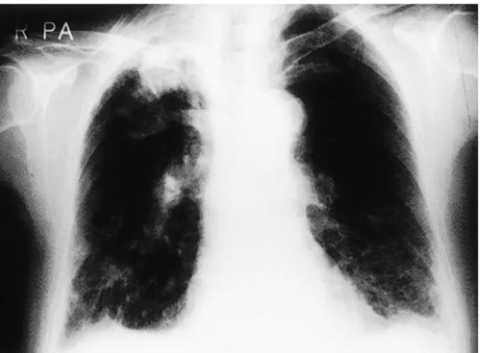
로 결핵약을 복용하였으며, 완치 판정을 받았다. 가족력 및 사회력에서 특이 소견은 없었다. 내원 당시 만성 병색을 보였 고 의식은 명료하였으며, 신체검진에서 혈압 140/90 mmHg, 맥박 85회/분, 호흡 20회/분, 체온은 36.6℃였다. 오른쪽 폐 기저부에서 나음이 청진되었고, 심와부 압통이 있었다.2)말 초혈액검사는 백혈구 4,760/mm3, 혈색소 12.6 g/dL, 혈소판 194,000/mm3였고, 프로트롬빈 시간 1.0 INR , αPTT 34.8초 였다. 생화학검사에서 AST/ALT 29/18 IU/L, 총 빌리루빈 0.6 mg/dL, 총 단백 7.0 g/dL, 알부민 4.2 g/dL, BUN 19.9 mg/dL, 크레아티닌 1.2 mg/dL이었다. 면역혈청학적검사에 서 HBs 항원, anti-HCV 항체는 음성이었고, anti-HBs 항체 는 양성이었다. 소변검사는 정상이었으며, 혈청 종양표지자 검사에서 CEA 22.76 ng/mL이었다. 객담도말검사에서 AFB 는 음성이었다. 내원시 시행한 단순 흉부방사선검사에서 우상엽과 우하엽 및 좌하엽에 섬유성 음영과 소결절 음영
Correspondence to: Jane C Oh, M.D.
Department of Internal Medicine, Sun General Hospital 10-7 Mok-dong, Jung-gu, Daejeon 301-725, Korea Tel: +82-42-220-8801, Fax: +82-42-335-1431 E-mail: [email protected]
면역조직화학검사로 확진된 소세포성 폐암의 위전이 1예
선병원 내과, 방사선과*
오재인·이계성·김재수·박 열·이성훈·김안나·이종민·김규순*
A Case of Gastric Metastasis from Small Cell Lung Carcinoma
Jane C Oh, M.D., Gye Sung Lee, M.D., Jae Su Kim, M.D., Yol Park, M.D., Sung Hoon Lee, M.D., Anna Kim, M.D., Jong Min Lee, M.D., and Kyu Soon Kim, M.D.*
Departments of Internal Medicine and Radiology*, Sun General Hospital, Daejeon, Korea
Gastric metastasis of lung carcinoma is a rare entity which is detected mostly at autopsy. Patients diagnosed as
having those on lifetime are extremely rare. In addition to our case, 54 cases of lung carcinoma metastasis to the
gastro-intestinal tract have been reported in the literature since 1961. We report a case of gastric metastasis
originated from small cell lung carcinoma. The patient was a 87-year-old man. He refused lung biopsy and further
treatment and died 2 months after the diagnosis. This is the case of gastric metastsis originated from lung
carcinoma, which was confirmed by immunohistochemical staining. (Korean J Gastroenterol 2004;44:168-171)
Key Words: Stomach; Lung; Small cell carcinoma; Metastasis; Immunohistochemistry
오재인 외 7인. 면역조직화학검사로 확진된 소세포성 폐암의 위전이 1예
Fig. 1. Chest X-ray. It shows nodulo-streaky infiltration with inflammatory granuloma in both lung parenchyma.
Fig. 2. Chest CT. It shows multifocal, mass-like lesions with irregular margin in right upper lung, right lower lung and left lower lung with enlargement of lymph nodes in left lower paratrachea area and AP window.
이 관찰되었다(Fig. 1). 흉부CT검사에서 우상엽과 우하엽 및 좌하엽에 다초점성이면서 비정형 경계를 가지는 종괴가 있었고 종괴 주위로 섬유화와 기종성 변화가 함께 동반되 었다. 좌측 기관지 주위 하부와 기관용골 상부에서 림프절 종대가 관찰되었다(Fig. 2). 전신골주사스캔검사에서 양측 늑골에 다수의 열소가 불규칙하게 분포하는 양상을 보였다.
상부위장관내시경검사에서 중체부 대만측과 상체부 대만측 에 중심부 함몰 궤양을 동반한 약 1 cm 크기의 융기형 점 막 병변이 2개 관찰되었다(Fig. 3). 위내 병변에서 시행한 조직검사에서 과염색되는 핵과 적은 세포질을 가지는 난원 형의 둥근 세포들이 밀집된 형태를 보이면서 CD56 면역조 직화학검사에서 진한 갈색으로 염색되는 양성 소견을 보였 다(Fig. 4). 환자는 폐조직검사 및 치료를 거부하였고 내원 2개월 후 사망하였다.
Fig. 3. Endosopy. It shows ulceraions of Bull's eye pattern in upper body and mid body of the stomach.
Fig. 4. Histologic findings of the gastric biopsy specimen. (A) It shows oval shaped cells with hyperchromatic nuclei in tumor nest pattern (H&E stain, ×100). (B) Immunohistochemical staining with CD56 shows stained cells in brown color (×100).
169
The Korean Journal of Gastroenterology: Vol. 44, No. 3, 2004
고 찰
악성 종양에 의한 혈행성 위 전이는 드물며, 부검으로 연 구된 보고들에서의 발생 빈도는 약 0.2-3%이다. 위 전이를 잘 일으키는 종양으로 유방암, 악성 흑색종, 폐암이 있으 며, 드물게 간암, 융모막암, 정상피종, 식도 편평세포암, 이 하선 점액표피양 종양이 일으키기도 한다.2,4 폐암의 전이 가 호발하는 부위는 뼈(26%), 간(24%), 골수(19%), 중추신 경계(9%)이며,5 폐암에 의한 위 전이는 발생 빈도가 약 0.2-9%에 불과하다.1,2 본 증례에서는 늑골 전이와 위 전이 가 병발하였다.
폐암에 의한 위 전이는 대부분 무증상이어서 부검 중 우 연히 발견되는 경우가 많으나, 드물게 병의 초기에 발생하 여 연하 곤란, 심와부 동통, 오심, 구토, 식후 복부 팽만감 외에도 토혈, 장폐색, 천공, 출혈 등의 증상이 나타날 수 있 다. 출혈로 인해 철 결핍성 빈혈이 나타날 수 있고, 천공이 나 폐색으로 인해 사망에 이르기도 한다.6-8본 증례의 환자 는 심와부 동통과 CEA의 증가로 위내시경과 대장내시경을 시행한 후 위 전이를 진단하였다.
혈행성 위 전이를 하는 암세포는 점막하층에서 기원하며 종괴가 커짐에 따라 점막층에 미란과 혈류 감소로 인한 궤 양을 형성한다.9 육안적으로 황소안(bull's eye), 표적 병변 (target lesion) 또는 화산 분화구 모양의 병변이 나타나며 본 증례의 환자는 다양한 크기의 황소안 병변을 보였다.9,10 원발성 위소세포암은 위선암과 유사하게 버섯형(fungating) 또는 궤양형의 단일 종괴로 보이며, 조직학적으로 선암, 편 평상피암이 많아 점막층에서 기원하는 것으로 생각된
다.11,12 원발성 위소세포암이라도 세포의 증식이 빠를 때,
점막하층으로 암이 침범하여 황소안 병변이 나타날 수 있 다.13 본 환자에서는 폐 병변에 대한 조직검사가 시행되지 않아 위소세포암이 전이성인지 원발성인지 여부를 확진하 지 못했다. 그러나 내시경에서 다양한 크기의 황소안 병변 을 보이고 흉부 CT에서 기관지 내벽을 따라 종괴가 관찰되 어 소세포성 폐암의 위 전이로 간주하였다.
소세포암은 작은 원형 또는 타원형의 세포로서 림프구와 형태적으로 비슷하며, 소세포암을 진단할 때 가장 결정적 인 요소는 H&E 염색한 조직의 특징적인 염색질과 핵소체 의 패턴을 광학현미경으로 보는 것이나, 많은 경우에서 H&E만으로 확진하기 힘들다. 추가로 전자현미경을 이용해 세포 미세 구조를 파악하거나, 면역조직화학염색을 통해 진 단하는 것이 필요하다.14 소세포암의 면역학적 표지자는 neurofilaments, Leu-7, chromogranin, synaptophysin, neuron- specific enolase (CD56) 등이 있으며, 이 중에서도 neuron- specific enolase (CD56)는 약 70%의 환자에서 양성 반응을
나타내는 유용한 표지자이다.15 본 증례에서는 림프종 세포 와 혼동되기 쉬운 소세포암을 면역형광염색법(CD56)으로 확진하였다.
1961년도 이후 국내외 문헌에서, 생존 당시 진단된 폐암 환자의 소화기관 전이는 54예가 있으며,16소장으로의 전이 가 가장 빈번하며, 위로의 전이도 다수 포함되어 있다. 조 직학적으로 편평상피암과 대세포암이 가장 흔하며, 이는 폐암의 조직형 중 편평상피암과 대세포암이 많은 것과 일 치한다.17 국내에 발생된 소세포성 폐암의 위 전이는 2예가 보고된 바 있다.3,18
본 증례의 환자에게 치료를 시행하지 않았으나, 폐암에 의한 위 전이에는 수술 및 항암화학요법이 효과가 있다. 천 공이 흔하고 항암화학요법에 따른 천공 사례도 보고되므로, 가능하면 항암화학요법 전 수술이 선행되는 것이 바람직하 다.7
소세포성 폐암의 위 전이는 드물며, 생존시에 진단하기 도 어렵다. 악성 종양 환자에서의 위장관 증상은 항암화학 요법의 부작용 또는 중추신경계 전이에 의한 것으로 대부 분 간주되나, 위 전이에 의한 가능성을 고려하여서 이에 대 한 검사를 시행할 것을 권유하며, 천공과 같은 치명적인 합 병증이 흔하므로 이 점을 숙지하여 환자의 치료에 임하여 야 할 것이다.
참고문헌
1. Antler AS, Ough Y, Pitchumoni CS, Davidian M, Thelmo W. Gastrointestinal metastses from malignant tumors of the lung. Cancer 1982;49:170-172.
2. Green LK. Hematogenous metastses to the stomach. A review of 67 cases. Cancer 1990;65:1596-1600.
3. Kim HS, Jang WI, Hong HS, et al. Metastatic involvement of the stomach secondary to lung carcinoma. J Korean Med Sci 1993;8:24-29.
4. Menuck LS, Amberg JR. Metastatic disease involving the stomach. Am J Dig Dis 1975;20:903-913.
5. Abrams J, Doyle LA, Aisner J. Staging, prognostic factors and special considerations in small cell lung cancer. Semin Oncol 1988;15:261-277.
6. Edwards R, Royle G. Metastatic carcinoma causing haematemesis. Br Med J 1975;14:598.
7. Suzaki N, Hiraki A, Ueoka H, et al. Gastric perforation due to metastasis from adenocarcimona of the lung. Anticancer Res 2002;22:1209-1212.
8. Maeda J, Miyake M, Tokita K et al. Small cell lung cancer with extensive cutaneous and gastric metastases. Intern Med 1992;31:1325-1328.
170
Oh JC, et al. A Case of Gastric Metastasis from Small Cell Lung Carcinoma
9. Rubin SA, Davis M. "Bull's eye" or "target" lesions of the stomach secondary to carcinoma of the lung. Am J Gastroenterol 1985;80:67-69.
10. Sandler RS, Sartor RB, Bozymaski EM. Endoscopic appearance of cancer metastatic to the stomach. J Clin Gastroenterol 1981;3:35-37.
11. Takaku H, Oka K, Naoi Y, Santoh N, Setsu Y, Mori N.
Primary advanced gastric small cell carcinoma. Am J Gastroenterol 1999;94:1402-1404.
12. Kusayanagi S, Konishi K, Miyasaka N, et al. Primary small cell carcinoma of the stomach. J Gastroenterol Hepatol 2003;18:743-747.
13. Hussein AM, Otrakji CL, Hussein BT. Small cell carcinoma of the stomach, case report and review of the literature. Dig Dis Sci 1990;35:513-518.
14. Juan Rosai. Ackerman's surgical pathology. 8th ed. Mosby.
1996.
15. Bergh J, Esscher T, Steinholts L, Nilsson K, Pahlman S.
Immunocytochemical demonstration of neuron-specific enolase (NSE) in human lung cancers. Am J Clin Pathol 1985;84:1-7.
16. Yamamoto M, Matsuzaki K, Kusumoto H, et al. Gastric metastasis from lung carcinoma. Case report. Hepatogastro- enterology 2002;49:363-365.
17. Schottenfeld D. Epidemiology of lung cancer. In: Pass HI, Mitchell JB, Johnson DH, Turrisi AT, ed. Lung Cancer. 1st ed. Philadelphia: Lippincott-Raven Publishers, 1996:305- 321.
18. Lee CH, Lee SJ, Kim YA, et al. A case of metastatic gastric cancer resulting from small cell lung cancer. Korean J Gastrointest Endosc 1998;18:755-760.
171