서 론
환
자가 폐암으로 진단되면 그 다음 단계로 환자의 병기 를 판정해야 한다. 폐암의 정확한 병기 판정은 ① 수 술 가능성 여부, ② 치료의 종류 선택 및 ③ 환자의 예후 예 측에 있어 매우 중요하다. 병기는 수술적 절제가 가능한 비 소세포성 폐암(non-small cell lung cancer, NSCLC)에 주 로 적용되며 소세포성 폐암(small cell lung cancer, SCLC) 의 경우는 방사선 치료가 가능한 방사선 조사 구간에 따라 limited disease와 extensive disease로 간단하게 병기를분류한다. 현재 폐암의 병기 판정은 1997년에 American Joint Committee on Cancer (AJCC)과 Union Interna- tionale Contre le Cancer (UICC)에서 개정한 TNM 분류 법을 사용한다(Table 1, 2). 여기에서 T는 원발종양, N은 림프절 전이, M은 원격전이를 의미한다(1, 2). 최근에 IASLC (International Association for the Study of Lung Cancer)에서 현재의 병기 체계를 개정하고자 5년 생존율에 근거하여 새로운 병기 권고안을 발표하였다(3, 4). 그러나 아직 확정된 안은 아니며 이에 대해서는 후에 간단히 언급 하도록 하겠다.
폐암의 병기 결정
Staging of Lung Cancer
김 혜 영 | 국립암센터 영상의학과·폐암센터 | Hyae Young Kim, MD
Department of Diagnostic Radiology, Center for Lung cancer, National Cancer Center E - mail : [email protected]
J Korean Med Assoc 2008; 51(12): 1118 -1124
A
ccurate staging of lung cancers is important to determine the treatment options and the prognosis of patients with a lung cancer. TNM system revised in 1997 by American Joint Committee on Cancer and the Union Internationale Contre le Cancer is widely used in staging of the lung cancer. The TNM system is an expression of the anatomic extent of diseases and is based on the assessment of three components; extent of the primary tumor (T), regional lymph node metastasis (N), and distant metastasis (M). Non-invasive staging of lung cancers is based primarily on chest computed tomography (CT), and if available, on positron emission tomography (PET). Chest CT scanning is useful in providing anatomic details, but the accuracy of the chest CT scanning in differentiating benign from malignant lymph nodes in the mediastinum is poor.PET scanning has a much better sensitivity and specificity than chest CT scanning for mediastinal lymph node staging, and distant metastatic diseases can be detected by PET scanning. With either test, abnormal findings must be confirmed by a tissue biopsy to ensure accurate staging. Invasive techniques for biopsy of mediastinal lymph nodes or pathologic tissue include transbronchial needle aspiration, transesophageal fine needle aspiration, and surgery.
Keywords: Lung; Lung cancer; Neoplasm staging 핵 심 용 어: 폐; 폐암; 병기
Abstract
폐암의 5년 생존율
임상적 병기로 폐암의 전체 5년 생존율은 약 14%이고 1A 병기가 약 61%, 1B 병기가 38%, 2A 병기가 약 34%, 2B 병 기가 약 24%, 3A 병기가 약 17%, 3B 병기가 약 5%, 4 병기 는 1%에 불과하다. 임상 병기 1기나 2기 및 3A 병기의 일부 는 수술을 고려하며, T4N0에 의한 3B 병기를 제외한 3B 병 기나 4 병기는 수술보다는 다른 치료 방법을 선택한다. 수 술로 치료한 폐암의 5년 생존율은 1A 병기는 약 67%, 1B 병기는 약 57%, 2A 병기는 약 55%, 2B 병기는 약 39%, 3A 병기는 약 23% 정도이다(2). 수술 전에는 병리 병기를 알 수 없으므로 치료는 임상적 병기에 의존한다.
폐암의 병기 결정을 위한 방법
폐암 환자의 병기 결정을 위해 고식적 방법으로 병력 청 취, 이학적 검사, 혈액 검사, 기관지 내시경 및 조영증강 흉 부 CT를 시행한다. CT는 폐암 병기 결정을 위해 가장 흔하
게 사용되는 비침습적인 방법으로 종격동 병기를 결정하는 데는 불완전한 방법이지만 흉부의 전반적인 해부학적 모양 을 보여주는 데는 가장 좋은 방법이다. 따라서 아직까지는 어느 림프절을 선택하여 조직검사를 시행할지 결정해 주는 방법으로 폐암 진단에 있어 매우 중요한 수단이다. 조영 증 강을 한 후 흉곽 입구에서부터 부신까지 포함시켜야 한다.
최근 PET-CT의 등장으로 그동안 사용되었던 공간 해상능 이 부족한 FDG-PET과는 달리 해부학적 위치 정보를 제공 함으로써 림프절 전이에 대한 진단의 정확도가 CT보다 우 수함이 보고되었을 뿐 아니라 수술이 가능하다고 생각했던 환자의 16% 가량에서 흉곽외 전이를 발견함으로써 폐암의 수술 전 필수 검사로 인식되고 있다(5).
종격동 림프절의 전이 여부를 판단하는 것은 림프절의 상 태가 대부분의 환자에서 수술 가능 여부를 판정하게 되므로 매우 중요하다. 이를 위하여 침습적인 방법으로 맹검 경기관 지세침흡입(blind transbronchial needle aspiration, TBNA), 기관지내시경 초음파(EBUS) 유도하의 TBNA, 경 식도 TBNA 등이 있다. 초음파 유도하에 기관지 혹은 식도 를 통한 림프절의 조직 검사로 최소한의 침습으로 거의 완벽 하게 종격동 병기를 결정할 수 있게 되었다. 그러나 초음파 유도하에도 검사가 어려운 부위에 위치한 림프절의 경우는 경부 종격동경 검사, video-assisted thoracoscopy나 mini- thoracotomy를 통한 방법으로 림프절 조직을 얻어야 한다.
원발성 종양 (T: Primary Tumor)
1. 비소세포암
T 병기의 일반적 사항은 Table 2에 정리되어 있다(1). 기 관지 점막에 국한된 작은 종양이 기관분기부(carina)에서 2cm 내에 위치하고 림프절 전이나 원위부 전이가 없는 경 우에는 5년 생존율이 80% 이상이므로 T1에 포함시킨다. 불 완전 간열(incomplete fissure)인 경우 종괴가 완전히 폐실 질 내에 위치하여 흉막과 접촉이 없으면 T1 병변이지만 불 완전 간열을 건너간 경우는 완전 간열이라면 간열을 침범한 것과 같으므로 흉막을 침범한 것으로 간주하여 T2 병변이 된다. 종양의 크기에 관계없이 장측 흉막을 침범하면 T2로
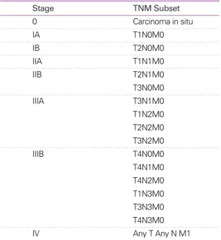
Table 1. Staging grouping - TNM subsets (1)
Stage TNM Subset
0 Carcinoma in situ
IA T1N0M0
IB T2N0M0
IIA T1N1M0
IIB T2N1M0
T3N0M0
IIIA T3N1M0
T1N2M0 T2N2M0 T3N2M0
IIIB T4N0M0
T4N1M0 T4N2M0 T1N3M0 T3N3M0 T4N3M0
IV Any T Any N M1
분류된다. 이는 장측 흉막을 침범하지 않은 3cm 이상 크기 의 T2 종양에 비해 예후가 더 나쁘다. 그 이유는 주변의 흉 막강 내로 파종되기 쉽고, 전신적 파급의 경로로 작용할 수 있기 때문이다.
T3 종양은 일반적으로 벽측 흉막, 흉벽, 횡격막, 심장, 상 구(superior sulcus), 근위부 주기관지 등을 침범한 경우를 말하지만, 종격동측 흉막, 종격동 지방, 심낭 및 신경조직을 침범한 경우에도 장기나 대혈관을 침범하지 않으면 T3 범 주에 포함시킨다. 이러한 종양은 수술적 절제가 가능하고 수술시 주변 조직을 함께 절제함으로써 완전 절제가 가능하 다. 폐동맥 혹은 폐정맥의 침범의 경우에는 심낭막 내부까 지 침범한 경우 T4이고 심낭 바깥에 국한되어 있으면 T3 병 변이다. 횡격막을 침범한 경우는 T3로 완전 절제가 가능하 고 횡격막 아래쪽의 장기가 종양에 의해 직접 침범된 경우
는 장기를 포함하여 절제가 가능하기도 하나 T4 병변이다.
상구종양(superior sulcus tumor)은 cupola의 벽측 흉막 을 침범하고, 인접한 흉벽과 신경혈관까지 침범할 수 있다.
늑골, 늑간근육, 교감신경고리, 성상신경절(satellite gang- lion), 상완신경총의 하신경섬유(lowest cord of brachial plexus)가 침범되었을 때는 T3에 포함된다. Pancoast 증후 군(Horner’s syndrome, pain in the C8-T1 distribution, atrophy of hand’s intrinsic muscle)을 동반한 경우는 상 완신경총이 심하게 침범되는 경우로 T4 병변으로 분류한 다. 척추체의 피질을 침범한 경우는 T4에 포함된다. 또한 costotransverse foramen을 침범해도 척수 때문에 완전절 제가 어려워 T4로 간주한다. 종양이 척추체에 붙어 있으나 골미란의 방사선학적 소견이 없고 흉막, 척추전근막 (prevertebral fascia), 골막 등의 침범이 있는 경우는 척추
Table 2.International staging system TNM classification (1) T0 No evidence of primary tumor
TX Primary tumor cannot be assessed, or tumor proven by the presence of malignant cells in sputum or bronchial washings but not visualized by imaging or bronchoscopy
Tis Carcinoma in situ
T1 Tumor ≤ 3cm in greatest dimension, surrounded by lung or visceral pleura, without bronchoscopic evidence of invasion more proximal than the lobar bronchus * (i.e., not in the main bronchus)
T2 Tumor with any of the following features of size or extent: > 3cm in greatest dimension; involves main bronchus; >2cm distal to the carina; invades the visceral pleura; associated with atelectasis or obstructive pneumonitis that extends to the hilar region but does not involve the entire lung
T3 Tumor of any size that directly invades any of the following: chest wall (including superior sulcus tumor), diaphragm, mediastinal pleura, parietal pericardium; or tumor in the main bronchus < 2cm distal to the carina, but without involvement of the carina; or associated atelectasis or obstructive pneumonitis of the entire lung
T4 Tumor of any size that invades any of the following: mediastinum, heart, great vessels, trachea, esophagus, vertebral body, carina: or tumor with a malignant pleural or pericardial effusion†: or with satellite tumor nodule(s) within the ipsilateral primary tumor lobe of the lung
NX Regional lymph node cannot be assessed N0 No regional lymph node metastasis
N1 Metastasis to ipsilateral peribronchial and/or ipsilateral hilar lymph nodes, and intrapulmnonary nodes involved by direct extension of the primary tumor
N2 Metastasis to ipsilateral mediastinal and/or subcarinal lymph node (s)
N3 Metastasis to contralateral mediastinal, contralateral hilar, ipsilateral or contralateral scalene, or supraclavicular lymph nodes (s) MX Presence of distant metastasis cannot be assessed
M0 No distant metastasis M1 Distant metastasis present‡
* The uncommon superficial tumor of any size with its invasive component limited to the bronchial wall, which may extend proximal to the main bronchus, is also classified T1.
†Most pleural effusions associated with lung cancer are due to tumor. However, there are a few patients in whom multiple cytopatho- logic examinations of pleural fluid show no tumor. In these cases, the fluid is non-bloody and is not an exudate. When these elements and clinical judgment dictate that the effusion is not related to the tumor, the effusion should be excluded as a staging element and the patient's disease should be staged T1, T2, or T3. Pericardial effusion is classified according to the same rules.
‡Separate metastatic tumor nodule (s) in the ipsilateral nonprimary-tumor lobe(s) of the lung also are classified M1.
체의 일부를 포함하여 절제 가능하므로 T3 병변으로 간주 한다. 그러나 병리학적으로 피질의 침범이 있으면 pT4가 된 다. 미주신경의 침범은 반회후두신경의 침범없이는 임상적 으로 알기 어려우며 해당 병변측의 종격동 림프절 전이나 종양에 의한 직접 침범에 기인하고 T4로 간주하며 수술 적 응증이 되지 못한다. 심낭막을 따라 주행하는 횡격막 신경 의 침범은 T3에 해당한다.
위성종양(satellite lesions)은 원발 종양과는 완전히 분 리되어 있는 경계가 분명한 부수적 종양 병소로 대엽절제 표본에서 약 7.6~19% 정도 발견된다. 원발 폐암과 같은 엽 에만 국한되어 있을 때는 T4로, 편측 폐의 다른 엽에 있을 때는 M1으로 간주한다. 흉막 삼출이 있는 종양은 흉막 삼출 이 양성으로 판명될 때까지는 T4 병변으로 간주한다. 흉막 삼출액이 양성으로 판명되기 위한 조건은 ① 적어도 연속적 인 2회의 흉부 천자에서 세포학 검사상 악성 세포 음성이고,
② 누출액(transudate)이며, ③ 혈액성이 아니어야 한다.
악성 흉막 삼출 등과 같이 벽측이나 장측 흉막에 종양이 파 급되었으면 T4 병변이다. 흉막 삼출액이 없더라도 흉막에 결절이나 열(fissure)의 비후 소견이 있으면 흉막 전이를 시 사하는 소견으로 T4 병변이다. 그러나 벽측 흉막 밖의 흉벽 이나 횡격막에 격리된 종양이 있으면 M1이 된다.
동기 원발성 폐암(synchronous primary tumors)은 전 체 폐암 환자의 0.26~1.7% 정도에서 발생한다. 이는 ① 두 종양이 떨어져 있고, ② 다른 분절, 엽, 혹은 폐에 존재해야 하며, ③ 같은 림프관 영역을 공유하지 않고, ④ 다른 조직 형을 보여야 한다. 병기는 각각의 종양에 대하여 병기 결정 한 후, 이 중 높은 병기를 환자의 병기로 결정한다. 생존율은 비슷한 병기의 한가지 폐암을 가진 환자보다 나쁘다. 3년 이 상 생존하는 폐암 환자의 10~25%에서 다발성 폐암이 발생 하며 이러한 병변은 후시성 종양, 전이성 병소 혹은 재발 때 문이다. 후시성 종양(metachronous tumors)은 ① 분리되 어 있는 다른 해부학적 위치에 생겨야 하며, ② 다른 세포유 형이어야 하며, ③ 원발 폐암이 stump에 남은 종양 없이 완 전 절제되었고, ④ 진단시 림프배액이 같은 부위나 폐이외 장소에 종양이 없으면서 이차적 원발암이 발견되어야 한다.
혹은 원발 장소가 밝혀지지 않으면 두 종양 사이에 2~3년
이상의 시간 차이가 있어야 한다. 병기 결정은 원발 폐암과 같은 방법으로 한다. 동기 폐암이나 후시성 종양의 예후는 전이암이나 재발암에 비하여 좋다.
세기관지 폐포암(bronchioloalveolar carcinoma, BAC) 은 43% 정도가 단일결절형으로 이는 TNM 체계로 병기를 나눌 수 있다. 나머지 경결형이나 다결절형의 경우는 기존 의 T 병기를 적용시키기는 어려우므로 TX로 분류하며 양측 성의 경우는 M1으로 한다.
2. 소세포암(Small Cell Lung Cancer)
간단하게 제한적(limited)과 광범위(extensive) 병기로 나눈다. 매우 드물게 폐에만 국한되어 수술적 절제가 가능 한 경우 TNM 체계로 병기를 결정할 수 있다. 제한적 병기 는 화학요법 및 방사선치료를 하고 광범위 병기는 화학요법 만 시행한다. 제한적 병기는 편측 혹은 반대측 종격동 림프 절이나 쇄골상 림프절에만 전이가 국한되어 있어 방사선치 료 영역에 포함시킬 수 있는 경우로 편측 흉막삼출과 상대 정맥 증후군도 제한적 병기에 포함된다. 심낭, 반대측 흉막 이나 흉부외 파급은 광범위 병기로 간주한다.
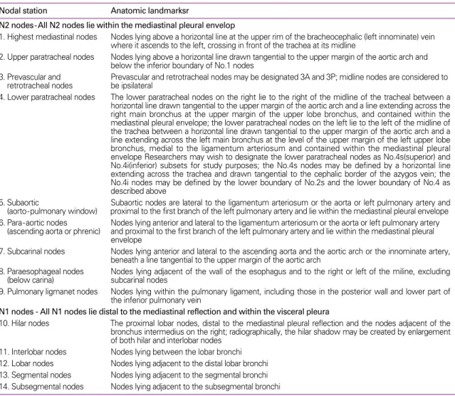
3. 림프절(N: Lymph Nodes)
림프절의 분류는 Table 3을 참조하기 바란다. CT를 촬영 하였을 때 림프절의 상태에 따라 폐암 환자를 네 그룹으로 나눌 수 있다(6). 이는 후에 침습적 림프절 검사의 필요 여 부를 결정하는 데 중요하다. 첫번째는 종격동 침범이 혈관 과 기도를 둘러싸고 있어 정확한 림프절 측정이 불가능한 경우로 종격동 침범을 영상으로만도 진단할 수 있고 이 경 우는 소세포암인지 비소세포암인지 구별을 위해 어떤 방법 으로 조직을 얻을지를 결정하면 된다. 두번째 그룹은 크기 를 정확히 측정할 수 있는 종격동 림프절 비대를 가진 환자 군으로 반드시 림프절 전이 여부를 확인해야 한다. 세번째 그룹은 종격동 림프절이 정상이지만 중심부 종괴이거나 N1 이 의심되는 경우 N2, 3의 가능성이 있으므로(20~25% 정 도) 림프절의 확진이 필요한 경우이다. 마지막 그룹은 임상 적 제 1병기인 변연부 종양인 경우 종격동 침범의 가능성이 매우 낮아 더 이상의 확진이 불필요하다. 림프절 전이의 진
단에 있어서 ① 장경 1cm 이상, ② 단경 1.5cm 이상, ③ 단 경 1cm 이상 및 중심부 괴사나 피막의 파괴, ④ 모양과 상 관없이 단경 2cm 이상 등의 기준이 쓰이기도 하지만 CT상 단경 1cm 이상인 경우를 전이로 간주하는 기준이 가장 보 편적으로 쓰인다. 림프절의 중심부가 저음영을 보이면 괴사 를 의미하고 전이의 좋은 지표이다. 림프절의 중심부에 지 방음영이 보이면 양성인 경우가 많다. 폐문부 림프절의 경 우는 폐와 만나는 부위가 모양이 둥글면 전이되었음을 시사 한다(7).
정상 크기의 림프절이 현미경적인 전이가 있는 빈도는 논
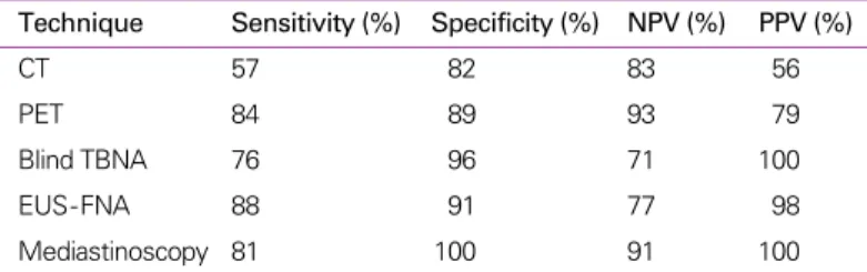
란이 많지만 종격동 전이가 있는 환자의 8~64%에서 보고 되고 있다. CT는 종격동 림프절 전이 발견에 높은 음성 예 측도를 보인다. 또한 CT상 음성으로 수술하여 전이가 있었 던 림프절을 가진 환자는 CT상 전이로 판단하였던 경우보 다 높은 생존율을 보인다. 따라서 CT상 림프절이 음성인 경 우 대개 종격동 경검사를 시행하지 않고 바로 개흉술을 시 행한다. CT상 양성인 림프절의 경우 종격동 내시경을 꼭 시 행하여야 하는데 많은 양성 림프절이 CT상 커져 보일 수 있 기 때문이다. 종격동 림프절 전이 진단에 있어 각 검사방법 에 대한 메타 분석 결과가 Table 4에 정리되어 있다(8).
Table 3.Lymph node mapping definition (1)
Nodal station Anatomic landmarksr
N2 nodes-All N2 nodes lie within the mediastinal pleural envelop
1. Highest mediastinal nodes Nodes lying above a horizontal line at the upper rim of the bracheocephalic (left innominate) vein where it ascends to the left, crossing in front of the trachea at its midline
2. Upper paratracheal nodes Nodes lying above a horizontal line drawn tangential to the upper margin of the aortic arch and below the inferior boundary of No.1 nodes
3. Prevascular and Prevascular and retrotracheal nodes may be designated 3A and 3P; midline nodes are considered to retrotracheal nodes be ipsilateral
4. Lower paratracheal nodes The lower paratracheal nodes on the right lie to the right of the midline of the tracheal between a horizontal line drawn tangential to the upper margin of the aortic arch and a line extending across the right main bronchus at the upper margin of the upper lobe bronchus, and contained within the mediastinal pleural envelope; the lower paratracheal nodes on the left lie to the left of the midline of the trachea between a horizontal line drawn tangential to the upper margin of the aortic arch and a line extending across the left main bronchus at the level of the upper margin of the left upper lobe bronchus, medial to the ligamentum arteriosum and contained within the mediastinal pleural envelope Researchers may wish to designate the lower paratracheal nodes as No.4s(superior) and No.4i(inferior) subsets for study purposes; the No.4s nodes may be defined by a horizontal line extending across the trachea and drawn tangential to the cephalic border of the azygos vein; the No.4i nodes may be defined by the lower boundary of No.2s and the lower boundary of No.4 as described above
5. Subaortic Subaortic nodes are lateral to the ligamentum arteriosum or the aorta or left pulmonary artery and (aorto-pulmonary window) proximal to the first branch of the left pulmonary artery and lie within the mediastinal pleural envelope 6. Para-aortic nodes Nodes lying anterior and lateral to the ligamentum arteriosum or the aorta or left pulmonary artery
(ascending aorta or phrenic) and proximal to the first branch of the left pulmonary artery and lie within the mediastinal pleural envelope
7. Subcarinal nodes Nodes lying anterior and lateral to the ascending aorta and the aortic arch or the innominate artery, beneath a line tangential to the upper margin of the aortic arch
8. Paraesophageal nodes Nodes lying adjacent of the wall of the esophagus and to the right or left of the miline, excluding (below carina) subcarinal nodes
9. Pulmonary ligmanet nodes Nodes lying within the pulmonary ligament, including those in the posterior wall and lower part of the inferior pulmonary vein
N1 nodes - All N1 nodes lie distal to the mediastinal reflection and within the visceral pleura
10. Hilar nodes The proximal lobar nodes, distal to the mediastinal pleural reflection and the nodes adjacent of the bronchus intermedius on the right; radiographically, the hilar shadow may be created by enlargement of both hilar and interlobar nodes
11. Interlobar nodes Nodes lying between the lobar bronchi 12. Lobar nodes Nodes lying adjacent to the distal lobar bronchi 13. Segmental nodes Nodes lying adjacent to the segmental bronchi 14. Subsegmental nodes Nodes lying adjacent to the subsegmental bronchi
최근 European Society of Thoracic Surgery (ESTS)에 서 제안한 종격동 림프절의 병기 결정의 지침을 보면 PET 혹은 PET-CT를 시행하여 음성인 경우(N0) 종격동경을 시 행하기도 하나 대부분은 수술을 시행하고 양성인 경우 (N2/N3)에는 비수술적 침습적 방법으로 조직검사하여 음 성이면 종격동경 검사까지 하고 음성인 경우 수술을 시행하 고 양성이면 복합요법으로 치료하고 비수술적 침습적 방법 으로 검사하여 림프절이 양성이면 바로 복합요법을 시행하 는 algorithm을 제안하였다(8).
4. 전 이(M: Metastasis)
비소세포암 환자에서 부신, 간, 뇌, 골, 림프절 전이는 진 단 당시에 존재할 수 있으나 이를 발견하기 위한 영상 방법 은 일정하지 않다. 비소세포암 T1-2, N0의 조기 환자의 경 우 눈에 보이지 않는 전이는 1% 미만이라고 한다. 또한 임 상적이나 laboratory 증거가 있는 환자에서 전이가 발견되 는 빈도는 그렇지 않은 경우보다 3배나 된다. 그럼에도 불구 하고 무증상의 환자에서 전이를 발견하기 위한 방사선 검사 가 지속되고 있다. 어떤 임상의는 선암인 경우, 전이가 좀 더 흔하기 때문에 전이 여부를 좀 더 빠른 시기에 검사해야 한 다고 하였다(7).
부신은 흔하게 전이되는 부위로 흉부 CT 촬영시 반드시 포함시켜야 하며 처음 진단시 약 20% 정도에서 부신 전이 가 발견된다. 부신 선종은 일반 인구의 약 2~10% 정도에서 발견될 정도로 흔하기 때문에 전이와 감별해야 한다. 조영 증강 전 CT에서 10HU 이하이거나 10분 지연 CT에서 50%
이상의 HU 감소가 있으면 양성(benign)으로 간주한다. 비 소세포암 환자의 18% 정도에서 뇌전이가 보고되고 있지만
대부분 신경학적 이상의 증후가 있기 때문에 routine CT를 권장하지 않는다. 생존율을 증 가시키기 위하여 부신 전이와 마찬가지로 뇌 전이도 단일성인 경우 절제하기도 한다. 골전 이가 있는 경우 대개 증상이 있거나 골전이를 의심할 수 있는 laboratory test의 이상 소견이 있다. 골주사 검사 및 MRI는 통증이 있거나 alkaline phosphatase의 상승이 있는 경우에 만 시행하고 있지만 FDG-PET이 대부분이 무증상인 골전 이를 약 13% 정도의 비소세포암 환자에서 발견하고 있기 때문에 PET 또는 PET-CT가 폐암 환자에서 전이의 진단에 필수적인 방법으로 인식되고 있다(5).
IASLC의 제안
International Association for the study of lung can- cer (IASLC)에서는 TNM 병기를 개정하고자 Lung Cancer Staging Project를 통하여 각각의 병기가 갖는 치료방침과 생존율에 미치는 영향을 조사하여 2007년 서울에서 개최된 세계폐암학회를 통해 제안하였다(3, 4). 아직 확정된 것은 아니지만 5년 생존율에 차이를 보여 몇 가지 변경하여 제안 한 사항을 기술하고자 한다. 현재 T1으로 분류된 3cm 이하 의 종양을 T1a 2cm 이하와 T1b 2~3cm으로 세분화할 것 을 권유했다. 또한 현재 크기 기준이 없는 T2를 7cm 이상의 종양은 T3로 T2를 3~5cm의 경우는 T2a로 5~7cm인 경우 는 T2b로 분류할 것을 권유했다. 현재 원발종양이 같은 엽 에 위치한 다른 결절이 있는 경우 T4로 분류하는 데 이는 T3 로 현재 전이로 구분되어 있는 동측에 위치한 다른 엽의 결 절의 경우도 M1 대신 T4로 분류할 것을 권유했다. 악성 흉 막삼출이 있는 경우는 M1으로 분류할 것을 권유했고 다른 기관에 전이가 있는 경우는 M1b로 폐나 악성 흉막삼출의 경우는 M1a로 분류할 것을 권유했다. 현재 사용중인 N체계 는 비교적 환자의 예후를 잘 반영하고 있는 것으로 나타나, 현재의 체계를 유지하는 것이 좋다고 보고했다. 그러나 위 분류가 아직 통용되는 되는 것은 아니므로 여기서는 간단히 언급하였다.
Table 4.Value of technique for staging metastatic mediastinal nodes (8) Technique Sensitivity (%) Specificity (%) NPV (%) PPV (%)
CT 57 82 83 56
PET 84 89 93 79
Blind TBNA 76 96 71 100
EUS-FNA 88 91 77 98
Mediastinoscopy 81 100 91 100
결 론
폐암의 병기 결정은 환자의 치료 방침 결정과 예후 결정 에 있어 매우 중요하다. 폐암의 병기를 잘 숙지하고 있고 좀 더 정확한 병기 진단을 위해 적절한 영상을 촬영하여 흉곽 외 전이를 발견하고 침습적 방법을 동원하여 종격동 림프절 의 전이 유무를 결정하는 것은 불필요한 수술을 감소시키고 환자의 치료 방침을 결정하는 데 매우 중요하다.
참고문헌
11. Mountain C. Revisions in the International System for Staging Lung Cancer. Chest 1997; 111: 1710-1717.
12. Silvestri GA, Tanoue LT, Margolis ML, Barker J, Detterbeck F.
The noninvasive staging of non - small cell lung cancer: the guidelines. Chest 2003; 123: 147S-156S.
13. Rami-Porta R, Ball D, Crowley J, Giroux DJ, Jett J, Travis WD, Tsuboi M, Vallières E, Goldstraw P, International Staging Committee, Cancer Research and Biostatistics, Observers to the Committee, Participating Institutions. The IASLC Lung
Cancer Staging Project: proposal for the revision of the T descriptors in the forth coming (seventh) edition of the TNM classification for lung cancer. J Thorac Oncol 2007; 2: 593- 602.
14. Rusch VW, Crowley J, Giroux DJ, Goldstraw P, Im JG, Tsuboi M, Tsuchiya R, Vansteenkiste J, International Staging Com- mittee, Cancer Research and Biostatistics, Observers to the Committee, Participating Institutions. The IASLC Lung Cancer Staging Project: proposal for the revision of the N descriptors in the forth coming (seventh) edition of the TNM classification for lung cancer. J Thorac Oncol 2007; 2: 603-612.
15. Lardinois D, Weder W, Hany TF, Kamel EM, Korom S, Seifert B, von Schulthess GK, Steinert HC. Staging of non-small-cell lung cancer with integrated position - emission tomography and computed tomography. N Engl J Med 2003; 348: 2500- 2507.
16. Silvestri GA, Gould MK, Margolis ML, Lardinois D, Weder W, Hany TF, Kamel EM, Korom S, Seifert B, von Schulthess GK, Steinert HC. Noninvasive staging of non-small cell lung cancer ACCP Evidence - based clinical practice guidelines, 2nd ed.
Chest 2007; 132: 178S-201S.
17. MacDonald SL, Hansell DM. Staging of non-small cell lung cancer: imaging of intrathoracic disease. Eur J Radiol 2003;
45: 18-30.
18. Weder W. Lung cancer: new opportunities - changing algo- rithm in staging. Ann Oncol 2008; 19: 28S-30S.
Peer Reviewers Commentary
본 종설은 1997년 American Joint Committee on Cancer (AJCC)와 Union Internationale Contre le Cancer (UICC)에서 제시한 폐암의 병기결정에 관하여 주로 CT와 PET의 소견을 중심으로 기술하여 폐암의 임상적 병기 결정 에 도움이 되는 내용을 기술하였다. 또한 각 영상과 진단방법의 정확성과 한계를 제시함으로써 각종 검사들의 상호보완 적인 관계를 이해하는데 도움을 주고 있다. 향후 영상기법의 발전과 함께 기관지내시경 초음파 또는 경식도 세침흡입술 의 발전으로 수술전 병기 결정은 정확성이 높아질 것으로 기대된다. 종설의 마지막 부분에서는 IASLC (International Association for the Study of Lung Cancer)에서 제시된 병기 개정에 대한 내용이 소개되었다. 이러한 폐암 병기 결정에 대한 내용의 숙지와 각종 검사방법에 대한 이해는 폐암 환자의 치료 방침 결정과 생존율을 예측하는데 도움을 줄 것으로 보인다.
[정리:편집위원회]