大韓放射線醫쩔會誌 第 23 卷 第
6
號 pp.973 - 980
,1987
Journal of Korean Radiological Society,
23(6) 973-980
,1987
Pulmonary Venous Wedge Angiogram 의 임 상응용
연세대학교 의과대학 방사선과학교실
최 규옥 • 설 준 희 * 이 승 규*조 범 구 **
- Abstract -
Clinical Application of Pulmonary Venous Wedge Angiogram
Kyu Ok Choe, Jun Hee Sul*, Seung Kyu lee*, Bum Koo Cho* *
Department of Radiology, College of Medicine, Yonsei University
Pulmonary venous wedge angiogram (PVWA) was performed in 23 patients with cyanotic congential heart disease due to decreased pulmonary blood flow, as the part of whole of pulmonary arterial system had not been opacified by conventional angiogram. The pulmonary venous wedge angiogram visualized pulmonary arteries
(1)
with completely c10sed feeder or with severe peripheral stenosis in16
patients with pulmonary atresia or TOF,
(2) with the interruption of one pulmonary a따ry in 3 patients,
and (3) in 4 patients with tight pulmonary stenosis where pulmonary arteries were not adequately delineated. PVWA has completed the map of entire p비 monary arterial system,
which was essential in the surgical management of these patients서 료g
폐혈류가 감소되어 청색증을 일으키는 선천 심질환에 서 폐동액계 형태 및 발달정도는 교정 수술의 가능성과 일정한 연관이 있다 1-7) 폐동맥 폐쇄증, 선천적 혹은 수 술후 합뱅증으로 생키는 폐동맥의 말초성 협착 등의 경 우, 심실이나 대통액의 분치를 통하여 관츄되던 폐혈류
*
연세대학교 의과대학 소아파학교실* Department of Pediatrics
,
College of Medicine,
Yonsei University
**
연세대학교 의과대학 흉부외과학교실*. Deoartment of Thoracic Surgery
,
College of Medicine,
Yonsei University
본 논문은 1987년도 연세대학교 의과대학 교수 연쿠 비의 보조로 이루어졌음.
이 논문응 1987 년 10 월 28일 접수하여 1987 년 12 월 16일에 채택되었음.
가 차단되연 페혈관계는 기판지 측부 혈판 등헤 의하여 공급되 며, 이들 측부 혈관은 precapillary JXl lmonary
arteriole 과 연결되기 때문에 통상 심혈관 조영술로는 좋은 영상의 펴l동액 조영상을 얻을 수 없다
저자는 이러한 환자 23 영에서 pulmonary venous wedge angiogram (PVWA) 을 시행함으로써 페 모세 혈 관을 역류한 조영제에 의하여 폐동액계의 좋은 영상을 얻을 수 있었으요로 그 결과를 보고하는 바이다.
대상 및 방법
1981
년 l 월부터 1986 년 7 월까지 연세대학교 부속세브란스영원에서 선천십기형 환자를 심도자 검사하연 서 펴|의 일부 혹은 전부위 폐 혈류가 감소되고 통상 섬 실 조영숭 혹은 대동액 조영술로 폐동액계 가 전혀 혹은 만족할만큼 조영되지 않은환자 23 명에서 PVWA를시 행하였다. 환자는 남자 18 명, 여자 5 명이며 연령 7 개 월부터 쟁세 (중앙값 4 세) 이었다. 모든 환자가펴|혈
973 -
大l함JiX~‘t짜챔핑짤:1:
:
第 23~ 행6
.9Æ1987-
류의 감소로 청색증을 동반한 싱기형 이었으며(Table
1 ) •
그중 폐동액 폐쇄증이 15 영으로 가장 않았고, 펀 측 폐동액차단 3 명.Waters ton
shunt후 합뱅증1
명,심한 폐동맥 협착 4 명이었다.
방볍은 통상 심도자 검사 및 심혈관 조영술을 시행한 후
video
monitor로 전부 혹은 일부 폐동액계 조영이 되지 않았을혜 첨공도자(end ho
lecatheter)
인cou-
rnand
catheter를 개 존난원공 흑은 싱 방중걱 결손을 통 히여 적당한폐정액에 꼭 끼게 밀어넣는데,이때 4 개의 폐정액중 폐동액계 조영이 안된 부위를선택하였으맥 l 개부터 3 개의 폐동액에서wedge
angiogram이 시행되 었다 o .3~O. 5
mgjkg 의 조영제(Hypaque
75)를 손 으로 주입하였다. 이해 방사선 투시로 폐운부 폐동액,Table
1.Subjects
Heart Anomaly Case Number
Pulmonary atresia 15
with VSD 8
A V discordance & VSD 3
single ventricle 3
Tricuspid atresia 1
In terruption of one PA 3
with TOF 2
DORV
Complication of Waterston
shuntl
Tight PS
4with TOF 1
Tricuspid atresia 2
TGV 1
Total 23
흑응 그보다 내즉의 중심부 폐동맥이 조영되는 것을 관 찰할 수 있었다. 이들 폐동액이 조영되지 않으연 주업
압력을 약간 올려서 재차 촬영하였다.
PVWA 에 의하여 조영된 페옹액계 영상 및 통상 심 혈판 조영상을 종합하여 심악강내 폐동액의 존재, 합류 및 발달정도, 폐문부 폐동액이 있을 경우 발달 정도, 좌 우폐동액이 합듀되어 있지 않을 경우 양 폐동액의 가장 내측에 촌재하는 폐동맥의 위치, 펴l 운부 혹은 엽 간 폐 동맥과 연결된 구역성 폐동액
(segmental pulmonar y
artery) 의 수효을 판찰하였다. 폐문부 폐동액의 직경 은 삽업된 도자의 직경과 비교하여 확대를 교정하였다결 과
1) 폐혈류 공급원이 차단된 폐동맥 폐쇄증
폐동액 폐쇄증 환자로서 얼부 혹응 전 폐혈류를공급 하는 동액이 완전허 흑은 거의 악힘으호써, 관류되던폐 동액계가 대동액 조영상으로 조영 되지 않는 9 명에서
PVWA
가 시행되 었다-이중 4 명은 대동맥조영상에 의하여 폐동액계 전체가 보이지 않고 비후된 기판지 동백만 보였는데,이들은 1 회의
PVWA
에 의히여 천체 펴1 동액계 즉, 싱막강내 퍼l 동액뿐 아니라 반대측 폐동맥도 조영되었으며,전부pu-
l
monary valvular
atresia 이고, 십 막강내 폐동액이18
개의 구역폐동액파 연결되 L 말초부 폐동액 협착이 없 는 소견을 보였다 (Table2)_
이들에서 펴l 운부 펴l동액 의 크기에 따라서 폐동액 발달이 냐쁜case 2
,3
,4
는 좌측Blalock- Taussig shunt
플 시 행 하였고 폐 동액 이발달되연 2 차 교정수술을 시행할 예정이여,반연 폐동 액 이 잘 발달된
case 1
은conduit
를 이용하여 일단계 교정수술을 시행하였다Table 2. Pulmonary Atresia with Closed Feeder. Nonvisualization of Entire Pulmonary Artery.
Case Age Max Med PA Dimension Connected Surgical
No (yrs) Rt(mm)
Lt(mm) Seg (No) Intervention
1 3 MPA 10.7 5.7 all Rastelli op
2 8 MPA 2 3.5 all Lt. B-T shunt
3 2 MPA 6
4all Lt. B-T shunt
4
7/ 12 MPA 3.5 8 all Lt. B-T shunt
B.T shunt: Blalock-Taussig shun
t.Max Med PA: maximal medial position of central pulmonary arteries
Connected seg (No): Number of segmental pulmonary artery. Connected to central pulmonary artery
974
A
B 최 규옥 외 :
Pulmonary Venous Wedge
Angiogram 의 임 상응용~
Fig.
1.7 month old
boy.Cardiac situs invesus
,atrioven- tricular discordance
,VSD and pulmonary atresia Right superior pulmonary vein wedge angiogram delineated pulmonary valvular atresia , the presence of intrapericardial pulmonary artery
,and all segmental pulmonary arteries connected to the in- trapericardial P A (Case
4in Table
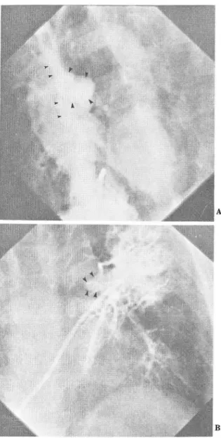
2)Fig 2. 8 mont
h old girl.Cardiac situs inversus , atrioven- tricular discordance
,VSD and pulmonary atresia.
A) Aortogram revealed the
leftinterlobar pulmonary artery supplied via
leftductus arteriosus , but right pulmonary arterial system was not opacified.
B) Right
inferior pulmonaryvenous wedge angiogram delineated right inte
r10bar pulmonary artery (arrow heads)
‘(Case
1in Table
3)나머지 5 영중 2 명은 대동액조영술상 대동액판에 의 하여 일측 심막강내 폐동액이 관류되고 있었으며,
3
영 은aortopulmonary collaterals
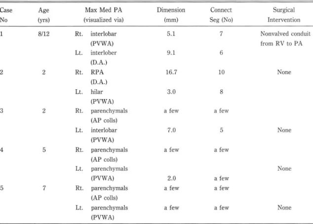
에 의 하여 일측 심 막 강내 흑은 펴|문부 폐동액이 각각 관류되었으냐, 냐머지 조영되지 않는 펴|동액계를 알기 위하여 PVWA 가 시행 되었다. 그결과 5 명 전부 대동맥 조영상£로 펴l동액이 보이지 않던 부위에서 폐실질내 펴l 동액이 말견되었으며,천부 양측 폐동맥의 합류는 없으며,
Case 1
및 2 는 양 측의 폐문부 폐동맥이 있고 이와 연결된 구역성 폐동액 의 합이10
개 이상이 었으나 냐머지3
예는 폐실질내 폐 동액만 손재하였다 (Table 3). 이중 폐문부 폐동액이발달펀
Case
1 응 일단계 교정수술이,좌측 페동액의 발육부전。l 심 한
Case
2는 좌측 단락 수술이 적응되냐 수 술에 응하지 않았고,Case
3 , 4 , 5 는 아무런 수술 처치 가 시행되지 않았다동액 협착에 의하여 반대측 폐동액계가 충분히 보이지 않아서
PVWA
가 시행되 었다(Table
4). 이들중 5 영 은 동백판에 의히여, 2 명은 각각Blalock-Taussig sh-
unt 및Wa terston
shunt 에 의하여 심막강내 폐동액이 조영되었다 4 명응죠}우폐동맥접합부에 (case 1,2, 말초성 폐동맥 협착을 동반한 폐동맥 폐쇄증및 활로 4 증후군 2)
다l 옹액조영 말초부펴1
m
μ
펴|동액 폐쇄증 환자 6 영과
TOF
1 명은 숭상 일측 심막강내 펴|동액은 조영되었으나-大짧放射線醫쟁會誌 : 第 23짱 第
6
~1987-
Table 3. Pulmonary Atn!sia with Closed Feeder. Nonvisualization of the Pa rt of Pulmonary Artery.
Case No
12
Age (yrs) 8/12
2
3 2
4 5
5 7
R
t.L
t.Rt.
Lt.
Rt.
L
t.R
t.Lt
R
t.L
t.Max Med PA (visualized
via)interlobar (PVWA) interlober (D.A.) RPA (D.A.) hilar (PVWA) parenchymals (AP colls) interlobar (PVWA) parenchymals (AP colls) parenchymals (PVWA) parenchymals (AP colls) parenchymals (PVWA)
Max Med PA
,connect seg (No); Legend same as Table 2.
D.A.: ductus arteriosus
AP colls: aorto-p ulmonary collaterals
Dimension Connect
(mm) Seg (No)
5.1 7
9 .1 6
16.7 10
3.0 8
a fe
wa few
7.0 5
a few a few
2.0 a few
a few a few
a few a few
Surgica l Intervention
Nonva lved conduit from RV to PA
None
None
None
None
3
,4).
2 영은폐문부에 (case5
,6) peripheral pul-
액의 죠}우직경외 합은 6 세인 두환자에서 각각4.8 monary stenosl s
가 있었으며.TOF
환자인 l 영 (ca-
se
7)은Waterston
shunt 를 시행한 후 shunt 컵합부 에 협착이 생겼다. 천예에서 조영되지 않았던 폐동액이PVWA 에 의하여 천부개좀되어 있고,좌우폐동액 합 류가 있£며, 죠}우폐의 모든 쿠역성 펴l 동액이 십막강내 폐동맥과 연결되어 있음을 알 수 있었으며,폐문부폐동 액의 발달정도에 따라 일단계 혹은 이단계 교정숭이 시 행되었거나 시행될 예정이다.
3)
편측성 폐동맥 차단3 영의 환자에서
interruption of one PA
가 있었 으며, 2 영은 TOF 와. 1영은 양동맥 우심실 기시증(DORV)
파 동반되어 있었고, 전부 죠덕에 있었다.PVWA 에 의하여 천부 좌측폐동액이 개종되어 있고,좌 폐의 모든 쿠역 펴1 동액과 연결되어 있으며,폐문부 폐동
mm
, 6.5mm 이며, 25세인 환자는 8.6mm 이었다 4) 심한 폐동맥 협착TOF 1
명, tricuspid atresia 2
명 transposition of great vesse l
1 명은누두부혹은 판악폐동액 협 착이 너우 심하여 심도자 검사중v ideo monitor
상 일 측 폐동액을 확인할 수 없어서PVWA
가 시행되었다 이들은전부죠뷰폐동맥이 합류하고폐실질내 펴1 동액이 비교적 잘 발달되어 있요며 모든 구역성 폐동액과 연결 되어 있음을 판철하였다.5)
합 병 증대부분의 환자에게 조영제 주입시 기침을 하였으나 그 외 중대한 합형증은 없었다.
- 976 -
-최 규욱 외 :
Pulmonary Venous Wedge
Angiogram의 임 상응용-A
B
Fig 3. 5 year old boy. Cardiac situs solitus , tricuspid atresia.
VSD ,overriding of aorta and pulmonary atresia.
A) Pulmonary angiogram via previous left
B1ack.
Taussing shunt delineated the entire shunt flow to ward left pulmonary artery and its branches
,and tight stenosis at the confluence of right.left pulmonary artery with faint wash out.
B) Right superior pulmonary venous wedge angiogram delineated the right pulmonary arterial system , the patent intrapericardial right pulmonary artery , connected with all segmen.
tal pulmonary arteries of right lung. (Case 4 in Table
4)Fig 4. 6 year old boy .
A) T
etralogy of fallot with the interruption of leftpulmonary artery
,delineated by right
ven- triculogram. Arrowheads indi
catenight pulmonary artenial tree.
B) Left superior pulmonary vein wedge angiogram revealed patent and well developed left hilar pulmonary artery (arrow heads).
- 977-
-大합liX射級짧學會註、 · 第 23卷 第
6
號1987-
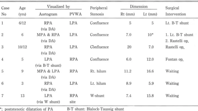
Table 4. Pulmonary Atresia with Peripheral Stenosis
Case No
Age Visualized by Peripheral Dimension Surgical
(yrs) Aortogram PVWA Stenosis Rt (mm)
Lt(mm) Intervention
6/12 RPA LPA Confluence 5 5 L
t.B-T shunt
(via DA)
2
6 MPA & RPA LPA Confluence 7.0 10'
1.L
t.B-T shunt
(via DA) 2. Rastelli op.
3 10112 RPA LPA Cònfluence 20 7.0 Rastelli op.
(via DA)
4 5 LPA RPA Confluence 6.0 12.0 Fontan oP.
(via B-T shunt)
5
9 MPA & LPA RPA R
t.hilum 1
1.2 16.6 Waiting
(via DA)
6 3 RPA LPA L
t.hilum 8.9 5.9 Waiting
(via DA)
7
13 LPA RPA W-shunt 7.4 15.8 Waiting
(via W shunt) s
Jte
•. poststenotic dilatation of P A D A: ductus arteriosus
B-T shun
t:Blalock-Taussig shunt W shunt: Waterston shunt
고 찰
펴|혈관계중 폐살질내 펴l 옹맥과 심막강내 폐동맥응 태 생학적 기원이 서로 다르며 폐 가 발달된 개체에서 폐실 결내 폐동액은 항상 존재한다8) 예를 들연소위
unila-
tera
labscence of one
PA 는 실제 ìnterruptionof
a
PA로서 나머지 폐살질내 폐동맥은 폐혈류 감소로 인한 이차적 말육부전 외에는 정상이다인 폐동액 폐쇄증 은 판악의 펴|쇄증。|나, 6 차 대동액궁에서 기시히는 십 막강내 폐동액의 일부 폭은 전부의 폐쇄로 올 수 있마.
이때 펴|혈류는 동액판 또는
aortopulmonary co Il a te-
ral에 의하여 공급되며, 이들 동맥은 정상개체에서 출 생전 흑은직후에 폐쇄되는 경파를 밟는 동액이다.폐동 액 폐쇄증의 경우 이들 동액이 개존되어 있어야 펴1 혈류 가 유지되나, 폐혈류가 너무 파도히여 폐혈판저항 증가 가 유발되는 수도 있고, 반대로 이들 통액의 점진척 폐 쇄로 페혈류가 정상보다 감소되는 경우가 머 흔한 소견 이다8) 이러한f
eeder 들이 완전히 흑응 거의 완전히 폐 쇄되연, 폐혈판계는 기관지 측부 혈판이나pleuro-par-
enchymal co
llater al
등에 의 하여 혈류를 공급받으며,이들 동액은
precapìll ary pulmonary arterìo
le 과 연결되기 혜문에 대통액 조영술시 말초부 폐동액으로 약 간 역류되어 보이기는 하나 영상이 불분명하며, 심악강 내 펴|동액에 관한 정확한 정보는 얻을 수가 없다. 선천 성 혹은 수술후 합뱅 증으로 오는
interruptìon of one
PA
혹은perìpheral pu1morary
stemsis의 경우도 마 찬가지이다.이와 같이 폐동액계의 형성 및 발육 부천이 있는 경우 수술 방법, 시기 등의 결정에 중요한 것은 섬기형, 폐동 맥계의 상태 및 환자의 연령으로써, 대동액, 폐동액 및 심 실조영상으로 폐 혈판계의 천부가 조영되지 않을 경우
pu1monary venous wedge angìogram( PVWA)
이 펄수 적이다. 펴l정백제는 판악이 없기 혜문에 폐정맥에서 모 세 혈판을 통하여 폐동액으로 역행 판류됨이 동물에서 증 명되었으며 I안 사링에서도 폐동맥계의 압력 및 혈류량 이 감소되어 있는 경우 페정 액에 도자를 꼭끼게 밀어 넣 고 적당한 앙력으호 조영제을 주업함으로써 역류에 의 하여 폐의 모세혈관과 직접 연결된 폐실질내 폐동맥을 조영시킬 수 있을 뿐만 아니라, 폐실질내 폐동액이 폐문 부 혹은 심막강내 폐동액과 연결되어 있으연 이들도 조 영시킬 수 있다11-14) 그러냐조영된 폐동맥이 발육 부 전이 심한 경우 폐동액 역류가 완전치 옷하여 실제는 좀재하는 중심부 폐통액이 조영되지 않는 경우가 운헌-
978-
-최 규옥 외 : Pulmonary Venous Wedge Angiogram의 임 상응용
상 보고되어 있으며씬 저자도 심막강내 폐동액이 조 영되지 않는 경우에는 1 회 이상재시도하고,조영된 가 장 내측 폐동액에 wash→ in 이 없을 때에만 중심부 폐동 액이 없마고 결론을 내렸다. 이애 한개 이상의 폐정액 wedge angiogram 을 필요로 하는 경우도 있으며, 개방 성 난원공 혹은 심앙 중격결손이 없으연 transseptal
puncture 에 의하여 혹은 죠}점실에서 좌심땅으로 특옐히 고안된 도자를 역류하여 넣는 땅법도 있마-
심한 폐동백 협착에 의하여 펴1 혈류가 심하게 감소되 어 심실 조영술로 펴l 동액제가 충분히 조영되지 않는 경 우 심실 누두부 흑은 선택적 주펴1 동액내 주업에 의하여 더 좋은 폐동액 조영상을 얻을 수도 있으나, 저자는 이 러한 4 영의 환자에서 PVWA 에 의하여 충분히 조영되 지 않는 부분의 폐동맥계을 조영할 수 있었다.합병증은 옐로 없으냐 대부분의 환자에서 기침이 있고, 일시석인 펴1 부종 15)’ 기판지경 련 16) 이 보고되어 있다.
교정수술이 가능하려연 섬악강내 펴l 동액 혹은적어도 폐문부 펴1 동맥이 있어야 하며, 이들 동액을 심싣고} 연결 시 펴l 실질 폐동맥위 절반 이상 즉 10개 。l상의 구역성 폐통액이 연결될 수 있어야 교정술후 펴1 동액압이 적정 선을 유지할 수 있으며, 양측 폐문부 폐동액 직경의 합 이 횡격막 수준에서의 하행 대동맥의 직정보다 커야만 완전교정 줄이 가능하다. 또한 펴1 혈판 저 항이 높지 않아 야 하는데 , 혈관 조영 상만으로는 판단키 어 려운 항목이 다1-9 ) 따라서 상기한 폐혈관계의 특정 및 환자의 연 령과 심기행을 고려하여 교정가능성, 일단계 혹응 。 l 단 계 수숭, 완전교정 수숭시기에 대한 결정을 내릴 수 있 으며, 폐혈류가 감소된 챙색증 선칠성 기형의 알부 환자 에서는 통상 심조영술외에 PVWA을 병행하여 폐동맥계 천체의 해부학적 구조을 파악함으로써 치료땅침에 결정 적인 역할을 할 수 있었다.
결 론
1 폐혈류 감소로 청색증을 보이며, 상도자 검사 및 통상 싱헬판 조영술로 펴1 통액계의 전부 흑응 일부가 조 영되지 않은 선천성 심기형 환자
23
영에서 PVWA 가 시행되었다2.
이러한 환자들에서는 PVWA 에 외하여 폐동맥계 전체의 형태, 발달을 관찰할 수 있었마.3.
PVWA는 안전하고 쉬운 시술로써, 큰 휴유증을 동반한예는 없었다.979
REFERENCES
1. McCoon O
c,
Baird OK, David CO: 5urgical management of large bronchial collateral arteries with pulmonary stenosis or atresia. Circulation 52:109-18, 1975 2. Cill Cc,
Moodie 05, McCoon OC: 5taged surgical management of pulmonary atresia with diminutive pulmonary arteries. }. Thorac Cardiovasc 5urg. 75:436, 1977 3. Thiene C, Frescura C, Bini RM, et al: Histology of
pulmonary arterial supply in pulmonary atresia with ven- tricular septal defect. Circulation, 60:1066, 1979 4. Haworth SC, Rees PC, Taylor jFN, et al: Pulmonary atresia
with ventricular septal defect and major aortopulmonary collateral arteries. Effect of systemic-pulmonary anastomosis. Br Heart J 45:153, 1981
5. Kirklin jW, Bargeron LM jr, Pacifico AO, et al.: Manage ment of the T etralogy of F allot with la영e aortopulmonary colateral arteries. In: Codman Ml, ed. Paediatric Car diologι Vol.4, Churchill Livingstone, New York, 483-491, 1981
6. Pacifico AO: Pulmonary atresia. Corrective 5urgery Pediatric Cardiology Vol. 5:137, 1983. Churchill Liv mgstone
7. Alfieri 0, Blackstone EH, Kirklin jW, et al.: 5urgical treat- ment of Teteralogy of Fallot with pulmonary atresia.
J
Thorac Cardiovasc 5urgery 76:321-35, 1978
8. Thiene C & Anderson RH: Pulmonary atresia with V50 anatom
y.
Pediatric Cardiology Vol 5:80, 1983. Churchill Livingstone9. Pool Pε Vogel jHκ Blount SC: Congenital unilateral absence of a pulmonary arter
y.
The importance of flow in pulmonary hypertension. AmJ
Cardio 10:706, 1962 10. Nadas As, & Fyler OC: Pediatric Cardiology.
3rd edPhi떠delphia, WB 5aunders Co. 575, 1972
11. Takamiya M, Taugel, Tadokoro M: Retrograde pulmonary arteriography: a new apporach to opacification of pulmonarya앤ry in pulmonary atresia 껴bs띠 In: Pro- ceedings of the 13th international Congress of Radiology Madrid. Amsterdam: Excerpta Medica, 1973:233. (Inter national Congress 5eries No. 301)
12. Singh SP, Rigby ML, Astley R: Oemonstration of pulmonary arteries by contrastinjection into pulmonary vein. Br Heart J 40:55-57, 1978
-大韓放射絲짧學會품‘ · 第 23흉 第6 號 1987-
13. Nihill MR, Mullins CE, McNamara DG: Visualization of the 51’936-941, 1983.
pulmonary arteries in pseudotruncus by pulmonary vein 15. Singh SP, Astley R: 5evere complication of pulmonary vein wedge angiography. Circulation 58:140-147
,
1978. angiography. Br HeartJ
41 :740,
197914. Freedom RM, Pongiglione ι Williams WG et al.: 16. Alpert BS, Culham JAG: A severe complication of Pulmonary vein Wedge angiography: Indications, results, pulmonary vein angiography. Br Heart
J
41:727-729, 1979andsu땅ical correlates in 25 patients. American