Received:June 6, 2014, Revised:August 1, 2014, Accepted:August 1, 2014
Corresponding to:Kyong-Hee Jung, Division of Rheumatology, Department of Internal Medicine, Inha University Hospital, 27 Inhang-ro, Jung-gu, Incheon 400-711, Korea. E-mail : [email protected]
pISSN: 2093-940X, eISSN: 2233-4718
Copyright ⓒ 2015 by The Korean College of Rheumatology. All rights reserved.
This is a Free Access article, which permits unrestricted non-commerical use, distribution, and reproduction in any medium, provided the original work is properly cited.
Case Report
지주막하 출혈로 내원한 호산구육아종증다발혈관염 환자 1예
오세환1ㆍ박 원1ㆍ권성렬1ㆍ임미진1ㆍ주고운1ㆍ이오현1ㆍ이하영2ㆍ오세양3ㆍ정경희1 인하대학교 의과대학 1내과학교실 류마티스내과, 2영상의학교실, 3신경외과학교실
A Case of Eosinophilic Granulomatosis with Polyangiitis
Presenting with Subarachnoid Hemorrhage and Mononeuritis Multiplex
Se Hwan Oh1, Won Park1, Seong Ryul Kwon1, Mie Jin Lim1, Ko Woon Joo1, Oh Hyun Lee1, Ha Young Lee2, Se Yang Oh3, Kyong-Hee Jung1
1Division of Rheumatology, Department of Internal Medicine, Departments of 2Radiology and 3Neurosurgery, Inha University School of Medicine, Incheon, Korea
Eosinophilic granulomatosis with polyangiitis (EGPA), previously called Churg-Strauss syndrome, is an anti-neutrophil cyto- plasmic antibody associated vasculitis, accompanied by asthma, hypereosinophilia, nonfixed pulmonary infiltrates, and sinusitis. Peripheral neuropathy is common in patients with EGPA; however, a few cases of EGPA with central nervous system (CNS) involvement have been reported. A 45-year-old female referred for right side weakness and posterior neck pain was diag- nosed as EGPA with subarachnoid hemorrhage and mononeuritis multiplex. She was effectively treated with a high dose gluco- corticoid, cyclophosphamide, and intravenous immunoglobulin. EGPA with CNS involvement is uncommon and causes sig- nificant morbidity and mortality. Therefore more rapid and accurate diagnostic evaluation may be required. EGPA should be considered in patients with neurological symptoms and hypereosinophilia. (J Rheum Dis 2015;22:190-194)
Key Words. Churg-Strauss syndrome, Subarachnoid hemorrhage, Mononeuropathies
서 론
호산구육아종증다발혈관염(eosinophilic granulomatosis with polyangiitis, EGPA)은 과거 Churg-Strauss syndrome 으로 불리던 질환으로 호산구의 조직 침착, 괴사성 혈관염 및 혈관 외 조직의 육아종 형성의 조직 병리학적인 특징과 천식이나 알레르기 비염, 호산구 증다증의 임상적 특징을 갖는 항호중구 세포질 항체(anti-neutrophil cytoplasmic antibody, ANCA) 관련 혈관염의 드문 형태이다. EGPA는 여러 장기의 침범이 가능한데, 약 65%~75%에서 말초신경
병증을 일으키고, 뇌 신경을 침범하거나 뇌경색 혹은 뇌내 출혈이나 지주막하출혈 등의 중추신경계를 침범하는 경우 는 매우 드물다[1].
EGPA에서의 중추신경계 침범은 진단이 늦어지는 경우 사 망할 수도 있는 합병증으로 조기 진단과 적절한 치료가 매 우 중요하다. 저자들은 천식 환자가 지주막하출혈로 내원하 여 EGPA로 진단되었고, 중추신경계 침범 및 심한 말초신경 염을 보였으나 조기 치료로 임상적 호전을 보인 1예를 경험 하였기에 보고한다.
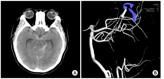
Figure 1. (A) Non-contrast- en- hanced brain computed tomog- raphy image demonstrated sub- arachnoid hemorrhage in basal cistern, suprasellar cistern, peri- mesencephalic cistern and pre- pontine cistern. (B) Volume-ren- dered cerebral angiography im- age shows mild segmental ste- nosis at the P2 segment of left posterior cerebral artery (arrow).
Figure 2. This chest computed tomography shows diffuse ground glass opacity, interlobular septal thickening. Small nodular lesion was seen in peripheral left upper lobe and right middle lobe (arrow).
증 례
45세 여자 환자로 내원 3주 전부터 양측 상하지의 저린 증 상 및 통증이 시작되었고, 내원 당일 뒷목의 통증과 우측 위 약감이 발생하여 응급실로 내원하였다. 20년 전부터 알르레 기성 비염과 14년 전부터 천식을 진단받고 치료 중이었으며, 5년 전부터는 고혈압으로 약물 복용 중이었다. 천식 치료로 는 류코트리엔 수용체 길항제(leukotriene receptor antago- nist), 흡입 스테로이드와 베타2 항진제(Seretide diskusⓇ; GlaxoSmithKline plc, Brentford, England)를 투여 중이었으 며, 7년 전에는 급성 천식 악화로 입원 치료를 받은 적이 있 었다. 외상의 과거력은 없었다.
신장은 160 cm, 체중은 83 kg, 내원 시 혈압은 113/77 mmHg, 맥박 91회/분, 호흡수 15회/분, 체온은 37oC였다.
양측 폐야에 천명음은 관찰되지 않았고, 복부 진찰에서 특 이 소견은 없었다. 양측 부비동의 압통이 있었고, 양측 하 지에 중심성 수포와 자색 구진을 동반한 반점이 관찰되었 다.
신경학적 검사 소견은 내원 당시 의식은 명료하였으며 양측 동공은 동일하였고, 대광 반사는 정상이었다. 좌측 상하지의 근력은 정상이었으나 우측 상지의 근력은 medi- cal research counsil (MRC) grade IV, 우측 하지는 MRC grade III로 저하되어 있었다.
방사선학적 검사 소견은 뇌전산단층촬영에서 뇌바닥수 조(basal cistern), 안장위수조(suprasellar cistern), 중뇌 주위조(perimesencephalic cistern) 및 전교뇌수조(pre- pontine cistern)의 지주막하출혈과 함께 제 4뇌실의 뇌실 내출혈도 동반되었으며(Figure 1A), 뇌혈관조영술에서는 후대뇌동맥의 협착 소견이 보였다(Figure 1B). 그러나 뇌 동맥류 파열, 동정맥기형 파열 등의 소견은 관찰되지 않았 고, 내원 10일째 재시행한 뇌혈관조영술에서도 지주막하 출혈의 원인이 될 만한 동맥류나 동정맥기형의 소견은 없 었다. 흉부 전산단층촬영에서 양측 폐의 전반적인 간유리 음영(ground glass opacity)과 함께 소엽간 비후, 좌상엽과
우중엽에 작은 결절들이 관찰되었다(Figure 2). 단순 부비 동 촬영에서는 만성 부비동염 소견이 보였다.
말초 혈액 검사에서 백혈구는 33,150/mm3 (호산구 62.7%) 이었고 적혈구 침강 속도는 63 mm/h, C-반응성 단백질은 1.99 mg/dL로 증가되어 있었다. 혈액요소질소는 8.8 mg/dL, 크레 아티닌은 0.73 mg/dL로 정상이었으나, 현미경 혈뇨 (11-15/high power field)가 있었고 24시간 단백뇨는 664 mg/d로 측정되었다. 간접면역형광법을 이용한 ANCA 검사에 서 cytoplasmic ANCA (c-ANCA)는 음성이었고 perinuclear ANCA (p-ANCA)는 1:320으로 강양성이었다. Enzyme- linked immunosorbent assay (ELISA)를 이용한 항골수세포 형과산화효소(anti-myeloperoxidase, anti-MPO)는 6.3 Index (참고지: 1.10 이상 시 양성)으로 양성이었고, 항단백분해효소 (anti-protease 3)는 음성이었다. 류마티스인자는 양성(41.2
Figure 3. The progress report of this patient. Eosinophil count, anti-neutrophil cytoplasmic antibody (ANCA) titer and periph- eral neuropathy are improved gradually after administration of high dose methylprednisolone, cylclophosphamide and intra- venous immunoglobulin (IVIG).
IU/mL)이었고, 항핵항체, 보체, venereal disease research laboratory, cryoglobulin은 음성이었다. 총 호산구 수는 2,499/
μL (참고치: 50~500/μL), 혈청 immunoglobulin E는 856 IU/mL (참고치: 0.00~183.00 IU/mL), eosinophil cationic pro- tein은 151 μg/L (참고치: 0.00~24.00 μg/L)로 증가되어 있 었다. 기생충에 대한 충란 검사나 혈청 ELISA는 음성이었다.
근전도 및 신경 전도 검사에서 우측 정중 신경병증, 우측 척골 신경병증, 양측 종아리 신경병증과 좌측 장딴지 신경 병증을 보이는 소견으로 다발성단일신경염(mononeuro- pathy multiplex)을 시사하였다.
병리학 소견으로는 양 하지의 자색 반점에서 시행한 피 부조직검사에서는 모세혈관 주위에 호산구가 침범하는 괴 사성 혈관염이 관찰되었으며, 육아종은 보이지 않았다. 지 주막하출혈로 중환자실 치료가 필요하여 신경조직검사는 시행하지 않았다.
환자는 천식의 과거력과 호산구 증가증, 만성 부비동염 과 말초신경염, 폐 침윤 소견, 피부조직검사에서 혈관 주 위에 호산구 침윤이 있는 혈관염을 보였고, 1990년 미국 류마티스학회에서 제시한 분류 기준에 따라 호산구육아종 증다발혈관염으로 진단하였다[2].
중추신경계를 침범하고, 중증의 말초신경병증을 동반한 EGPA에 대해 methylprednisolone 1 g을 3일간 정맥 투여 한 후 prednisolone (1 mg/kg)을 경구로 투여하였다. Cy- clophosphamide 1,200 mg (15 mg/kg)을 4주 간격으로 총 여섯 차례 주사하였다. 첫 번째 cyclophosphamide를 주사 후, 정상이었던 좌측 상지의 근력이 MRC grade II 로 감소 하면서 말초신경병증이 악화되는 소견을 보이고, ANCA
titer 수치도 1:320에서 1:80까지 감소하였다가 1:160으로 증가하였다. 이에 대해 정맥 면역글로불린(intravenous im- munoglobulin [IVIG], 400 mg/kg/d)을 5일간 주사하였으 며, 이후 말초신경병증도 호전이 되었고, ANCA titer도 감 소하였다(Figure 3). 현재 경구 prednisolone을 5 mg으로 감량하였고, azathioprin을 유지하고 있다. 적극적인 재활 치료를 병행하면서 환자는 양측 상지의 MRC grade IV/IV, 양측 하지의 MRC grade IV/IV로 호전되었고 보조 기구 없 이 보행이 가능한 상태이다.
고 찰
EGPA는 주로 모세혈관이나 소혈관을 침범하는 혈관염 으로 약 40% 환자에서 ANCA 양성 소견을 보여, 베게너 육아종증, 미세다발혈관염과 함께 ANCA 연관 혈관염으 로 분류된다[3]. EGPA의 진단에는 1990년 미국 류마티스 학회에서 제시한 분류 기준이 가장 흔하게 사용되고 있는 데, 천식, 말초혈액의 백혈구 중 10% 이상의 호산구 증가, 단발성 또는 다발성 신경병증, 영상 검사에서 발견된 이동 성 또는 일시적 폐 침윤, 부비동의 이상 소견, 호산구성 혈관염의 조직 검사의 여섯 가지 소견 중 네 가지 이상을 만족하는 경우에 진단할 수 있다[2].
EGPA는 일반적으로 세 단계의 임상 경과를 보이게 되는 데 먼저 천식, 비염의 증상이 나타나는 전구기를 거친다.
이 시기에는 96%~100% 환자가 천식으로 진단받게 되고 평균 3~9년, 길게는 30년 후에 말초혈액과 조직에 호산구 의 증가가 발생하는 두 번째 시기를 거치게 된다. 세 번째 시기에는 전신성 혈관염이 발생하게 되며, 65%~75% 환 자에서 신경계 침범이 일어나 주로 다발성단일신경염이 나타나게 된다. 이 시기에 드물게 중추신경계 침범이 생기 는데, 주로 뇌 경색을 일으키기도 하고 허혈성 시신경염과 같은 뇌신경의 마비나 뇌출혈은 매우 드물게 동반되는 것 으로 보고되고 있다[3,4].
EGPA가 중추신경계를 침범하는 경우는 드물지만 일단 동반되면 매우 치명적인 합병증으로 현재까지 뇌경색 13 예, 뇌내출혈 9예가 보고되었고, 지주막하출혈이 동반된 경우는 지금까지 전 세계적으로 9예가 보고되었을 정도로 드물다[5]. 국내에서는 2009년에 처음으로 자발내뇌출혈 이 보고되었고, 2012년에 지주막하출혈이 동반된 환자의 첫 보고가 있었다[6,7]. 앞선 증례들 중 EGPA에 대한 진 단이 늦게 이루어지는 경우 스테로이드나 cyclophospha- mide 등의 면역 억제제 투여 시기가 늦어져 환자가 사망 하거나 치명적인 신경학적 결손을 남긴 보고가 있었다.
EGPA 환자에서 심장 침범, 소화기 질환, 신장기능 이상, 단백뇨, 중추신경계 침범은 불량한 예후를 시사하는 다섯 가지 요인으로 이러한 합병증이 동반된 경우 재발률과 사 망률이 크게 증가하는 것으로 알려져 있다[8]. 따라서 본 환자의 중추신경계 침범은 적극적인 치료가 필요하다고
판단하였고, 비교적 빠른 시기에 스테로이드와 cyclo- phosphamide의 병합 치료를 시행할 수 있었다. 그러나 일 시적인 증상 호전 후 말초신경병증이 다시 악화되는 소견 을 보여 IVIG를 추가로 투여하였고, 이후 환자의 말초신경 병증이 호전되면서 운동 신경과 감각 신경이 회복을 보였 다. EGPA 환자에서 말초신경병증에 IVIG의 투여가 효과 를 보인 증례는 1991년에 처음 보고가 되었으며, 이후 반 복적인 고용량 IVIG의 투여로 성공적인 치료가 된 증례들 이 보고되었다[9].
EGPA 환자에서 지주막하출혈이 동반된 예전의 증례들 을 정리해보면(Table 1), 대부분의 환자가 여자였고, 호산 구증다증과 p-ANCA 양성 소견이 관찰되었다. 혈관조영 술에서 동맥류가 관찰된 환자는 2예가 있었다. 지주막하 출혈로 내원한 천식 환자에서 동맥류가 관찰되지 않고 호 산구 증가 및 ANCA 양성 소견이 있다면, EGPA의 빠른 의심이 진단 및 치료에 도움이 될 수 있겠다[7,10-13]. 본 환자처럼 질환의 활성도가 증가되어 있으면서 중증의 합 병증인 중추신경계 침범이 있고, 특히 심각한 말초신경병 증까지 동반되어 있다면 고용량 스테로이드와 면역 억제 제 및 IVIG 투여도 고려해야겠다. 하지만 지주막하출혈이 동반되었더라도 질환의 활성도가 안정되어 있는 EGPA 환 자의 치료에 대해서는 신중을 기할 필요가 있다. 뇌혈관조 영술이 정상인 지주막하출혈 환자의 경우 중뇌주위조 지 주막하출혈(perimesencephalic non-aneurysmal sub- arachnoid hemorrhage)과 같이 비동맥류성 출혈에 의한 경우가 있는데 이러한 경우는 거의 재출혈이 없고 임상적 인 경과도 양호한 것으로 알려져 있다[14]. 따라서 EGPA 환자에서 생긴 지주막하출혈 치료의 결정에는 중뇌주위조 지주막하출혈과 같은 비동맥류성 출혈은 아닌지와 EGPA 의 예후 요인 및 질환의 활성도를 모두 감안하여 결정해야 할 것이다.
ANCA가 질병의 발생 기전에 어떻게 관여하는지는 아직 명확하지 않으나, ANCA가 활성화된 호중구의 항원에 결합 하면 호중구의 탈과립이나 활성 산소의 발생을 초래하고, 염증 매개 물질의 생성을 증가시켜 직접 혈관 손상을 일으 키는 것과 관련이 있다고 한다. 약 40%의 EGPA 환자에서 ANCA 양성이 보고되는데, ANCA 유무에 따라 임상적 활 성도나 질병의 양상이 달라진다는 주장도 있다[15]. 본 환 자에서 진단 시 높은 MPO-ANCA titer 수치를 보인 것과 한 차례 증상 악화 시에 다시 그 수치가 상승된 소견은 MPO-ANCA가 질병의 활성도와 연관이 있다고 생각되며, 이는 추후 다기관의 연구 분석이 필요할 것으로 보인다.
EGPA의 중추신경계 침범은 치명적인 합병증 중 하나로 조기 진단이 중요하나, 보통 신경학적 증상을 동반하기 전 에는 천식과 부비동염 이외의 다른 이상 소견은 동반하지 않는 경우가 많아 진단이 쉽지 않다. 따라서 호산구 증다 증이 있는 천식 환자가 신경학적 증상으로 응급실을 방문 하는 경우, EGPA의 중추신경계 침범을 의심하고, 빠른 진 Table 1. Summary of reports of subarachnoid hemorrhage in eosinophilic granulomatosis with polyangiitis patients CaseAge (yr)SexInitial manifestationEosinophil count (%)ANCAAngiographyTreatmentOutcome Case 1 [7]39MTonic clonic seizures19P-ANCA: positiveLeft vertebral artery dissectionMP (1 mg/kg/d), CYC, followed by daily PDN 1 mg /kgDeath Case 2 [10]47FSevere headache46.6C-ANCA: positiveVasculitis in the basilar artery without aneurysm or arteriovenous malformation
PDN (1 mg/kg/d) and CYC (2 mg/kg/d)Remission Case 3 [11]36FSevere headache, vomiting38.4ANCA: negativeVasculitis in the basilar artery and intracranial dissecting aneurysmCoil embolization and PDNRemission Case 4 [12]37FSevere headache17.4P-ANCA: positiveVasculitisMPNot descriptive Case 5 [13]64FHeadache, nuchal pain, vomiting10.9P-ANCA: positiveA left PICA aneurysm associated with a focal narrowing of the ipsilateral proximal vertebral artery
CYC (2 mg/kg/d)Remission Our case45FRight side weakness62.7P-ANCA: positiveMild segmental stenosis at the P2 segment of left PCAMP (1 g/d), CYC (15 mg/kg), IVIG (400 mg/kg/d), followed by PDN (5 mg/d) and AZA Remission ANCA: anti-neutrophil cytoplasmic antibody, AZA: azathioprine, CYC: cyclophosphamide, F: famale, IVIG: intravenous immunoglobulin, M: male, MP: methylprednisolone, PCA: posterior cerebral artery, PDN: prednisolone, PICA: posterior inferior cerebellar artery.
단적 접근 및 적극적인 약물 치료의 고려가 필요하겠다.
요 약
저자들은 지주막하출혈로 응급실을 내원한 천식 환자에 서 EGPA를 진단하고 고용량의 스테로이드와 cyclo- phosphamide 및 IVIG를 투여하여 중증의 중추신경계 침 범과 말초신경병증의 호전을 보인 1예를 경험하였기에 문 헌 고찰과 함께 보고하는 바이다.
감사의 글
This work was supported by Inha University Research Grant.
CONFLICT OF INTEREST
No potential conflict of interest relevant to this article was reported.
REFERENCES
1. Noth I, Strek ME, Leff AR. Churg-Strauss syndrome. Lancet 2003;361:587-94.
2. Masi AT, Hunder GG, Lie JT, Michel BA, Bloch DA, Arend WP, et al. The American College of Rheumatology 1990 cri- teria for the classification of Churg-Strauss syndrome (allergic granulomatosis and angiitis). Arthritis Rheum 1990;33:1094-100.
3. Uhm WS. ANCA associated vasculitis. J Korean Rheum Assoc 2010;17:108-32.
4. Baldini C, Talarico R, Della Rossa A, Bombardieri S. Clinical manifestations and treatment of Churg-Strauss syndrome.
Rheum Dis Clin North Am 2010;36:527-43.
5. Cheng MJ, Huang PH, Liao PW, Chen JT, Chiang TR.
Multiple cerebral and cerebellar infarcts as the first clinical
manifestation in a patient with Churg-Strauss syndrome:
case report and literature review. Acta Neurol Taiwan 2012;21:169-75.
6. Nam TS, Jung HJ, Kim JT, Park MS, Kim BC, Kim MK, et al.
Churg-Strauss syndrome complicated with intracerebral hemorrhage. J Korean Neurol Assoc 2009;27:257-9.
7. Go MH, Park JU, Kang JG, Lim YC. Subarachnoid and intra- cerebral hemorrhage in patients with Churg-Strauss syn- drome: two case reports. J Cerebrovasc Endovasc Neurosurg 2012;14:255-61.
8. Mouthon L, Dunogue B, Guillevin L. Diagnosis and classi- fication of eosinophilic granulomatosis with polyangiitis (formerly named Churg-Strauss syndrome). J Autoimmun 2014;48-49:99-103.
9. Vaglio A, Buzio C, Zwerina J. Eosinophilic granulomatosis with polyangiitis (Churg-Strauss): state of the art. Allergy 2013;68:261-73.
10. Calvo-Romero JM, del Carmen Bonilla-Gracia M, Bureo-Dacal P. Churg-Strauss syndrome presenting as spontaneous subarachnoid haemorrhage. Clin Rheumatol 2002;21:261-3.
11. Sakamoto S, Ohba S, Eguchi K, Shibukawa M, Kiura Y, Okazaki T, et al. Churg-Strauss syndrome presenting with subarachnoid hemorrhage from ruptured dissecting aneur- ysm of the intracranial vertebral artery. Clin Neurol Neurosurg 2005;107:428-31.
12. Sheerin UM, Barreto J, Brown MM, Brew S, Losseff NA.
Subarachnoid haemorrhage as the first clinical manifes- tation of Churg-Strauss syndrome. J Neurol 2008;255:
607-8.
13. Menditto VG, Di Rienzo A, De Nicola M, Balzano L, Polonara S. Subarachnoid haemorrhage from PICA aneur- ysm rupture in a Churg-Strauss patient: a case report and a review of the literature. Clin Neurol Neurosurg 2013;115:
197-9.
14. van Gijn J, van Dongen KJ, Vermeulen M, Hijdra A.
Perimesencephalic hemorrhage: a nonaneurysmal and be- nign form of subarachnoid hemorrhage. Neurology 1985;35:493-7.
15. Abril A. Churg-Strauss syndrome: an update. Curr Rheuma- tol Rep 2011;13:489-95.