1 책임저자:서영록
130-701, 서울시 동대문구 회기동
경희대학교 의과대학 약리학교실 의과학연구소 Tel: 02-961-0674, Fax: 02-963-0674
E-mail: [email protected] 김종석
560-180, 전주시 덕진구 금앙동 산2-20번지 전북대학교 의과대학 생화학교실
Tel: 063-270-3085, Fax: 063-274-9833 E-mail: [email protected]
접수일:2009년 3월 10일, 게재승인일:2009년 3월 19일
Correspondence to:Young Rok Seo
Department of Pharmacology, Biomedical Science Institute, College of Medicine, Kyung Hee University, Heogi-dong, Dongdaemun-gu, Seoul 130-701, Korea
Tel: +82-2-961-0674, Fax: +82-2-963-0674 E-mail: [email protected]
Co-Corresponding author:Jong-Suk Kim
Department of Biochemisty, Chonbuk National University, San 2-20 Geumam-dong, Deokjin-gu, Jeonju 560-180, Korea
Tel: +82-63-270-3085, Fax: +82-63-274-9833 E-mail: [email protected]
암의 분자적 기전 연구를 위한 3차원적 세포 배양 시스템 도입의 유용성: 방사선 치료 연구를 중심으로
1경희대학교 의과대학 약리학교실 의과학연구소, 2전북대학교 의과대학 생화학교실
권지영1ㆍ김종석2ㆍ서영록1
3-Dimensional Cell Culture System: A Potential Model for Monitoring Effects of Radiotherapy
Jee Young Kwon1, Jong-Suk Kim2 and Young Rok Seo1
1Department of Pharmacology, Biomedical Science Institute, College of Medicine, Kyung Hee University, Seoul 130-701,
2Department of Biochemistry, Chonbuk National University Medical School, Jeonju 561-180, Korea
Recently, one in four Koreans dies of cancer. Cancer is one of the most serious diseases faced not only by Koreans but also by the whole world. Elucidation of molecular and cellular mechanism of cancer is required for minimization of carcinogenesis and maximization of remedial values. In tumor, multiple factors exist simultaneously, which together may significantly influence on radiation-induced apoptosis.
Therefore, in order to develop new model it would be needed to consider the effects of combined factors to assess tumor response more realistically. Hypoxia has been known to be a limiting factor in tumor therapy for a long time, but effective solutions haven’t been yet developed though a huge attention has been given to this problem. In addition, other barriers including glucose depletion and acidosis have been raised; it may be needed to be solved to maximize curative values. Nowadays, 3-dimensional (3D) cell culture system has an invaluable role in tumor biology providing the significant insight into radiotherapy.
They provide a well defined environment for experimental design similar to an in vivo model. Here, we discussed on the potentiality of 3-dimensional cell culture system in a field of cancer research including radiotherapy. (Cancer Prev Res 14, 1-6, 2009)
Key Words: 3-dimensional cell culture system, Cancer, Radiotherapy
서 론
암 세포는 균일하지 않은 혈관 공급으로 인해 낮은 산
소율과 glucose 농도 그리고 lactic acid와 같은 생성물의 축적으로 세포 외부의 낮은 pH를 가지고 있으며 이를 해결하고자 암세포는 끊임없이 지속적인 혈관확장을 통 해 그 영역을 확장하는 생활사를 이뤄나가고 있다.1) 결
력이 임상의학계 뿐 아니라 기초의학계에도 일어나고 있다.
특히, 부분적인 암 치료에 효과적으로 알려져 있는 방 사선 치료는 치료 효율이 비교적 높고 암의 초기 단계 시 제한적인 범위의 방사선 조사로 인하여 암 세포사멸 을 유도할 수 있기에 항암 치료 중 대중적으로 사용되어 지고 있는 요법이다. 하지만 동일한 방사선 치료에 대해 서도 개개인에 따라 다양한 반응과 결과를 보이고 있으 며 치료 효율도 달라 암 치료에 어려움을 겪고 있는 것 이 실 사정이다.3) 이는 좀 더 인체와 유사한 시스템을 도입한 방사선 치료 연구의 필요성을 상기시켜 주고 있 으며 방사선 기초의학을 통해 밝혀진 기전들이 실제 임 상에서 효율적으로 활용되어 환자들의 항암치료에 유용 하게 적용되기 위해 기초의학계에서 고려되어야 할 연 구 분야 중 하나이다.
실제 종양에서 나타나는 세포내의 반응들을 in vitro system에서 잘 반영하기 위해서는 일반적인 monolayer cell culture system에서는 한계가 있기에 인체와 유사한 sphe- roid 형태의 3-dimensional cell culture system이 주목을 받고 있다. 3차원으로 배양된 spheroid는 산소분포도, pH, 영양 공급 상태 등과 같은 암 세포의 미세 환경이 인체와 유 사하여 방사선 치료 시 암 세포가 가지는 방사선에 대한 민감도 또는 저항성에 대한 기전연구에 있어 필수적으 로 수반되어야 할 모델임을 말해 주고 있다.4) 이러한 장 점을 가지고 있는 3차원적인 세포 배양 시스템은 다양한 분야에 접목되어 사용되어지고 있다. 특히, 암 치료 연구 에 있어 다양하게 사용되어지고 있다. 대표적으로 다양 한 항암제의 감수성을 평가하기 위한 방법으로 많은 연 구가 이루어지고 있으며5) 방사선치료에 저항성을 보이 는 암세포 치료 기전연구에 사용되어지고 있다.4)
일반적인 2-dimensional (2D) cell culture system와 3-dimensional (3D) cell culture system의 차이점
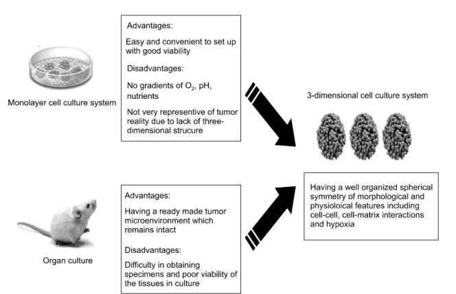
일반적으로 사용되고 있는 monolayer cell culture system 은 배양법이 비교적 용이하고 세포 생존 능력이 탁월하 다는 장점을 가지고 있어 생물학 분야에 많이 이용되고 있다. 하지만 인체 조직과 같은 3차원적인 미세 환경 조
구에 있어서는 세포의 미세 환경 조건을 고려하지 않은 채 연구되어왔다.7) 특히 방사선을 이용한 항암치료의 경 우 암 세포의 방사선에 대한 민감도는 산소의 분획도, 영양분의 공급 상태, 세포들 간의 신호전달 상호작용 및 방사선 조사 선량에 따라 달라지므로 방사선 치료 연구 의 임상적 적용 범위를 확대시키기 위해서는 실제 암 세 포와 유사한 시스템이 도입된 기전 연구가 필요하다.
이러한 monolayer cell culture system의 한계점을 보완할 수 있는 모델로서 3-dimensional (3D) cell culture가 주목을 받고 있다(Fig. 1). 3D cell culture system의 이점은 형태학 상 실제 암 세포와 매우 유사한 구조를 가지고 있다는 것이다. 세포의 spheroid 구조는 단지 형태학적 의미 뿐 아니라 그가 지니고 있는 기능과도 매우 연관성이 깊은 것으로 알려져 있기에 다양한 세포에서 형성될 수 있는 spheroid의 구조가 세포 생활사에 미치는 영향을 고려할 수 있다.7) 또한 세포와 세포사이의 상호작용 뿐 아니라 세포와 주변의 미세 환경과의 상호작용으로 인한 세포 내 신호전달이 가능하기에 항암제 및 방사선과 같은 암 치료에서 나타나는 세포의 성장, 분화, 죽음을 조절하는 기전을 monolayer에서 보다 정교하게 구연할 수 있다.6,8)
3차원적인 세포 배양 시스템의 유용성 1. Tumor microenvironment
암 세포는 빠르게 성장하는 특성에 비해 영양소 및 산 소를 공급하는 혈관 공급이 불충분하다.9) 이러한 혈액의 흐름은 혈관 주위의 산소공급 및 균등한 영양공급에 있 어 원활한 소통을 이루지 못한다. 또한, 암세포 주변 환 경은 glycolysis의 과다한 활성과 저산소증 및 암세포에서 방출되는 H+에 의하여 lactic acid가 생성되어지고 산성 화되는 경향이 있다.10) 이러한 미세 환경 조건은 암세포 의 방사선 저항성을 조절하는 중요한 인자로서 다양한 요인이 함께 복합적으로 작용하여 세포 반응을 일으키 는 가능성에 초점을 맞추고 연구되어지고 있다. 특히, 암 세포의 미세 환경 조건은 세포사멸에 영향을 주는 것으로 알려져 있는 proliferation,11) repair,12) energy metabolism, signal transduction13)과 같은 세포 반응을 통하여 간접적인 세포 사멸을 조절하는 것으로 알려져 있다. 이러한 세포 반응
Fig. 1. Comparison with three different kinds of cell culture system. 2-dimensional cell culture models are easy and convenient to set up with good viability of cells in culture; however, they lack the 3-dimensional microenvironment of intact tissue including pH, oxygen, nutrient and cell-cell communication. Spheroid cultures mimic both the 3D organization and differentiated function of intact tissues to a much greater extent than 2D cell monolayers.
들을 유도하는 암 세포 외부의 미세 환경 조건은 세포 내의 유전자 발현에 영향을 줄 수 있으며14) 세포와 미세 환경 사이의 부적절한 변화는 비정상적인 세포 반응을 유도하여 암의 발생과정에도 기여할 수 있다.15)
2. Tumor-stromal interactions
정상적인 stroma 세포는 암 세포 형성을 예방하거나 그 형성 시기를 지연시킬 수 있다는 것과는 달리,16∼18) 비정상적인 stroma 세포는 암의 생성을 촉진시킬 수 있 을 뿐 아니라 암의 형성에 매우 중요한 역할을 한다는 것이다. Stroma는 종양형성에 직접적인 영향을 주는 것 으로 알려져 있으며 stromal element는 종양 성장에 가장 적합한 환경을 제공해 줌으로써 종양의 확산과 침윤을 야기하는 것으로 알려져 있다.19) 또한 암과 stromal 세포 의 상호작용은 암 세포의 형태학적 변화와 extracellular matrix distribution과도 연관이 있을 수 있다는 연구가 보 고되었다.20) 이는 암 세포로부터 나오는 tumor signal은 정상세포로 전달되어지며 이는 non-malignant 세포를 악 성종양으로 바꿀 수 있는 가능성을 가지고 있음을 의미 한다. 더 나아가 정상세포가 종양세포로 변환되기에는
장시간의 오랜 신호전달이 아닌 일시적인 순간의 자극으 로도 충분하다. Stroma는 matrix matalloproteinases, recruit- ment of inflammantory, stromal signal의 변화의 복합적인 작 용을 통하여 종양발생을 촉진하는 것으로 예측 된다.19)
3. Cell-cell adhesion and signaling
3D cell culture system에서 stroma 세포와의 상호작용 외에 중요하게 고려되어져야 할 부분이 cell signaling과 adhe- sion이다. Adhesion 분자들을 통한 세포들 간의 신호전달 상호작용이 세포의 생장을 유지시키는데 매우 중요한 요소임이 밝혀지고 있다. 그 예로, cell-cell interaction 분자 중 하나인 E-cadherin의 발현은 여러 종류의 암 세포주의 전이와 분화를 억제하는 것으로 알려져 있다.21) 또 다른 cadherin 분자는 desmoplasia, 암의 전이 및 침윤 부위에서 그 발현이 증가되는 것으로 알려졌다.22) 최근 human model system에서 세포표면의 receptor (integrins)의 기능적 인 연구에서는 세포의 구조적인 안정성이 integrin- mediated cell- ECM의 상호작용에 의해 조절될 수 있으며 이는 정상세포의 기능 뿐 아니라 조직의 구조적인 변화 를 유도할 수 있으며 결과적으로 암의 생성과정을 야기
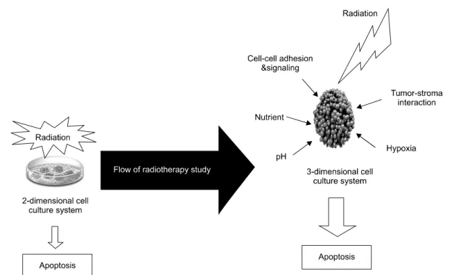
Fig. 2. Benefits of the 3-dimensional cell culture system including radiotherapy. Radiation-induced apoptosis in tumors may be inhibited due to alteration of apoptotic elements or tumor microenvironmental conditions. In order to represent cellular response to radiation, 3-dimensional cell culture system should be applied to investigate the effects of combined factors to assess tumor response more realistically.
할 수 있다는 연구결과들이 나오고 있다.23∼27)
Spheroid의 형태로 자라는 세포는 adhesion의 기전 뿐 아니라 adhesion 분자들과 hormones, cytokines, growth fac- tors와 같은 성장 조절 인자들과의 상호작용에 의해 조절 되기에 3차원적인 세포 배양 시스템을 이용한 연구들이 지속적으로 발전하고 있다. 이를 뒷받침해 주는 근거 연 구로서 인간 암 세포에서 EGRF (Epidermal growth factor) 와 β1-integrin 사이의 bidirectional cross modulation28)과 FAK (focal adhesion kinase) 신호전달 중 tyrosine phosphory- lation이 2D monolayer와는 다르게 3D co-culture에서 down regulation되는 것이 보고되고 있다.29) 이는 세포 생리학 적 측면에서 세포 반응이 2D와 3D에서 다르게 나타난다 는 것을 의미한다.
4) Hypoxia: 인간의 암 세포에서 흔히 나타나는 저산
소증(hypoxia)은 방사선에 저항성을 가지는 것으로 보고 되고 있다.30) Leukemia,31) lung carcinoma,32) lymphona33)를 이용한 in vitro 연구에 따르면 방사선 노출에 따른 세포 사멸이 저산소증에 의해 저해된다는 보고가 있으며 일 부의 연구에서는 anti-apoptotic bcl-2 단백질의 과발현 및 pro-apoptotic bax단백질의 저발현이 만성적인 저산소증 의 암에서 발생되었음이 보고되기도 하였으나34) 이와는
반대로 bcl-2의 발현과는 관련이 없다는 연구결과도 보 고되었다. 이는 저산소증 상태에서 일어나는 방사선 저 항성이 모든 암 세포에서 적용되지 않다는 것을 의미하 며 단순한 분자적 기전에 따라 나타나는 현상이 아님을 시사해 주고 있다. 분자적 기전 외에 저산소증에 대한 방사선 저항성의 조절 요인으로 제시되고 있는 것이 natural selection으로 이는 유전적으로 불안정한 암세포에 서 natural selection 과정을 거쳐 저산소증에 의한 방사선 저항성이 나타난다는 연구 결과이다.35) 또 다른 요인으 로, 암 세포의 저산소증은 방사선에 의해 유도되는 SAPK 활성을 억제함으로서 세포사멸의 저항성을 유발 한다는 보고가 있다.36) 이렇게 직접적으로 방사선 저항 성에 영향을 주는 요인들 뿐 아니라 세포사멸에 관여하 는 반응을 조절함으로서 방사선 저항성을 유도한다는 보고들도 있다. 근거로서, 20 Gy 정도의 높은 선량의 방 사선이 세포에 조사되었을 경우 세포에서는 pre-mitotic apoptosis의 과정이 일어나지만 5 Gy 이하의 낮은 선량이 조사되었을 경우 mitosis과정을 지나 post-mitotic apoptosis 의 과정이 일어나게 된다. 그러므로 세포 분열은 방사선 에 의해 유도된 세포사멸을 간접적으로 예방해주는 역 할을 한다고 할 수 있다.37)
지금까지 밝혀진 많은 연구들을 통하여 암 세포내에 서 발생하는 hypoxia는 암의 분자적 기전 연구를 위해 반 드시 고려되어야 하는 요소임이 밝혀졌으나 대부분의 in vitro 연구는 실험자가 조절할 수 있는 범위 내에서 이루 어진 결과이다. 하지만 실제 암 세포에서 생성되는 저산 소증은 산소 고갈 상태 동안의 다양한 변화가 일어나기 에 자연적인 저산소증 상태를 유도할 수 있는 3D sphe- roid 모델이 필요하다(Fig. 2).
결 론
암 세포의 발생을 억제하며 항암제와 방사선을 이용 한 치료요법의 극대화를 유도하기 위한 기전연구는 기 존의 2차원적인 세포 배양 시스템에서는 한계가 있으며 이를 극복하기 위한 일환으로 실제 인체와 유사한 3차원 적인 세포 배양 시스템의 중요성이 부각되어지고 있다.
특히 암 세포와 stroma의 상호작용, 세포 내의 adhesion 분자들의 상호작용 및 이들과 상호작용하는 hormones, cytokines, growth factors과의 상호작용, spheroid 종양 내에 서 빈번하게 발생하는 저산소증을 자연적으로 유도할 수 있기에 방사선을 이용한 암 치료 연구에서 뿐 아니라 종양 형성을 예방하는 기전연구에서도 유용히 사용될 모델로서의 가능성을 가지고 있다고 사료된다.
감사의 글
본 연구는 과학재단(2007-01365, 2008-01694, R13-2002- 020-03001-0 (2008))의 지원을 받아 수행된 연구임.
참 고 문 헌
1) Vaupel P, Kallinowski F, Okunieff PG. Blood flow, oxygen and nutrient supply, and metabolic microenvrionment of human tumors. Cancer Res 49, 6449-6465, 1989.
2) Kunz-Schughar LA, Groebe K, Mueller-Klieser W. Three dimensional cell culture induced novel proliferative and meta- bolic alterations associated with oncogenic transformation. Int J Cancer 66, 578-586, 1996.
3) Jung YS, Cho YU, Suh YJ, Kim JS, Oh SJ, Lim CW, Kim MB, Park HK. Can the histoculture drug response assay predict the clinical results of chemotherapy in breast cancer?
J Breast Cancer 10, 193-198, 2007.
4) Dubessy C, Merlin JL, Marchal C, Guillemin F. Spheroids in radiotherapy and photodynamic therapy. Crit Rev Oncol Hemat 36, 179-192, 2000.
5) Furukawa T, Kubota T, Hoffman RM. Clinical applications
of the histoculture drug response assay. Clin Cancer Res 1, 305-311, 1995.
6) Kim JB, Stein R, O'Hare MJ. Three-dimensional in vitro tissue culture models of breast cancer-a rivew. Breast Cancer Research and Treatment 85, 281-291, 2004.
7) Kim JB. Three-dimensional tissue culture models in cancer biology. Semin Cancer Biol 15, 365-377, 2005.
8) Bartholomȁ, Gorjup E, Monz D, Reininger-mach A, Thielecke H, Robitzki A. Three-dimensional in vitro reaggregates of embryonic cardiomyocytes: a potential medel system for mo- nitoring effects of bioactive agents. J Biomol Screen 10, 814-822, 2005.
9) Vaupel P. Tumor microenvironmental physiology and its implications for radiation oncology. Semin Radiat Oncol 14, 198-206, 2004.
10) Stubbs M, McSheehy PM, Griffiths JR, Bashford CL. Causes and consequences of tumour acidity and implications for treatment. Mol Med Today 6, 15-19, 2000.
11) Amellem O, Pettersen EO. The role of protein accumulation on the kinetics of entry into S phase following extreme hypoxia. Anticancer Res 11, 1083-1087, 1991.
12) Yuan J, Narayanan L, Rockwell S, Glazer PM. Diminished DNA repair and elevated mutagenesis in mammalian cells exposed to hypoxia and low pH. Cancer Res 60, 4372-4376, 2000.
13) Belka C, Jendrossek V, Vink S, Verheij M, Budach W.
Apoptosis-modulating agents in combination with radiothe- rapy-current status and outlook. Int Radiat Oncol Biol Phys 58, 542-554, 2004.
14) Bissell MJ, Hall HG, Parry G. How does the extracellular matrix direct gene expression. J Theor Biol 99, 31-68, 1982.
15) Rǿnnov-Jessen L, Petersen OW, Bissell MJ. Cellular changes involved in conversion of normal to malignant breast: im- portance of the stromal reaction. Physiol Rev 76, 25-69, 1996.
16) Shekhar MP, Werdell J, Santner SJ, Pauley RJ, Tait L. Breast stroma plays a dominant regulatory role in breast epithelial growth and differentiation: implications for tumour develop- ment and progression. Cancer Res 61, 1320-1326, 2001.
17) Hayashi N, Cunha GR. Mesenchyme-induced changes in the neoplastic characteristics of the dunning prostatic adenocarci- noma. Cancer Res 51, 4924-4930, 1991.
18) Cooper M, Pinkus H. Intrauterine transplantation of rat basal cell carcinoma as a model for reconsersion of malignant to benign growth. Cancer Res 37, 2544-2552, 1977.
19) Rǿnnov-Jessen L, Petersen OW, Koteliansky VE, Bissell MJ.
The origin of the myofibroblasts in breast cancer. Recapitu- lation of tumor environment in culture unravels diversity and implicates converted fibroblasts and recruited smooth muscle cells. J Cli Invest 95, 859-873, 1995.
20) Kunz-Schughart LA, Heyder P, Schroeder J, Knuechel R. A heterologous 3D co-culture model of breast tumor cells and fibroblasts to study tumor-associated fibroblast differentiation.
Exp Cell Res 266, 74-86, 2001.
23) Alford D, Taylor-Papadimitrou J. Cell adhesion molecules in the normal and cancerous mammary gland. J Mam Gland Biol Neoplasia 1, 207-218, 1996.
24) Jones JL, Critchley DR, Walker RA. Alteration of stromal protein and integrin expression in breast-a marker of pre- malignant change. J Pathol 167, 399-406, 1992.
25) Howlett AR, Bailey N, Damsky C, Petersen OW, Bissell MJ.
Cellular growth and survival are mediated by B1 integrins in normal human breast epithelium but not in breast carcinoma.
J Cell Sci 108, 1945-1957, 1995.
26) Koukoulis GK, Virtanen I, Korhonen M, Litinen L, Quaranta V, Gould VE. Immunohistochemical localization of integrins in the normal, hyperplastic and neoplastic breast: correlations with their function as receptors and cell adhesion molecules.
Am J Pathol 139, 787-799, 1991.
27) Zutter M, Mazoujian GM, Santoro SA. Decreased expression of integrin adhesive protein receptors in adenocarcinoma of the breast. Am J Pathol 137, 863-870, 1990.
28) Wang F, Weaver VM, Petersen OW, Larabell CA, Dedhar S, Briand P, Lupu R, Bissell MJ. Reciprocal interactions between beta1-integrin and epidermal growth factor receptor in three-dimensional basement membrane breast cultures: a different perspective in epithelial biology. Proc Natl Acad Sci USA 95, 14821-14826, 1998.
29) Cukierman E, Pankov R, Stevens DR, Yamada KM. Taking cell-matix adhesions to the third dimension. Science 294, 1708-1712, 2001.
Biol 78, 267-274, 2002.
32) Weinmann M, Jendrossek V, Güner D, Goecke B, Belka C.
Cyclic exposure to hypoxia and reoxygenation selects for tumor cells with defects in mitochondrial apoptotic pathways.
Faseb J 18, 1906-1908, 2004.
33) Weinmann M, Marini P, Jendrossek V, Betsch A, Goecke B, Budach W, Belka C. Influence of hypoxia on TRAIL-induced apoptosis in tumor cells. Int J Radiat Oncol Biol Phys 58, 386-396, 2004.
34) Cuisnier O, Serduc R, Lavieille JP, Longuet M, Reyt E, Riva C. Chronic hypoxia protects against gamma-irradiation- in- duced apoptosis by inducing bcl-2 up-regulation and inhi- biting mitochondrial translocation and conformational change of bax protein. Int J Oncol 23, 1033-1041, 2003.
35) Graeber TG, Osmanian C, Jacks T, Housman DE, Koch CJ, Lowe SW, Giaccia AJ. Hypoxia-mediated selection of cells with diminished apoptotic potential in solid tumours. Nature 379, 88-91, 1996.
36) Samuni AM, Kasid U, Chuang EY, Suy S, Degraff W, Krishna MC, Russo A, Mitchell JB. Effects of hypoxia on radiation-responsive stress-activated protein kinase, p53, and caspase 3 signals in TK6 human lymphoblastoid cells. Cancer Res 65, 579-586, 2005.
37) Ljungkvist AS, Bussink J, Kaanders JH, Wiedenmann NE, Vlasman R, van der Kogel AJ. Dynamics of hypoxia, pro- liferation and apoptosis after irradiation in a murine tumor model. Cancer Res 165, 326-336, 2006.