Korean J Gastroenterol Vol. 77 No. 2, 92-94 https://doi.org/10.4166/kjg.2019.150 pISSN 1598-9992 eISSN 2233-6869
IMAGE OF THE MONTH
Korean J Gastroenterol, Vol. 77 No. 2, February 2021 www.kjg.or.kr
옥돔 가시에 의해 발생한 막창자의 방선균증
이동현, 현창림1, 유정래2
제주대학교 의학전문대학원 외과학교실, 병리학교실1, 내과학교실2
Fish Bone of Branchiostegus Japonicas Causing Actinomycosis in the Cecum in Male on Jeju Island
Donghyoun Lee, Chang Lim Hyun1 and Jeong Rae Yoo2
Departments of General Surgery, Pathology1 and Internal Medicine2, Jeju National University School of Medicine, Jeju, Korea
CC This is an open access article distributed under the terms of the Creative Commons Attribution Non-Commercial License (http://creativecommons.org/licenses/
by-nc/4.0) which permits unrestricted non-commercial use, distribution, and reproduction in any medium, provided the original work is properly cited.
Copyright © 2021. Korean Society of Gastroenterology.
교신저자: 유정래, 63241, 제주시 아란13길 15, 제주대학교 의학전문대학원 내과학교실
Correspondence to: Jeong Rae Yoo, Department of Internal Medicine, Jeju National University School of Medicine, 15 Aran 13-gil, Jeju 63241, Korea. Tel:
+82-64-717-2283, Fax: +82-64-717-1131, E-mail: mdyoojr@gmail.com, ORCID: https://orcid.org/0000-0002-5488-7925 Financial support: None. Conflict of interest: None.
Case: A 57-year-old male patient whose initial diagnosis was acute appendicitis visited at the outpatients department of general surgery. He has been suffered recurrent pain in the right lower quadrant abdomen for 2 years. Acute abdominal pain in the right upper and lower quadrants had increased 5 days ago. On admission, he was febrile with stable vital signs. Serum white blood cell count was 13,600 cells/µL (normal range 4,000-10,000 cells/µL) and CRP level was 14.95 mg/dL (reference range 0.0-0.3 mg/dL). Abdominal CT re- vealed inflammatory lesions, including appendicitis and peri- appendiceal abscess (Fig. 1). On admission day 3, he under- went right hemicolectomy for perforated cecum (Fig. 2).
Intraoperatively, we found a 4-cm sized abscess with a fish bone in the cecum and the small bowel mesentery, copious dirty fluid in the pelvic cavity, and inflammation in the distal ileum and proximal ascending colon. BacT/Alert blood (bioMerieux, Marcy-l’Etoile, France) and anaerobic cultures of the surgical specimen identified each gram-positive rods.
Histopathology revealed chronic active inflammation, edema, and microscopic actinomycotic colonies (Fig. 3). We diagnosed
a cecal actinomycosis caused by a fishbone. Intravenous cef- triaxone was prescribed for 4 weeks, followed by oral amoxicillin 1.5 g for 6 months.
Diagnosis: Fishbone causing actinomycosis in the cecum and perforation of cecal abscess
Actinomycosis, a rare infectious disease caused by Actinomyces spp., an anaerobic gram-positive bacteria, com- monly affects the appendix, cecum, and colon in the abdo- men, especially in patients with poor dental hygiene, women with intrauterine devices, and fish bone related lesions.1 In addition, this is one of the greatest challenges for diagnosis because of mimicking malignant neoplasm. The organisms cause disease only when the normal mucosal barrier is bro- ken, followed by abscess formation, fistula, or mass lesion.2 The current role of imaging in the diagnosis of intra-abdominal actinomycosis is poorly defined. Of some of studies have sug- gested a feature of CT imaging, the colon was involved in the gastrointestinal tract, CT scans showed solid masses or cystic mases with thickened walls.3,4 Actinomycosis should be
Lee D, et al. Fish Bone of Branchiostegus Japonicas Causing Cecal Actinomycosis 93
Vol. 77 No. 2, February 2021 A
B
Fig. 1. (A) Abdominal computed tomography showing a 6-cm sized irregular peripheral enhancing low density lesion abutting the cecum with perilesional infiltration (yellow arrowheads) and a 2.5-cm sized linear radiopaque lesion (fishbone; red arrowhead) within the mass. (B) Coronal view.
Fig. 2. Excised specimen showing a perforated abscess (white arrows) and a sharp fishbone was retrieved from the abscess (not shown).
A
B
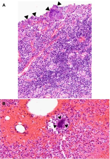
Fig. 3. (A) Photomicrograph of histopathological examination revealed inflammatory cells (mainly plasma cells and lymphocytes) were also noted, and demonstrates colonies of Actinomycetes (sulfur granules, black arrowheads) (hematoxylin and eosin stain, original magnification, ×200). (B) Sulfur granules, black arrowheads (hematoxylin eosin, original magnification, ×400)
included in the differential diagnosis when CT scans show bowel wall thickening and regional pelvic or peritoneal mass with extensive infiltration.4 However, the features are often insufficient to help distinguish the other diseases. Physicians should be considered a history of patients with intrauterine contraceptive devices in female and foreign body such as fish bone. Bacterial culture and pathology are the cornerstones of diagnosis.5 However, prolonged bacterial culture in anaero- bic conditions may be necessary, and most clinicians remain reluctant to consider cytological or tissue samples to avoid the possibility of tumor seeding before a specific diagnosis has been made. Intra-abdominal actinomycosis is uncommon, especially immunocompetent male. Imaging study and culture examination are shown to be useful but their roles in estab- lishing definitive diagnosis are yet to be defined. Although,
94 이동현 등. 옥돔 가시에 의한 막창자 방선균증증
The Korean Journal of Gastroenterology
treatment of long term parenteral then oral antibiotics of be- ta-lactam antibiotics were well curative, the patient addition- ally should be avoided an eating habit of fishbone for prophy- laxis, because of the patient gave a long-term history of habit- ually consuming fish bone with grilled Branchiostegus japonicas.
Herein, we report a case of fishbone associated actinomycosis causing perforated abscess in the cecum which was initially misdiagnosed as perforated appendicitis or colon cancer in immunocompetent male.
REFERENCES
1. Boyanova L, Kolarov R, Mateva L, Markovska R, Mitov I.
Actinomycosis: a frequently forgotten disease. Future Microbiol 2015;10:613-628.
2. Berardi RS. Abdominal actinomycosis. Surg Gynecol Obstet 1979;149:257-266.
3. Ha HK, Lee HJ, Kim H, et al. Abdominal actinomycosis: CT findings in 10 patients. AJR Am J Roentgenol 1993;161:791-794.
4. Lee IJ, Ha HK, Park CM, et al. Abdominopelvic actinomycosis in- volving the gastrointestinal tract: CT features. Radiology 2001;
220:76-80.
5. Wong VK, Turmezei TD, Weston VC. Actinomycosis. BMJ 2011;
343:d6099.