Effect of Experimental Muscle Fatigue on Muscle Pain and Occlusal Pattern
Jae-Chang Kim, D.D.S.,M.S.D.,Ph.D., Hyun-Dae Lim,D.D.S.,M.S.D., Jin-Kyu Kang, D.D.S.,M.S.D., You-Mee Lee, D.D.S.,M.S.D.,Ph.D.
Department of Oral Medicine, College of Dentistry, Wonkwang University
This study aimed to make an analysis of the occlusion in the state of muscle fatigue produced by excessive mouth opening and clenching during the dental treatment to control the dental pain and to evaluate the sensory nerve in the muscle pain state.
Most of the reasons why patients visit the dental office result in pain — either conceivably the dental origin pain or the non-dental origin pain. The dental offices have many therapeutic actions to produce the masticatory muscle fatigue for the treatment. Dental treatment with long minutes of mouth opening can cause some headaches, masticatory muscle pain and mouth opening difficulties. Patients with mastication problems who visits a dental office to alleviate pain run against another unexpected pain with other aspects.
This study uses T-scan Ⅱ system (Tekscan Co., USA) for the evaluation on the occlusal pattern in the experimental muscle fatigue after clenching, opening the mouth excessively and chewing gum. The occlusal contact pattern is analyzed by the contact timing, namely first, intercuspal, maximum and end point of contact.
This inspection was performed at frequencies of 2000Hz, 250 Hz and 5 Hz before and after each experimental muscle pain was produced to 24 subjects who had normal occlusion without the orthodontic treatment or a wide range of the prosthesis by using neurometerⓇ CPT/C (Neurotron, Inc. Baltimore, Maryland, USA). The measuring sites were mandibular nerve experimental muscle fatigue respectively.
This study could obtain the following results after the assessment of occlusion and sensory nerve of the experimental muscle fatigue.
1. There were the fastest expression after the excessive mouth opening in muscle fatigue and after tooth clenching in muscle pain. In the visual analog scale that records the subjective level, there was the highest scale after the clenching in the muscle fatigue in jumping off the point of pain.
2. Tooth contact time, contact force, relative contact force on the point of the first contact had no difference, and there were decreases in the contact force after the excessive mouth opening on intercuspal position point, after the excessive mouth opening and the gum chewing on the point of the maximum, and in the contact time after all the experimental muscle fatigue state on the point of the end contact.
3. There was no statistic significance in the current perception threshold before and after the experimental muscle fatigue.
4. There was no significant difference in the contact number, the maximal contact number on the point of the first contact, and the contact number after the mouth opening and gum chewing on the point of the intercuspal position and the contact number after the experimental muscle fatigue on the maximum point, and showed significant decreases.
In conclusion, it was found that the occlusal pattern can cause the changes on the case of the clinical muscle weakness by intra-external oral events. It was important that the sedulous attention to details is required during dental treatment in case of excessive mouth opening, mastication and clenching.
Key words: Muscle Fatigue, Muscle Pain, Occlusal Pattern, current perception threshold
1)
Ⅰ. INTRODUCTION
Temporomandibular joint disorder, TMD, is an acute or chronic disease that mainly developing joint pain, joint sound and limitation of mandibular movement on temporomandibular joint and associated structure including masticatory muscle.
Interlocking with a social phenomena, the more an aspect of modern society gets complicated, the more TMD causes or symptoms become complicated, which requires multi-lateral TMD diagnosis and treatments approaches.
1)There are many factors that place symptoms on the TMJ: occlusion, trauma, emotional stress, deep pain input. Parafunctional activities or habits such as tooth grinding or tooth, mandibular movement such as mouth opening or lateral excursion can produce joint pain and sometimes accelerate and aggravate TMD. This aggravating joint pain affects jaw movements, which limits or problems in opening the jaw, and excessive mouth opening or too tight tooth contact exacerbates already existing TMD symptoms, which repeat vicious cycle.
2)Pain is a defense mechanism that people with pain try to get rid of causes of pain or avoid, which includes uncomfort feeling and suffering caused in the peripheral nerve ending by stimulation.
Especially, orofacial pain is produced due to pain related to the head, the face and the mouth which shows a variety of disorders from teeth, gingival disease to TMD, neuropathic pain.
3)Likewise, orofacial pain is the leading cause for
Corresponding Author: You-Mee Lee
Department of Oral medicine, College of Dentistry, Wonkwang University,
344-2 Shinyoung-dong, Iksan, Jeonbuk 570-749, Korea Tel: 82-63-859-2914
Fax: 82-63-857-4002
E-mail: [email protected] Received: 2008-06-20
Revised: 2008-07-27 Accepted: 2008-08-13
* This paper was supported by Wonkwang University in 2007.
dental patients to visit dental offices, and such pain can be either dental origin or non-dental origin.
While controling pain of dental origin, considering its characteristics, long minute of opening the jaw or clenching can cause masticatory muscle fatigue, and then such fatigue provoke problems like headache, pain in the masticatory muscle, mouth opening difficulty and mastication problems after the treatment, which patients face another kinds of unexpected pains.
Of the unexpected pains, muscle-originated pains are starting from skeletal muscle, tendons and fascia. If protective muscle splinting, local muscle soreness and myofascial pain are starting acutely without proper treatment, it may end up with a chronic, troublesome muscle pain disorder. When an ordinary, healthy muscle was malfunctionally stimulated from, for instance, prosthetics, or it was injured from damages to tissue caused by dental anesthesia or excessively long minutes of mastication, tooth clenching, excessive opening the jaw, protective muscle splinting gets created and then form mechanism to protect itself from the damages. At that time, if function of muscle is rehabilitated, the muscle can come back to a ordinary state. However, if it is not restored, neurotransmitter that causes pain like bradykinin, prostaglandin, substance P can be locally extricated in ischemia area of muscle, and as a result, it can move on to the state of local muscle soreness. Due to the muscle pain that weak muscle when it functions, it was reported that masticatory ability is decreased from 33% to 50%.
Stimuli inside or around the oral cavity cause a series of suppression, which is called exteroceptive suppression, ES, especially causes ES1 and ES2.
These two activates interneuron and control
activity neuron in mouse closing muscle.
5,6)Even
though it was not officially acknowledged influence
on muscle fatigue in an exteroceptive suppression
response, it was known that when ES response
was applied to human beings, the result indicates
that it was affected by a number of different
experimental stimuli such as experimental jaw
muscle pain, headache, mental stress, and clinical stimuli, which are intermediated by the stages before synapse.
14)In order to evaluate the muscle condition at the moment of muscle fatigue, previous scholars have kept on research on such muscle pain. So as to induce experimental muscle fatigue, a number of methods have employed: temperature, electric stimulation, exercise, ischemia, injection of hypertonic saline. This study employed overly long minutes of opening the jaw, gum chewing, tooth clenching and induced muscle fatigue.
Previous scholars propose results from electromyography, but there are not enough researches on muscle weakness during experimental muscle fatigue, and its related objective assesment is not performed enough on a peripheral nerve that can assess sensory changes at the point of muscle pain induced at this point.
25-28)This study measured sensory nerve ending of the trigeminal nerve at muscle pain by using neurometer that use electric stimulation. Current perception threshold, CPT can evaluate neurological disorder around the relevant areas, when the minimum amount of electricity that subjects can feel was turned on. It also can measure the changes in sensory nerve by analyzing individually Aβ, Aδ, C fibers because it is applicable in responsive threshold value of trigeminal nerve.
29)As far as dental treatment is concerned, occlusion is taking a very important role, and if long treatment incurred muscle pain and the pain caused disorder, it cannot be negligent. Mastication problems after dental treatment are protective muscle splinting local muscle pain, fascia pain, so the rest of muscular spasms and central mediating pain are fairly rare. Protective muscle splinting is the first reaction from masticatory muscle when a stimulus was flowed in, and the muscle maintains the state of slight contraction. There is no academic evidence that protective muscle splinting causes the muscle weakness, but patients complain of it and it looks like patients have pain increased when the muscle function. When pain was incurred,
masticatory muscle is malfunctioning, and this malfunctioning muscle accelerates muscle fatigue and muscle pain.
30)In this research, in order to evaluate the changes on occlusal state caused by muscle weakness, and malfunction of masticatory muscle at the points of muscle pain, T-scan was used and then recorded the number of contact and contact force. T-scan is useful to observe in real time at the moment of occlusion in a computer screen and makes a kinetic evaluation and is highly reproducible. T-scan II system can analyze lapse of occlusal time when a row of maxillo-mandible contact, and contact force on each contact point.
31)As stated above, in this study Neurometer was applied in trigeminal nerve in order to assess sensory changes stimulated by some methods that can cause pain in the area of TMJ after long time of opening the jaw while patients who visited dental office due to dental-origin pain. Also we studied whether jaw opening for a long time or tooth clenching can make any influence on occlusion during dental treatment.
Ⅱ. MATERIALS AND METHODS 1. Subjects
Among non-patient volunteers and patient volunteers who visited Wonkwang University Dentistry Hospital, 24 subjects(average age of 25.7)were selected. They had not TMD symptoms and normal range of occlusion. Those who had orthodontic treatment and got prosthesis treatment more than 3 were excluded.
2. Methods
1) Clinical examination and Temporomandibular disorders charting
To begin with this research, all the subjects were
tested to fit in this study: TMD charting sheet was
recorded for an evaluation of clinical symptoms like
joint pain, joint sound, mouth opening limitation.
Among them, 24 subjects with no TMJ symptoms were finally selected. Before the test, they were fully understood the purpose of and the procedure of this research and gave consents by writing their names on the given forms. Research has started with authorization from Institutional Review Boards of dental hospital at Wonkwang University.
2) Experimental inducement of muscle fatigue Long minute of dental treatment can cause muscle fatigue, so subjects were told to clench their teeth, open their jaw and masticate gum to induce muscle fatigue until they feel pain.
In detail, subjects were to clench their teeth with the maximum force and to open the jaw enough 3 fingers to stick in. As far as chewing gum is concerned, subjects picked one kind of gum and chewed with either side and made the gum more smoothly. After a break, they were told to masticate it again with preferred chewing side with their ordinary speed. In a bid to minimize interaction in each experiment, subjects were given enough break and selected in random in tooth clenching, opening the jaw and chewing gum on one side.
Subjects were heard to keep the record of muscle fatigue in every minute using 10 cm visual analog scale (VAS) and mark the level of pain when they detected first pain.
The point of muscle fatigue and the point of pain after each event were kept on record, and the following is the definition of time when to produce muscle fatigue on each event.
The maximum point of tooth clenching is when the level of clenching force of maxillo-mandible is different from the first maximum level. At the test of staying the mouth open is when the point of level of opening the jaw even 3 fingers cannot get in enough, and in test of side chewing preference when the masticatory speed decreases or while chewing gum it takes more effort than the first.
3) Measurement of current perception threshold This is for assesment for datum point of muscle fatigue right before it was produced and the level of
experimental pain when first pain was caused right after clenching, staying with jaw open and chewing side preference. So, current perception threshold value was measured in mandibular division of subjects' chewing side preference using Neurometer
®
CPT/C (Neurotron, Baltimore, Maryland, USA).
Subjects were fully informed about test procedure to minimize experiment error. When they raised questions, we gave explanations repeatedly, and double blind test was employed not to make testers and testee see the test results.
Neurometer
®CPT/C has a pair of 1-cm diameter gold electrodes separated by a 1.7 cm. Goldtrode
®
(Neutron, Baltimore, Maryland, USA) coated with a thin layer of chloride-free electroconductive gel and then taped. R-CPT (rapid-current perception threshold) was measured at 3 frequencies — 2000Hz, 250Hz, 5Hz. Each frequency shows same response 2 times and was recorded current perception threshold value with automatic control mode. It marks the average of 3 minimum values.
Each electric frequency responds to Aβ fiber, Aδ fiber and C fiber.
Before inducing muscle fatigue, pulse oximeter [ARGUS OXM lus
Ⓡ(Medicalsupply Co., Seoul, Korea)] was taped to the left index finger and we observed changes in pulse during inducement.
4) occlusal analysis using T-scan
T-scan II system (Tekscan Co., USA) recorded occlusal contact points before subjects induced experimental muscle fatigue and after they induced the fatigue after clenching, opening the jaw for a long time and chewing gum with the preferred side.
T-scan II system consists of sensor, sensor support, scanning handle, hardware, and computer with a program compatible with micro software for T-scan system. The sensor is 60 ㎛ thick covered by a silver thread grid.
Its sensor is durable to tear and hole, it is elastic
so deformation can be minimized, and it can be
used multiple times for the same patient. Not to
make a few error, after drying oral cavity and the
sensor, did subjects bite on a thin sensor until
maximum contact, and marked data was sent and stored in a computer. Following categories were calculated: the first contact time, first contact force,
%contact force and the time of maximum contact, the force level exerted on that tooth during occlusion, %contact force , time and force of maximum contact point, %contact force , the time of end contact point.
5) Statistics Analysis
Using SPSS ver 11.5 collected data was analyzed the number of contact points and contact force current perception threshold, and the result was recorded on average and standard deviation. By performing paired t-test, datum point and contact number, contact force, current perception threshold was examined.
Time (minute) Fatigue VAS Pain VAS
Fatigue Pain Fatigue Pain
After clenching 3.43±8.17 3.40±3.08 4.29±1.65 5.46±1.89 4.58±2.28
After opening 2.34±1.35 5.16±3.01 3.50±1.53 5.13±1.57 3.63±1.66
After gum chewing 3.59±1.40 7.42±4.07 3.54±1.32 4.63±1.56 3.46±1.96
VAS ; visual analog scale
Fatigue VAS; fatigue level of masticatory muscle Pain; fatigue level in jump off point of pain Pain VAS; pain level in point of feeling pain
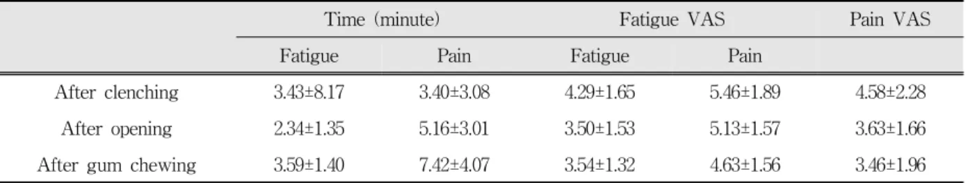
Table 1. The time and VAS of muscular fatigue in jump off point of pain and pain after each event
n=24, mean±S.D.
Time (Sec) p-value Force % p-value Force p-value
Before 0.00675±0.017266 19.84 ± 33.23 1778.17±4317.59
After clenching 0.00354±0.013436 .483 15.32 ± 31.58 .541 2219.75±6568.10 .525
After opening 0.0±0.0 .068 15.40 ± 26.14 .379 1318.13±3267.48 .128
After gum chewing 0.0±0.0 .068 14.07 ± 27.31 .423 783.21±1799.31 .215
Table 2. Comparison of contact time, contact force and contact force % (100%=maximum contact force) after each event on first contact point n=24, mean±S.D.
Ⅲ. RESULTS
The time of muscle fatigue marked an average of 3.43±8.17 minutes after clenching tooth, 2.34±1.35 minutes after opening the jaw, 3.59±1.40 minutes after chewing gum. The time of pain in masticatory muscle was 3.40±3.08 minutes, which showed up quicker than the fatigue after clenching. It took 5.16±1.53minutes for subjects to feel muscle pain after opening the jaw and 7.42±4.07 minutes after chewing gum. Muscle fatigue was first showed up after long minutes of opening the jaw, and muscle pain was the first that subjects claimed after clenching, which muscle fatigue followed later.
Fatigue VAS, visual analog scale that records
subjective muscle fatigue, marked 4.29±1.89, which
is highest right after clenching, and in fatigue VAS
pain caused by clenching was 5.46±1.89, which was highest as well. Pain VAS, that records the level of pain also marked highest after clenching(Table 1).
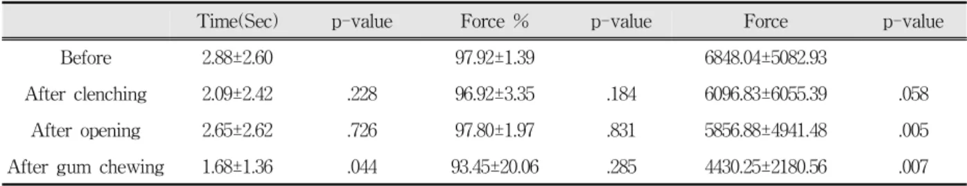
Contact time, contact force, and % contact force on the first contact point did not show significant difference, but after each event on intercuspal position contact point contact force marked 5711.11
±5215.14 after opening the jaw and 5214.33±4791.09 after chewing gum, which meaningfully decreased at the level of P<0.01 compared with 6971.63±
5122.49, which is contact force before it triggered experimental muscle fatigue(Table 2,3).
Time(Sec) p-value Force % p-value Force p-value
Before 2.88±2.60 97.92±1.39 6848.04±5082.93
After clenching 2.09±2.42 .228 96.92±3.35 .184 6096.83±6055.39 .058
After opening 2.65±2.62 .726 97.80±1.97 .831 5856.88±4941.48 .005
After gum chewing 1.68±1.36 .044 93.45±20.06 .285 4430.25±2180.56 .007
Table 3. Comparison of contact time, contact force and %contact force (100%=maximum contact force) after each event on intercuspal position contact point n=24, mean±S.D.
Time (Sec) p-value Force % p-value Force p-value
Before 2.95±2.79 100±0.0 6971.63±5122.49
After clenching 1.81±1.76 .068 99.95±0.24 .328 6233.13±6067.90 .069
After opening 2.86±2.58 .906 100±0.0 5711.11±5215.14 .005
After gum chewing 2.14±1.79 .230 95.80±20.41 .324 5214.33±4791.09 .009
Table 4. Comparison of contact time, contact force and %contact force (100%=maximum contact force) after each event on maximum contact point n=24, mean±S.D.
Time (Sec) p-value Force % p-value Force p-value
Before 8.37±1.79 83.59±13.16 5870.21±4442.42
After clenching 7.84±1.87 .000 68.95±25.82 .016 4543.38±5084.89 .052
After opening 7.49±2.56 .029 78.75±22.18 .351 4959.13±4442.42 .079
After gum chewing 7.14±1.86 .002 71.26±28.07 .045 3422.29±2043.51 .004
Table 5. Comparison of contact time, contact force and %contact force (100%=maximum contact force) after each event on end contact point n=24, mean±S.D.
Contact force notably decreased when muscle fatigue is started on maximum point contact after a long moment of opening the jaw and after chewing gum (P<0.01). Contact time (p=0.029, P<0.05) decreased on end contact point after clenching and after long minutes of opening the jaw, and contact time (p=0.002, P<0.001) after chewing gum, and
%contact force slided down after clenching and contact force declined after gum chewing(p=.004, p<0.01) (Table 4,5).
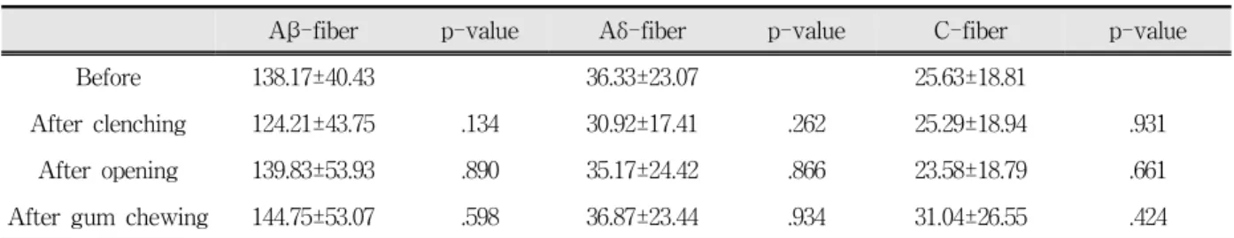
In current perception threshold value to assess
sensory changes where experimental muscle
fatigue was induced, there were not much different before and after the measurement of Aβ fiber, Aδ fiber, C fiber in which subjects can feel muscle fatigue and pain(Table 6).
Aβ-fiber p-value Aδ-fiber p-value C-fiber p-value
Before 138.17±40.43 36.33±23.07 25.63±18.81
After clenching 124.21±43.75 .134 30.92±17.41 .262 25.29±18.94 .931
After opening 139.83±53.93 .890 35.17±24.42 .866 23.58±18.79 .661
After gum chewing 144.75±53.07 .598 36.87±23.44 .934 31.04±26.55 .424
Table 6. Measurements of R-CPT after each event n=24, mean±S.D.
Contact teeth number Max point number
Left p-value Right p-value Both p-value Left p-value Right p-value Both p-value
Before 13.50±
28.80
17.17±
37.03
30.67±
65.61
0.38±
1.01
0.46±
1.35
0.83±
2.26 After
clenching
14.33±
36.87 .827 15.75±
39.30 .681 30.08±
76.14 .934 0.96±
3.06 .212 0.83±
2.78 .447 1.79±
5.83 .310 After
opening
13.50±
32.60 1.00 12.63±
25.60 .271 26.13±
56.59 .389 0.42±
1.35 .664 0.21±
0.83 .110 0.63±
2.16 .135 After gum
chewing
9.50±
22.64 .451 8.63±
15.58 .216 18.13±
37.61 .290 0.13±
0.34 .207 0.17±
0.48 .284 0.29±
0.69 .233 Table 7. The number of contact point and maximum point contact after each event on first contact point
n=24, mean±S.D.
Contact teeth number Max point number
Left p-value Right p-value Both p-value Left p-value Right p-value Both p-value
Before 57.29±
31.98
51.83±
29.84
109.13±
59.60
2.08±
2.45
1.96±
2.35
4.04±
4.38 After
clenching
50.54±
38.13 .063 46.29±
34.41 .027 96.83±
70.47 .016 2.71±
3.28 .239 1.88±
2.86 .845 4.58±
5.90 .481 After
opening
54.00±
41.24 .351 43.21±
24.74 .026 97.21±
61.92 .001 1.92±
3.32 .761 1.21±
1.32 .056 3.13±
4.20 .136 After gum
chewing
43.96±
25.51 .001 35.92±
20.10 .001 79.88±
41.86 .000 1.46±
1.53 .189 0.71±
1.12 .014 2.17±
2.37 .039 Table 8. The number of contact point and maximum point contact after each event on intercuspal position contact point
n=24, mean±S.D.
There were not much significant difference
between the number of contact on first contact
point and on maximum contact point from first
contact, and the contact number in left side declined
significantly after chewing gum on intercuspal position(P=0.001 P<0.01), and contact number on both sides showed significant decreases after opening the jaw for a long time and chewing gum(Table 7,8).
On maximum point contact the number of contact point in left side shows considerable decline after clenching tooth. The number of contact point in right teeth and the both sides decreased more than the muscle fatigue that was induced before(p<0.01).
The number of contact point on maximum point declined after opening the right jaw and after chewing gum. Contact number on end point showed decline after clenching both sides and right or left
Contact teeth number Max point number
Left p-value Right p-value Both p-value Left p-value Right p-value Both p-value
Before 59.75±
32.87
53.75±
33.26
113.50±
63.44
2.08±
2.24
2.13±
2.46
4.21±
4.31 After
clenching
51.88±
38.06 .039 48.08±
34.57 .009 99.96±
70.56 .008 2.58±
2.95 .274 2.04±
2.88 .845 4.63±
5.55 .542 After
opening
56.13±
43.17 .397 44.83±
26.47 .006 100.96±
65.63 .000 1.96±
2.94 .788 1.25±
1.51 .032 3.21±
3.98 .081 After gum
chewing
49.96±
40.90 .105 37.58±
22.34 .001 87.54±
58.74 .002 2.00±
2.98 .872 0.83±
1.27 .005 2.83±
3.87 .075 Table 9. The number of contact point and maximum point contact after each event on maximum contact point
n=24, mean±S.D.
Contact teeth number Max point number
Left P-value Right P-value Both P-value Left P-value Right P-value Both P-value
Before 53.29±
31.73
48.83±
31.50
102.13±
60.13
1.71±
2.29
1.25±
1.94
2.96±
3.78 After
clenching
40.08±
34.79 .004 35.75±
31.31 .000 75.83±
63.67 .000 1.75±
2.83 .950 1.33±
2.65 .892 3.08±
5.21 .916 After
opening
48.42±
42.32 .308 39.25±
25.09 .009 87.67±
64.02 .004 1.13±
2.01 .216 0.92±
1.28 .295 2.04±
2.71 .000 Aftergum
chewing
36.54±
22.45 .000 28.33±
18.00 .001 64.88±
36.48 .000 1.25.±
1.54 .332 0.58±
1.25 .107 1.83±
2.46 .186 Table 10. The number of contact point and maximum point contact after each event on end contact point
n=24, mean±S.D.
side. When subjects had muscle fatigue due to long minutes of jaw opening, the contact number in right and both sides dropped, and after chewing gum in both sides showed significant decrease(Table 9, 10).
The subject who felt muscle fatigue and chewed
gum in left side showed not much difference on
first contact number, intercuspal position contact
point, maximum occlusal contact, contact number
on end point and contact number on maximum
contact. Rt. side chewing group had significantly
more numerical values in left on intercuspal contact
position, on maximum occlusal contact and on end
contact point before muscle fatigue was induced
(p>0.05). The number of maximum contact point on
intercuspal contact position showed more numbers in left after clenching (p>0.05), but the number of first contact point was higher on right after a long
First contact Intercuspal
position Maximum contact End
Contact point no Max point no Contact point no Max point no Contact point no Max point no Contact point no Max point no
Lt. Rt. Lt. Rt. Lt. Rt. Lt. Rt. Lt. Rt. Lt. Rt. Lt. Rt. Lt. Rt.
Before 6.13±
12.87 7.20±
12.17 0.07±
0.26 0.07±
0.26 58.47±
31.38 49.07±
21.79 1.60±
2.20 1.07±
1.33 59.60±
31.51 49.33±
20.67 1.67±
2.02 1.33±
1.76 54.67±
29.82 44.20±
18.33 1.47±
2.17 0.80±
1.08
p-value .142 1.000 .037 .334 .039 .511 .039 .164
After clenching
5.73±
12.47 6.40±
13.81 0.13±
0.52 0.07±
0.26 50.60±
31.67 42.73±
19.78 2.13±
1.55 1.13±
1.64 51.93±
31.35 44.73±
20.19 2.07±
1.49 1.33±
1.80 41.13±
31.53 33.60±
22.10 1.47±
1.46 0.87±
1.41
p-value .290 .334 .146 .038 .180 .143 .179 .178
After opening
4.67±
9.04 6.87±
11.21 0 0 52.20±
32.13 42.93±
21.15 1.13±1.
36 0.87±
0.99 53.60±
30.00 45.27±
21.17 1.20±
1.32 0.93±
1.10 48.00±
30.48 38.80±
22.93 0.80±
1.15 0.60±
0.91
p-value .048 .058 .301 .065 .301 .023 .424
After gum chewing
6.20±
15.66 7.67±
12.15 0.07±
0.26 0.13±
0.35 46.80±
25.70 38.93±
19.98 1.53±
1.73 0.80±
1.26 48.20±
25.68 39.73±
20.47 1.53±
1.73 0.80±
1.37 38.87±
23.17 31.20±
18.16 1.40±
1.88 0.73±
1.49
p-value .419 .582 .107 .052 .086 .052 .106 .136
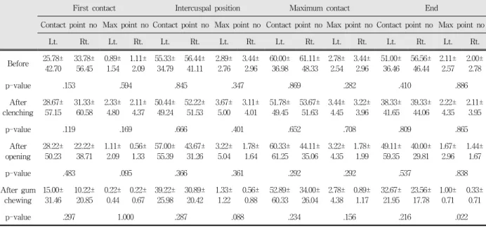
Table 11. The number of contact point and maximum point contact after each event on specific contact point in Rt. side chewing group n=15, mean±S.D.
First contact Intercuspal position Maximum contact End
Contact point no Max point no Contact point no Max point no Contact point no Max point no Contact point no Max point no
Lt. Rt. Lt. Rt. Lt. Rt. Lt. Rt. Lt. Rt. Lt. Rt. Lt. Rt. Lt. Rt.
Before 25.78±
42.70 33.78±
56.45 0.89±
1.54 1.11±
2.09 55.33±
34.79 56.44±
41.11 2.89±
2.76 3.44±
2.96 60.00±
36.98 61.11±
48.33 2.78±
2.54 3.44±
2.96 51.00±
36.46 56.56±
46.44 2.11±
2.57 2.00±
2.78
p-value .153 .594 .845 .347 .869 .282 .410 .886
After clenching
28.67±
57.15 31.33±
60.58 2.33±
4.80 2.11±
4.37 50.44±
49.24 52.22±
51.53 3.67±
5.00 3.11±
4.01 51.78±
49.45 53.67±
51.63 3.44±
4.45 3.22±
3.96 38.33±
41.65 39.33±
44.06 2.22±
4.35 2.11±
3.95
p-value .119 .169 .666 .401 .652 .708 .809 .865
After opening
28.22±
50.23 22.22±
38.71 1.11±
2.09 0.56±
1.33 57.00±
55.39 43.67±
31.26 3.22±
5.04 1.78±
1.64 60.33±
61.25 44.11±
35.06 3.22±
4.35 1.78±
1.99 49.11±
59.35 40.00±
29.81 1.67±
2.96 1.44±
1.67
p-value .483 .095 .366 .361 .292 .292 .537 .838
After gum chewing
15.00±
31.46 10.22±
20.85 0.22±
0.44 0.22±
0.67 39.22±
25.98 30.89±
20.42 1.33±
1.22 0.56±
0.88 52.89±
60.33 34.00±
26.04 2.78±
4.38 0.89±
1.17 32.67±
21.95 23.56±
17.78 1.00±
0.71 0.33±
0.71
p-value .297 1.000 .287 .088 .234 .156 .216 .022
Table 12. The number of contact point and maximum point contact after each event on specific contact point in Lt. side chewing group n=9, mean±S.D.
time of opening the jaw (P<0.05). On the end
contact point, left had more contact numbers
(p<0.05) (Table 11,12).
Ⅳ. DISCUSSIONS
Parafunctional habits of tooth clenching and grinding, and excessive load or excercise on masticatory muscle such as long minutes of jaw opening can result in orofacial pain. In a bid to assess such pain, a variety of methods were used to intrigue experimental muscle fatigue on specific part on which then electromyography was applied and kept study on masticatory muscle. Previous scholars used and studied to induce muscle fatigue by applying a warm stimulus, radiation using intensified argon laser, electric stimulation, pressure cylinder controlled by a computer, stinging with a needle, hypertonic saline.
15-24,32-34)On the other hand, in this study subjects were told to open their jaw wide enough 3 fingers to stick in and stay until mouth started to get closer, and to chew gum in preferred side so as to step up to muscle fatigue status. As far as gum chewing is concerned, subjects chewed with either side of teeth and made the gum more smoothly enough.
After a break, they were heard to masticate it again with their preferred side with their ordinary speed until the subjects feel the speed dropping or need more attempt than in first. In evaluating muscle on clenching, subjects clenched their teeth with the maximum contact force and stayed until it feels differently from the first force. In order to minimize interaction in each experiment, subjects were given enough break and applied tooth clenching, opening the jaw and chewing gum in one side in random.
Subjects were told to keep the record of muscle fatigue in every minute using 10 cm visual analog scale (VAS) and mark the level of pain when they detected first pain.
The time of muscle fatigue marked an average of 3.43±8.17 minutes after clenching tooth, 2.34±1.35 minutes after opening the jaw, 3.59±1.40 minutes after chewing gum. The time of pain in masticatory muscle was 3.40±3.08 minutes, which showed up quicker than the fatigue after clenching. It took 5.16±1.53minutes for subjects to feel muscle pain after opening the jaw and 7.42±4.07 minutes after
chewing gum. Muscle fatigue was first showed up after long minutes of opening the jaw, which it is assumed that tooth clenching cause more damage on masticatory muscle. Fatigue VAS, visual analog scale that recorded subjective muscle fatigue, also proved that subjects felt more fatigue after clenching the teeth and at the point of pain. Pain VAS marked 4,58±2.28, highest after clenching, which showed tooth clenching affected the most on subjective muscle fatigue or muscle pain (Table 1).
In addition, in order to observe changes of sensory nerve on muscle pain, current perception threshold was recorded using Neurometer
®CPT/C (Neurotron, Baltimore, Maryland, USA). We measured datum point of muscle fatigue right before it was produced and the level of experimental pain when first pain was caused, say, right after clenching, staying with jaw open and chewing side preference. So, current perception threshold was measured in mandibular division of subjects' chewing side preference. In this measurement, subjects were chosen for each event in random to minimize error caused by subjects' expectation. Current perception threshold, CPT can measure almost all the part of the body with the minimum amount of electricity that subjects can feel, and is used to observe orofacial pain. This study was done in the trigeminal nerve on subjects' chewing side preference.
There were no significant difference on CPT of Aβ fiber, Aδ fiber and C fiber before and after muscle fatigue was induced in the area that subjects feel muscle fatigue and pain (Table 6). As a result there were no change found in sensory nerve that cognate pain. This probably result in temporary experimental muscle fatigue, but comparative research on acute and chronic muscle disorder should be done later.
There are a variety of ways to claim muscular
pain, but patients often have hard time to express
the pain exactly to dentists. Sometimes it feels like
something sharp is poking muscle seriously, and
sometimes it is dull and vague so that it
accompanies with the feeling of pulling off or
pushing in. Sometimes pain results in movements of jaw or a series of stimuli. Pain in masticatory muscle brings muscle weakness, which can cause lack of chewing ability, and it may drop proprioceptive feed-back mechanism and decrease the accuracy to perform.
Long minutes of opening the jaw and clenching the teeth during dental treatment cause muscle fatigue, and when the fatigue is not clearly treated, pain follows. Induced muscle pain may obstruct the proper function of masticatory muscle. When it does not work, the muscle aggravates muscle fatigue, which worsen pain, and as a result, it cannot escape from a vicious cycle.
Vicious Cycle Theory
35)proposes any event with structure, posture, jaw movement, and stress causes pain and malfunction, and then pain leads lasting muscular overactivity, which such cycle, in the end, gets worse. According to Pain Adaptation Model,
36,37)other pain caused not by muscle and muscular overactivity can modify muscle activity on masticatory muscle and cause problems on jaw movement so that it protects and accelerates the treatment from no more damage. In this model modification of muscle activity presents the increase in antagonist muscle and decrease in agonist muscle.
Stimuli flowed into skeletal muscular tissue intermediate class III (a-δ fiber) and class Ⅳ (C fiber), which cause slow, dull pain in the fairly wide area of muscle. Muscle fatigue, on one hand, carried by class III and IV afferent nerve fiber, decreases monosynaptic reflex activity controled by Ia fiber excitation. On the other hand, low level of muscle fatigue increases monosynaptic reflex activity.
38,39)Therefore we are not sure yet whether mouse closing muscle activity either increases or decreases in experimental muscle fatigue induced by tooth clenching.
Descending inhibitory of midbrain and brain stem takes a significant role in controling endogenous pain,
40,41)and stimulus in this area result in harm receptive and non-harm receptive neuron such as the jaw opening reflex in animal experimen- tation.
42-44)Central descending inhibition affects jaw
reflex including ES reaction. Even though this modification on ES2 reflects harm receptive inhibitory pathway, this mechanism is still unknown.
Wang
46)said experimental induced pain decreases exteroceptive reflex, Torisu
47)said that excercise which causes muscle fatigue increases exteroceptive reflex. According to their opinions, what increases exteroceptive reflex is muscle fatigue rather than muscle pain. In this pathway suppression of muscle fatigue controls activity, and again the activity influence ES before synapse inhibitory activity.
48,49)ES2 is considered to reflect harm receptive inhibition in brain stem, which shows decreases in descending inhibitory activity after muscle fatigue.
Masticatory fatigue influence on descending inhibition not only spreads on orofacial area but also affects deeper part of the body.
T. Torisu
18)researched muscle fatigue produced by low level of tooth clenching with an electromyogram. Muscle fatigue caused by low level of clenching, headache, and similar symptom realized in positive, in VAS research. Male subjects were recorded significantly high, and in this study when subjects clenched their teeth, muscle fatigue was marked high VAS. In active electromyogram, female marked high on masticatory muscle, and when experimental muscle pain was induced by injection of glutamate, there were no difference between male and female. MKA van Selms
19)experimented with muscle fatigue produced by injecting isotonic saline or hypertonic saline in the muscle. The group with injected isotonic saline did not have pain, but the group with hypertonic saline and the subjects with the muscle fatigue produced by chewing gum reported they had pain. As a result, experimental muscle fatigue cannot mediate jaw stretch reflex, but muscle fatigue produced by chewing gum accelerates this reaction. In this study VAS recorded pain after chewing gum was 3.46±1.96, which was lower than VAS after clenching and long minutes of opening the jaw.
Sae-Lee D
33)told to mark VAS after experi-
mental muscle fatigue was produced by injecting 4.5% hypertonic saline on right masticatory muscle and operated an electromyogram test on lateral pterygoid muscle during jaw movement, that is protrusion, lateral, opening and closing, and during chewing, and then studied muscle pain.
Graven-Nielsen T
34)injected 5% hypertonic saline and studied experimental muscle pain and its referred pain, and as a result a visible sensory changes were found where referred pain appeared.
Previous scholars mentioned above usually electomyogram to study muscle state during experimental muscle fatigue produced. Due to not enough researches on occlusal changes during muscle fatigue or muscle pain, this study used T-scan II system to access muscle weakness that patients felt muscle fatigue and analyzed contact factors like the number of contact points and contact force on first contact point, intercuspal position, maximum contact point, and end contact point and then observed its relevance.
In the course of occlusion T-scan Ⅱ system can render dynamic analysis, since it is interlocked with microsoft windows, it allows real-time observation though computer screen. It is also used in evaluation on the state of occlusion during experimental induced muscle state, along with evaluation before and after treatment. In the studies that compared and evaluated a number of methods for status of occlusion, it was reported that T-scan II system is useful in measuring contact position, contact force, contact time.
In this study, in order to assess the level of muscle weakness subjects felt during muscle fatigue, it was categorized into contact time, contact force, %contact force, and was measured and analyzed. As a result, contact time, contact force, and %contact force on the first contact point did not show significant difference, but after each event on intercuspal position contact point, contact force meaningfully decreased after opening the jaw for a long time (Table 2,3). On maximum contact point, contact force dropped significantly during muscle fatigue produced after opening the jaw and
chewing gum (P<0.01). Contact force was decreased until it reached on maximum point contact after clenching.
%Contact force slided down on end contact point after clenching and contact force significantly decrease after gum chewing (Table 4,5).
There were not much significant difference between the number of contact on first contact point and on maximum contact point from first contact, and the contact number on intercuspal position showed significant decreases after opening the jaw and chewing gum (Table 7,8). The contact number in left side decreased on maximum contact position after clenching, and the contact number in right and both sides decreased as well after all three of events compared with before muscle fatigue was produced.(p<0.01).
What it means is, in the state of muscle fatigue produced, it can unconsciously avoid occlusal contact as a defence to avoid pain, or contract the muscle so as to temporarily change its function.
When experimental protective muscle splinting was caused, it can be the reason for patients to claim muscle weakness and occlusal change. If this status continues and develops to a chronic muscle disorder, the occlusal pattern should be kept an close eye on.
The number of contact point in right side on maximum point declined after opening the jaw and after chewing gum. Contact number on end point showed decline after clenching both sides and right or left side. When subjects had muscle fatigue due to jaw opening, the contact number in right and both sides dropped, and after chewing gum in both sides showed significant decrease (Table 9, 10).
In left side chewing group showed no difference
between right and left sides on first contact
number, intercuspal position, maximum contact,
contact number on end point, and contact number
on maximum point, but in right side chewing group
had more contact number in left side on intercuspal
position before muscle fatigue was induced, contact
number on maximum contact, which the number of
contact in right side is less than in left. In this
view, further study should be pursued in that right side chewing group has less contact number on right side, while left side chewing group has no difference between left and right side. Following research on the occlusal pattern was not accompanied after experimental muscle fatigue was rehabilitated, and on this, the evaluation on occlusal pattern of acute and chronic muscle disorder is unsatisfactory so that further study should be done.
There is high probability that pain caused in masticatory muscle is usually protective muscle splinting that first appears in masticatory muscle disorder when patients move the muscle, they claim pain increasing and jaw problem, but it does not accompany structural changes in muscle.
Sometimes along with these symptoms, acute malocclusion comes and brings about changes on tooth contact due to changes in mandibular rest position. When such changes in occlusal pattern follows temporarily, it can be cured after dental treatment. However, it is required to be careful with this before the occlusal change is confirmed whether it is temporary or permanent when to perform irreversible occlusal treatment to a patient who claims changes on occlusal pattern along with muscle weakness after dental treatment.
Ⅴ. CONCLUSIONS
In order to evaluate occlusal patterns on how muscle fatigue from excessive tooth clenching, jaw opening and gum chewing affect, and whether changes in sensory nerve influence muscle pain, 24 subjects at the average age of 25.7 participated in this research. They were non-TMD patients, and had ordinary occlussion. Those who had orthodontic treatment or got prosthesis treatment more than 3 were excluded. To induce experiment muscle fatigue, subjects were told to clench their teeth, open their jaw for a long time and masticate gum with preferred side.
For assesment of datum point of muscle fatigue right before it was produced and the level of experimental pain when first pain was caused right
after clenching, staying with jaw open and chewing side preference, electricity cognitive threshold value was measured in mandibular division of subjects' chewing side preference using Neurometer
®CPT/C (Neurotron, Baltimore, Maryland, USA).
T-scan II system (Tekscan Co., USA) recorded occlusal contact points before subjects induced experimental muscle fatigue and after they induced the fatigue after clenching, opening the jaw for several minutes and chewing gum with the preferred side. Following categories were researched: the time of first contact, force of first contact, %contact force and the time of maximum contact, the force level exerted on that tooth during occlusion, %contact force, time and force of maximum contact point, %contact force, the time of end contact point. Here are results.
1. Muscle fatigue was first felt after opening the jaw. The time of muscle pain was fastest after clenching. When subjects feel muscle fatigue and pain, the level of them were highest after clenching tooth.
2. There were not much difference on contact force at the first contact time, but contact time decreased after chewing gum, and contact force also dropped after long minutes of jaw opening and gum chewing on intercuspal position contact point. Contact force decreased after a long time of opening the jaw and after chewing gum.
Every experimental muscle fatigue on end contact point had contact time decreased and contact force decreased after chewing gum.
3. There was no change found in the muscle where experimental muscle fatigue was induced.
4. The number of contact point show the phase of decrease on first contact point, maximum contact point, and end contact point.
5. According to comparison of the number of
chewing side preference, left side chewing group
had not much difference with the number of
contact point on left and right side, but right
side chewing group had more in left.
In conclusion, a series of events in and out of oral cavity proved that when patients claim experimental muscle weakness, it causes damages on the occlusal pattern. These events that long minute of jaw opening, clenching, excessive chewing, can affect on bad oral system confirmed that especially during dental treatment dentists should be careful with the occlusal changes.
REFERENCES
1. Sundqvist B. Individual Prediction of Treatment Outcome in Patients with Temporomandibular disorders A quality improvement model. Swed Dent J 2007;186(s):1-42.
2. Okeson JP. Management of Temporomandibular Disorders and Occlusion. 6th ed., St. Louis, 2008, Mosby, pp.130-163.
3. de Leeuw R. Orofacial pain: Guidelines for Assessment, Diagnosis, and Management/ the AAOP 4th ed, 2008, Chicago, Quintessence Publishing Co.
Inc., pp.1-59.
4. Okeson JP. Bell's Orofacial Pains The Clinical Management of Orofacial Pain 6th ed, Chicago, 2005, Quintessence Publishing Co. Inc., pp.287-328.
5. Cruccu G, Agostino R, Inghilleri M et al. The masseter inhibitory reflex is evoked by innocuous stimuli and mediated by A beta afferent fibers. Exp Brain Res 1989;77:447–450.
6. Cruccu G, Ongerboer de Visser BW. The jaw reflexes. The International Federation of Clinical Neurophysiology. Clin Neurophysiol Suppl 1999;52:
243–247.
7. Wang K, Svensson P, Arendt-Nielsen L. Modulation of exteroceptive suppression periods in human jaw- closing muscles by local and remote experimental muscle pain. Pain 1999;82:253–262.
8. Svensson P, McMillan AM, Graven-Nielsen T, Wang K, Arendt-Nielsen L. Modulation of an inhibitory reflex in single motor units in human masseter by tonic painful stimulation. Pain 1999;83:441–446.
9. Bendtsen L, Jensen R, Brennum J, Arendt-Nielsen L, Olesen J. Exteroceptive suppression of temporal muscle activity is normal in chronic tension-type headache and not related to actual headache state.
Cephalalgia 1996;16:251–256.
10. Schoenen J, Jamart B, Gerard P, Lenarduzzi P, Delwaide PJ. Exteroceptive suppression of temporalis
muscle activity in chronic headache. Neurology 1987;37:1834–1836.
11. Tataroglu C, Kanik A, Sahin G, o¨zge A, Yalc¸inkaya D, Idiman F. Exteroceptive suppression patterns of masseter and temporalis muscles in central and peripheral headache disorders. Cephalalgia 2002;22:
444–452.
12. Cadden SW, van der Glass HW, van der Bilt A.
Modulation of jaw reflexes by remote noxious stimulation and mental state: possible association with psychological measurements of mental stress and occupation. J Oral Rehabil 1999;26:952–961.
13. van der Glas HW, Cadden SW, van der Bilt A.
Mechanisms underlying the effects of remote noxious stimulation and mental activities on exteroceptive jaw reflexes in man. Pain 2000;84:193–202.
14. Wang K, Svensson P, Arendt-Nielsen L. Modulation of exteroceptive suppression periods in human jaw-closing muscles by local and remote experimental muscle pain. Pain 1999;82:253–262.
15. Svensson P. What can human experimental pain models teach us about clinical TMD? Archive oral biol 2007;52:391-394.
16. Graven-Nielsen T, Arendt-Neilsen L, Sevensson P, Jensen TS. Experimental muscle pain: a qunatitative study of local and reffered pain in humans following injection of hypertonic saline. J Musculoskel Pain 1997;5:49-69.
17. Jubeau M, Zory R, Gondin J, Martin A, Maffiuletti NA. Effect of electrostimulation training–-detraining on neuromuscular fatigue mechanisms. Neuroscience Letters 2007;424:41-46.
18. Torisu T, Wang K, Svensson P, Laat AD, Fujii H, Arendt-Nielsen L. Effects of muscle fatigue induced by low-level clenching on experimental muscle pain and resting jaw muscle activity: Gender differences.
Exp Brain Res 2006;174:566–574.
19. van Selms MKA, Wang K, Lobbezoo F et al. Effects of masticatory muscle fatigue without and with experimental pain on jaw-stretch reflexes in healthy men and women. Clinical Neurophysiology 2005;116:1415–1423.
20. Maton B, Rendell J, Gentil M, Gay T. Masticatory muscle fatigue: endurance times and spectral changes in the electromyogram during the production of sustained bite forces. Archs oral Biol 1992;37:
521-529.
21. Palla S, Ash MM. Power spectral analysis of the surface electromyogram of human jaw muscles
during fatigue. Archs oral Biol 1981;26:547-553.
22. Naeije M. Correlation between surface electromyo- grams and the susceptibility to fatigue of the human masseter muscle. Archs oral Biol 1984;29:865-870.
23. Kroon GW, Naeije M, Hansson TL. Electromyo- graphic power-spectrum changes during repeated fatiguing contractions of the human masseter muscle.
Archs oral Biol 1986;31:603-608.
24. Leonard CT, Kane J, Perdaems J. et al. Neural modulation of muscle contractile properties during fatigue: afferent feedback dependence. Electroenceph clin neurophysiol 1994;93:209-217.
25. Graven-Nielsen, T, Arendt-Nielsen, L, Svensson, P, Jensen TS. Stimulus-response functions in areas with experimentally induced referred muscle pain- A psychophysical study. Brain Res 1997;744:121-128.
26. Graven-Nielsen T, Fenger-Gröon LS, Svensson P et al. Quantification of deep and superficial sensibility in saline-induced muscle pain: A psychophysical study.
Somatosens Mot Res 1998;15:46-53.
27. Svensson P, Graven-Nielsen T, Arendt-Nielsen L.
Mechanical hyperesthesia of human facial skin induced by tonic painful stimulation of jaw muscles.
Pain 1998;74:93-100.
28. Seo Yong-Sun, Park Moon-Soo, Jung Jin-Yoo.
Influence of muscle fatigue on perception threshold in masseter muscle and on masseteric silence period.
Korean Academy of Oral Medicine 2002;27(1):
107-116.
29. Kim HS, Kho HS, Kim YK, Lee SW. Reliability and characteristics of current perception thresholds in the territory of the infraorbital and inferior alveolar nerves. J Orofac Pain 2000;14:286-292.
30. Okeson JP. Management of Temporomandibular Disorders and Occlusion. 6th ed., St. Louis, 2008, Mosby, pp.164-331.
31. Baba K, Tsukiyama Y, Clark GT. Reliability, validity, and utility of various occlusal measurement methods and techniques. J Prosthet Dent 2000;83:83-9.
32. Ellrich J, Hopf HC, Treede RD. Nociceptive masseter inhibitory reflexes evoked by laser radiant heat and electrical stimuli. Brain Research 1997;764:214–220.
33. Sae-Lee D, Wanigaratne K, Whittle T et al. A method for studying jaw muscle activity during standardied jaw movements under experimental jaw muslce pain.
J Neurosci Methods 2006;157:285-293.
34. Graven-Nielsen T, Arendt-Nielsen L, Svensson P, Jensen TS. Stimulus-response functions in areas with experimentally induced referred muscle pain-a
psychophysical study. Brain Research 1997;744:
121-128.
35. Travell JG, Rinzler S, Herman M. Pain and disability of the shoulder and arm. Treatment by intramuscular infiltration with procaine hydrochloride. J Am Med Assoc 1942;120:417–422.
36. Lund JP, Donga R, Widmer CG, Stohler CS. The pain-adaptation model:a discussion of the relationship between chronic musculoskeletal pain and motor activity. Can J Physiol Pharmacol 1991;69:683–694.
37. Svensson P, Graven-Nielsen T. Craniofacial muscle pain: Review of mechanisms and clinical manifestations. J Orofac Pain 2001;15:117–145.
38. Tayor JL, Butler JE, Gandevia SC. Changes in muscle afferents, motorneurons and motor drive during muscle fatigue. Eur J Physiol 2000;83:106-115.
39. Kalezic I, Bugaychenko LA, Kostyukov AI, Pilyavskii AI, Ljubisavljevic M, Windhorst U, Johansson H Fatigue-related depression of the feline monosynaptic gastrocnemius-soleus reflex. J Physiol 2004;556:283–
296.
40. Cruccu G, Deuschl G. The clinical use of brainstem reflexes and handmuscle reflexes. Clin Neurophysiol 2000;111:371–387.
41. Cruccu G, Iannetti GD, Marx JJ et al. Brainstem reflex circuits revisited. Brain 2005;128:386–394.
42. Oliveras JL, Woda A, Guilbaud G, Besson JM.
Inhibition of the jaw opening reflex by electrical stimulation of the periaqueductal gray matter in the awake, unrestrained cat. Brain Res 1974;72:328–331.
43. Oliveras JL, Redjemi F, Guilbaud G, Besson JM.
Analgesia induced by electrical stimulation of the inferior centralis nucleus of the raphe in the cat. Pain 1975;1:139–145.
44. Oliveras JL, Hosobuchi Y, Redjemi F, Guilbaud G, Besson JM. Opiate antagonist, naloxone, strongly reduces analgesia induced by stimulation of a raphe nucleus (centralis inferior). Brain Res 1977;120:221-229.
45. Ba¨r KJ, Greiner W, Letsch A, Ko¨bele R, Sauer H.
Influence of gender and hemispheric lateralization on heat pain perception in major depression. J Psychiatr Res 2003;37:345–353.
46. Wang K, Svensson P, Arendt-Nielsen L. Modulation of exteroceptive suppression periods in human jaw-closing muscles by local and remote experimental muscle pain. Pain 1999;82:253–262.
47. Torisu T, Wang K, Svensson P, Laat AD, Fujii H, Arendt-Nielsen L. Effect of low-level clenching and subsequent muscle pain on exteroceptive suppression
and resting muscle activity in human jaw muscles.
Clinical Neurophysiology 2007;118:999–1009.
48. Pettorossi VE, Della Torre G, Bortolami R, Brunetti O.
The role of capsaicin-sensitive muscle afferents in fatigue-induced modulation of the monosynaptic reflex in the rat. J Physiol 1999;515:599–607.
국문요약
실험적으로 유발되는 근피로가 근통증 및 교합양상에 미치는 영향
원광대학교 치과 대학 구강내과학 교실
김재창․임현대․강진규․이유미
치과에 내원하는 주된 이유 중의 하나는 통증이며 이런 통증 치료시에 장시간의 개구는 저작근에 근육문제를 야기할 수 있다. 장시간의 근피로를 유발할 수 있는 치과진료실내에서의 치료로 인하여 두통, 저작근의 통증, 개구 장애, 저작 곤란 등 을 유발할 수 있으며, 통증을 치료하기위하여 내원한 환자로서는 예상하지 못한 다른 국면의 통증에 맞부딪치게 된다.
물론 이악물기등의 악습관 그리고 과도한 껌저작등으로 인해서도 임상적으로 근쇠약감이 호소하며 이에 대해서는 실험적 근피로유도후에 근전도등을 이용하여 연구가 이루어져왔다. 근쇠약감 및 근피로로 인하여 교합양상의 변화를 주관적으로 호소하며 이 상태에서의 근육의 근전도 평가가 이루어져 왔던 것에 비하여 교합의 변화에 대해서는 평가가 미진하였다.
과도한 이악물기나 개구 및 껌 저작으로 인한 근피로가 교합양상에 미치는 변화를 평가하고 감각신경 변화로 인하여 근통 증에 영향을 미치는 조사하고자 측두하악장애 증상이 없고 정상 범주의 교합을 가졌으며 교정치료나 3개 이상의 보철 치료 를 받지 않은 지원자 총 24명(평균나이 25.7세)을 대상으로 하였다. 근 피로를 실험적으로 유발하기 위하여 이악물기, 장시간 개구 상태 유지 , 주저작측으로 껌 저작을 시행하여 근피로를 느끼도록 하였다.
본 연구에서는 전기적 자극을 이용하는 뉴로미터를 이용하여 근통증시에 주저작측 삼차신경의 감각신경을 측정하였고, 근통증을 느끼는 시점에 근쇠약감이나 저작근 기능 장애로 인한 교합상태의 변화를 평가하기 위하여 T-scan Ⅱ system을 이용하여 교합접촉, 교합력 등을 조사하여 다음의 결과를 얻었다.
1. 근피로는 장시간 개구 후에 가장먼저 느꼈으며 근통증을 느끼는 시간은 이악물기 후에 가장 빨리 나타났다. 근피로, 통증 을 느끼는 시점에서의 근피로, 통증의 정도는 이악물기 후에 가장 높았다
2. 최초접촉시의 접촉시간 접촉력은 차이가 없었으며 교두간 접촉위에서 껌저작후 접촉시간, 장시간 개구 후와 껌저작후에 접촉력이 감소하였다. 최대접촉위에서는 장시간 개구후와 껌저작후에 접촉력이 감소하였다. 최종접촉위에서는 모든 실험 적 근피로후에 접촉시간이 감소하였고 접촉력은 껌저작후에 감소하였다.
3. 실험적 근피로가 유발된 근육에서의 감각 변화는 보이지 않았다.
4. 접촉수는 실험적 근피로 유발후에 최초접촉위 최대 접촉위 그리고 최종접촉위에서 감소되는 양상을 보였다.
5. 접촉수의 주저작측에 따른 비교에서 좌측으로 껌을 저작한 군은 접촉수에 좌우측 차이를 보이지 않았으며, 우측으로 껌을 저작한 군에서는 좌측이 많았다.
결론적으로 구강내외에 발생하는 일련의 사건은 임상적으로 근쇠약감을 호소하는 경우 교합양상에 변화를 초래 할 수 있음을 확인 할 수 있었으며, 장시간의 개구나 이악물기, 과도한 저작등의 악구강계에 영향을 미칠 수 사건은 특히 치과치료 시에 교합 변화에 주의를 요함을 확인할 수 있었다.
주제어: 근피로, 근통증, 교합양상, 전류인지역치
49. Brunetti O, Della Torre G, Lucchi ML, Chiocchetti R, Bortolami R, Pettorossi VE. Inhibition of muscle spindle afferent activity during masseter muscle fatigue in the rat. Exp Brain Res 2003;152:251–26