Introduction
Iliopsoas is a compound muscle which consists of the psoas major and iliacus. The psoas major is placed lateral to the vertebral column. It begins at the thoracic vertebrae 12 and the vertebral body of the lateral surface of the lumbar vertebrae 5 and extends to the transverse process of the lumbar vertebrae 1 to 5, and finally attaches to the femur lesser trochante and the linea aspera medial. The iliacus has several points of origin; it starts with the iliac crest, anterior
inferior iliac spine, iliolumbar ligament, and anterior sacroiliac ligament, and eventually attaches to femur lesser trochante and linea aspera medial [1]. In modern times, most of the day to day activities like work or study involves constant sitting or standing with limited bodily movements. Such conditions in the long-term can have a negative effect on the iliopsoas muscle and often lead to shortening of adaptations [2]. Since the iliopsoas is constantly active while sitting or standing, it plays an important role in stabilizing the pelvis and lumbar region along with the erector spinae and https://doi.org/10.14474/ptrs.2021.10.2.225
eISSN 2287-7584 pISSN 2287-7576
Phys Ther Rehabil Sci 2021, 10(2), 225-234
www.jptrs.org
Effect of Proprioceptive Neuromuscular Facilitation Stretching on Pain, Hip Joint Range of Motion, and Functional Disability in Patients with Chronic Low Back Pain
Beomryong Kim
a, Taewoo Kang
b, Dahee Kim
ca
Department of Physical Therapy, Design Hospital, Jeonju, Korea
b
Department of Physical Therapy, College of Health and Welfare, Woosuk University, Wanju, Korea
c
Department of Physical Therapy, Howon University, Gunsan, Korea
Objective: We aimed to identify the effects of proprioceptive neuromuscular facilitation (PNF) stretching on pain, hip range of motion, and functional disability in patients with chronic low back pain.
Design: Randomized controlled trial
Methods: In total, 45 patients with chronic low back pain were randomly divided into a conventional stretching group (n=22) and a PNF stretching group (n=23). Both interventions were performed three times per week for 6 weeks. Assessments were made using the visual analog scale, Flexion‐Abduction‐External Rotation test, modified Thomas test, prone hip extension test, and Oswestry disability index before and after the 6-week intervention period. We conducted a paired t-test to compare the within-group findings before and after the intervention. An independent t-test was used to compare the between-group differences.
The statistical significance level was set at α=0.05, for all variables.
Results: Both groups showed significant improvements in pain, hip range of motion, and functional disability after the intervention (p<0.05). A significant difference was observed in pain, hip range of motion, and functional disability in patients belonging to the PNF stretching group (p<0.05).
Conclusions: This study provides evidence that the application of PNF stretching can effectively reduce pain and improve hip range of motion and functional disability in patients with chronic low back pain.
Key Words: Chronic low back pain, Functional disability, Hip range of motion, Proprioceptive neuromuscular facilitation
Received: Jun 14, 2021 Revised: Jun 19, 2021 Accepted: Jun 20, 2021
Corresponding author: Dahee Kim (ORCID https://orcid.org/0000-0001-5455-5616) Department of Physical Therapy, Howon University
64, Howondae 3-gil, Impi-myeon, Gunsan-si, Jeollabuk-do, Republic of Korea [54058]
Tel: +82-63-450-7790 Fax: +82-63-450-7799 E-mail: [email protected]
This is an Open-Access article distributed under the terms of the Creative Commons Attribution Non-Commercial License (http://creativecommons.org/licenses/
by-nc/4.0) which permits unrestricted non-commercial use, distribution, and reproduction in any medium, provided the original work is properly cited.
Copyright © 2021 Korean Academy of Physical Therapy Rehabilitation Science
2 Phys Ther Rehabil Sci 10(2)
quadratus lumborum [3]. Hence shortening or straining of the iliopsoas can cause excessive pelvic anterior tilt or increased spine extension during hip joint motion, thereby acting as a risk factor for low back pain [4].
Numerous studies have focused on the relaxation and elongation of the iliopsoas using relaxation techniques like massages and stretching. In a recent interventional study for shortened iliopsoas in patients with low back pain, Lee and Song [4] demonstrated that passive and active stretching can significantly rescue the length of iliopsoas, thereby reducing low back pain. Volpato et al. [5] had reported a reduced low back pain and improved flexibility while applying iliopsoas intervention with stabilization exercises. Lee et al. [6] showed significant changes in the thickness, muscle tone, and pelvic angle of the iliopsoas after deep muscle massage, passive stretching, and muscle energy technique intervention in patients with nonspecific low back pain.
Proprioceptive neuromuscular facilitation (PNF) is a form of flexibility exercises used to resolve muscle shortening and strain [7,8]. The "PNF stretching" is called
"muscle energy techniques", "active musculature relaxation techniques", "rapid resistance duction", "active stretching", and "PNF stretching" depending on the group used or called [9]. Jeong and Kim [10] observed that application of PNF stretching significantly reduces low back pain and dysfunction in patients with chronic low back pain.
Birinci et al. [11] reported significant improvements in elbow pain, range of motion (ROM), and arm function upon the application of PNF stretching in patients with elbow stiffness. Gunn et al. [12] reported that PNF stretching is more effective for hamstring flexibility than static stretching. Malai et al. [13] reported that PNF stretching immediately reduced low back pain, decreased lumbar lordosis angle, and increased transversus abdominis activation.
Hence PNF stretching has been identified as an effective method for increasing flexibility and reducing pain and dysfunction. And it can be easily used to treat patients who experience chronic low back pain as a result of limited hip joint motion. Nevertheless, previous studies using PNF stretching for chronic low back pain fail to address hip ROM and iliopsoas dysfunction. In addition, it has been addressed in previous studies, many studies conducted with increased reliability through randomization of long-term intervention of 6
weeks are hard to find. In this study, we investigated the effect of PNF stretching on pain, hip ROM, and dysfunction in patients with chronic low back pain and presented our findings as base data for intervention of patients with chronic low back pain.
Materials and methods
Research design
This was a single-blinded, randomized clinical trial that included 50 participants who were randomly assigned to two groups: The PNF and/or conventional stretching groups. The visual analog scale (VAS), Flexion‐Abduction‐External Rotation test (FABERT), modified Thomas test (MTT), prone hip extension test (PHET), and Oswestry disability index (ODI) results were identified as the primary results of this study.
Concealed allocation was performed using GraphPad software prior to data collection by a qualified examiner.
The examination was performed by an independent physiotherapist who was blinded to the identity of each group as well as patient’s clinical information.
And the observations were made at the beginning of the intervention and 6 weeks after the intervention.
The examiner was provided with a separate examination form for documenting the results. Both PNF and conventional stretching interventions were applied by the same physiotherapist and were excluded from the outcome assessment (Figure 1).
Participants
The appropriate number of participants for this study was analyzed using the G-Power software program (G-Power software 3.1.2, University of Kiel, Germany).
The number of samples required to maintain an actual power of 0.87 at a significant α level (0.05), a large effect size (0.8), and power (0.8) in the independent t-test was found to be 26 participants in each group.
This study was conducted at Design Hospital in Jeonju city between December 2019 and March 2020.
Forty-five patients with chronic low back pain (CLBP)
agreed to participate in the study. These participants
were randomly assigned to the PNF and/or conventional
stretching groups. The inclusion criteria for participation
were as follows: (a) previously diagnosed with CLBP,
(b) pain in the lumbar spine for a period longer than 3 months, (c) pain with a severity level > 3 as per VAS (0–10 cm; 0 no pain, 10 severe pain); and (d) a MTT result with an angle of 5° or more. The exclusion criteria were as follows: (1) history of spinal and hip surgery; (2) previous record of spinal and hip fractures;
(3) episodes of spinal and hip inflammation; (4) history of spondylolisthesis or spondylolysis; (5) history of rheumatoid arthritis, ankylosing spondylitis, or others;
(6) persistent severe and acute pain; (7) history of neurological, respiratory, and cardiac disorders; (8) pregnancy; (9) osteoporosis; (10) continuous use of pain medications; and (11) mental problems or reduced cognitive ability. General characteristics of the participants’
are presented in Table 1.
Outcome measurements
The degree of LBP was evaluated using the VAS, which provides a visual representation of the patient's pain. The degree of pain that the patient experienced was subjective and scaled from 0 to 10. With 0 being the state of no pain at all and 10 being the state of extreme pain. The VAS has a high intra-rater reliability (intra-class correlation coefficient [ICC]=1.00) and inter-rater reliability (ICC=0.99) [14].
The participants were in the supine position for the FABERT. For this exercise, the left hip was placed between the right thigh and knee to allow flexion, abduction, and external rotation of the left hip joint.
The examiner stabilized the pelvis by applying pressure Figure 1. Flow chart of the study.
PNF: proprioceptive neuromuscular facilitation.
4 Phys Ther Rehabil Sci 10(2)
to the anterior-superior iliac spine. The left knee of the participant was slowly lowered toward the table by a passive force. The examiner measured the angle by placing a digital clinometer at the distal end of the medial epicondyle. The FABERT has a high intra-rater reliability (ICC=0.91) [15].
The MTT measurement allowed the participant to sit on the end of the table and then hold the knee and lie back. The examiner then instructed the participant to flex the knee and pull it closely to the chest area and hold it tightly. The leg to be measured was manually lowered to the table. The examiner then placed the knee joint in a flexion position of approximately 90°
and confirmed that the thigh was fully relaxed. The digital clinometer was then placed at the mid-point of the thigh, and the angle of the hip extension was measured. The MTT has a high inter-rater reliability (ICC=0.89-0.92) [16].
The PHET measurement was performed with the participant in a prone position with the arm placed next to the torso. The examiner then instructed the participant to actively maximize the extension of the hip joint. A digital clinometer was placed on the thigh, above the popliteal and the angle of hip extension was
measured. PHET has inter-rater reliability (ICC=0.76), sensitivity (0.18-0.27), and specificity (0.63-0.78) [17].
The ODI was proposed to measure the degree of disability that occurs due to LBP. It addresses 10 questions (pain intensity, personal care, lifting, sitting, standing, walking, sleeping, sex life, and social life). Depending on the potential of the task, scores are provided between 0 to 5 : the higher the score, the larger the disability [18]. The ODI is calculated by dividing the complete score by the number of questions and then multiplying by 100.
Intervention and procedure
All groups received 10-minutes of trunk stabilization exercise and 40 minutes of general physical therapy.
The PNF stretching group received an additional 20 minutes of iliopsoas stretching using PNF techniques.
The conventional stretching group received an additional 20-minutes of iliopsoas stretching using conventional techniques. All interventions were conducted three times per week for 6 weeks.
General physical therapy consisted of 15 minutes of hot pack treatment (two to three sheets of towels at 80℃), 15 minutes of interferential current therapy (100 Hz of
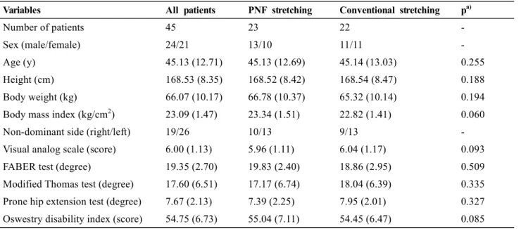
Variables All patients PNF stretching Conventional stretching p
a)Number of patients 45 23 22 -
Sex (male/female) 24/21 13/10 11/11 -
Age (y) 45.13 (12.71) 45.13 (12.69) 45.14 (13.03) 0.255
Height (cm) 168.53 (8.35) 168.52 (8.42) 168.54 (8.47) 0.188
Body weight (kg) 66.07 (10.17) 66.78 (10.37) 65.32 (10.14) 0.194
Body mass index (kg/cm
2) 23.09 (1.47) 23.34 (1.51) 22.82 (1.41) 0.060
Non-dominant side (right/left) 19/26 10/13 9/13 -
Visual analog scale (score) 6.00 (1.13) 5.96 (1.11) 6.04 (1.17) 0.093
FABER test (degree) 19.35 (2.70) 19.83 (2.40) 18.86 (2.95) 0.509
Modified Thomas test (degree) 17.60 (6.51) 17.17 (6.74) 18.04 (6.39) 0.335
Prone hip extension test (degree) 7.67 (2.13) 7.39 (2.25) 7.95 (2.01) 0.327
Oswestry disability index (score) 54.75 (6.73) 55.04 (7.11) 54.45 (6.47) 0.085 Values are presented as mean (SD)
PNF: proprioceptive neuromuscular facilitation, FABER: Flexion‐Abduction‐External Rotation.
a)
Shapiro-Wilk test
*p<0.05
Table 1. General characteristics of the participants (n = 45)
constant current), and 10 minutes of ultrasound (0.75 MHz, continuous wave) [19]. The trunk stabilization exercise was performed by 10 sets of bracing and bridge exercises, respectively with 30-second hold and 10-seconds rest [20].
PNF stretching was performed using contract-relax techniques of agonists in supine, prone, side-lying, and half-kneeling positions [21]. Contract-relax was applied with a 6-second contraction with 80% force of the maximal isometric contraction on the iliopsoas followed by a 15-second passive static stretching in the opposite direction of the iliopsoas [7]. Three sets of stretching were performed for each position. The PNF iliopsoas stretching is shown in Figure 2.
Conventional stretching was also performed by passive static stretching in the supine, prone, side-lying, and half-kneeling positions. Passive static stretching of the iliopsoas was performed for 30 seconds at the maximum stretch position [20]. Three sets of stretching were conducted for each position and are demonstrated in Figure 3.
Statistical analysis
Statistical analysis was performed using the statistical program for Windows; the SPSS/PC Statistics software
(version 23.0, IBM Co., USA). The Shapiro–Wilk test was used to check the normal distribution of the data.
A paired t-test was performed to compare differences in pain and hip ROM within the groups before and after the intervention. An independent t-test was performed to compare the differences between the PNF and conventional stretching groups. Statistical significance was set at a P-value of less than 0.05.
Results
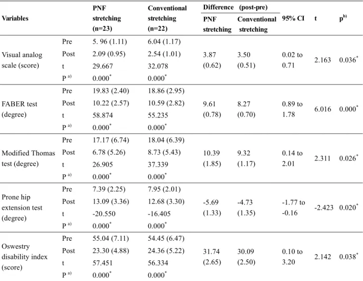
Comparison of the VAS score changes
Within-group changes in the VAS scores showed significant differences between the PNF (t=29.667, p<0.05) and conventional stretching groups (t=32.078, p<0.05).
The between-group changes in the VAS scores after intervention also showed significant differences between the PNF and conventional stretching groups (t=2.163; p<0.05; 95% confidence interval [CI], 0.02 to 0.71) (Table 2).
Comparison of FABERT score changes
Within-group changes in the FABERT scores were significantly different between the PNF (t=58.874, p<0.05) Figure 2. Proprioceptive neuromuscular facilitation iliopsoas stretching. (A) supine. (B) prone. (C) side-lying. (D) half-kneeling positions.
Figure 3. Conventional iliopsoas stretching. (A) supine. (B) prone. (C) side-lying. (D) half-kneeling positions.
6 Phys Ther Rehabil Sci 10(2)
and conventional stretching groups (t=55.235, p<0.05).
The between-group changes in the FABERT scores after intervention showed significant differences between the PNF and conventional stretching groups (t=6.016;
p<0.05; 95% CI, 0.89 to 1.78) (Table 2).
Comparison of the MTT score changes
Within-group changes in the MTT scores showed significant differences between the PNF (t=26.905, p<0.05) and conventional stretching groups (t=37.339, p<0.05). The between-group changes in the MTT scores after intervention showed significant differences
between the PNF and conventional stretching groups (t=2.311; p<0.05; 95% CI, 0.14 to 2.01) (Table 2).
Comparison of the PHET score changes
Within-group changes in the PHET scores were significantly different between the PNF (t=-20.550, p<0.05) and conventional stretching groups (t=-16.405, p<0.05).
The between-group changes in the PHET scores after intervention showed significant differences between the PNF and conventional stretching groups (t=-2.423; p<0.05;
95% CI, -1.77 to -0.16) (Table 2).
Variables
PNF stretching (n=23)
Conventional stretching (n=22)
Difference (post-pre)
95% CI t p
b)PNF stretching
Conventional stretching
Visual analog scale (score)
Pre 5. 96 (1.11) 6.04 (1.17)
3.87 (0.62)
3.50 (0.51)
0.02 to
0.71 2.163 0.036
*Post 2.09 (0.95) 2.54 (1.01)
t 29.667 32.078
P
a)0.000
*0.000
*FABER test (degree)
Pre 19.83 (2.40) 18.86 (2.95)
9.61 (0.78)
8.27 (0.70)
0.89 to
1.78 6.016 0.000
*Post 10.22 (2.57) 10.59 (2.82)
t 58.874 55.235
P
a)0.000
*0.000
*Modified Thomas test (degree)
Pre 17.17 (6.74) 18.04 (6.39)
10.39 (1.85)
9.32 (1.17)
0.14 to
2.01 2.311 0.026
*Post 6.78 (5.26) 8.73 (5.43)
t 26.905 37.339
P
a)0.000
*0.000
*Prone hip extension test (degree)
Pre 7.39 (2.25) 7.95 (2.01)
-5.69 (1.33)
-4.73 (1.35)
-1.77 to
-0.16 -2.423 0.020
*Post 13.09 (3.36) 12.68 (3.30)
t -20.550 -16.405
P
a)0.000
*0.000
*Oswestry disability index (score)
Pre 55.04 (7.11) 54.45 (6.47)
31.74 (2.65)
30.09 (2.50)
0.10 to
3.20 2.142 0.038
*Post 23.30 (4.88) 24.36 (5.22)
t 57.451 56.334
P
a)0.000
*0.000
*Values are presented as mean (SD)
PNF: proprioceptive neuromuscular facilitation, CI: confidence interval.
a)
paired t-test,
b)independentt-test,
*