Infliximab Partially Alleviates the Bite Force Reduction in a Mouse Model of Temporomandibular Joint Pain
Temporomandibular joint (TMJ) disorder is clinically important because of its prevalence, chronicity, and therapy-refractoriness of the pain. In this study, we investigated the effect of infliximab in a mouse model of TMJ pain using a specially-engineered transducer for evaluating the changes in bite force (BF). The mice were randomly divided into three groups (7 mice per group): the control group, the complete Freund’s adjuvant (CFA) group, and the infliximab group. BF was measured at day 0 (baseline BF). After measuring the baseline BF, CFA or incomplete Freund’s adjuvant was injected into both TMJs and then the changes in BF were measured at days 1, 3, 5, 7, 9, and 13 after the TMJ injection. For measuring the BF, we used a custom-built BF transducer. Control, CFA, and infliximab groups showed similar baseline BF at day 0. From day 1, a significant reduction in BF was observed in the CFA group, and this reduction in BF was statistically significant compared to that in the control group (P < 0.05). This reduction in BF was maintained until day 7, and BF started to recover gradually from day 9. In the infliximab group also, the reduction in BF was observed on day 1, and this reduction was maintained until day 7. However, the degree of reduction in BF was less remarkable compared to that in the CFA group. The reduction in BF caused by injection of CFA into the TMJ could be partially alleviated by the injection of anti-tumor necrosis factor alpha, infliximab.
Keywords: Bite Force; Infliximab; Temporomandibular Joint Pain Sang-Hyon Kim,1,2* Chang-Nam Son,1*
Hyo-Jung Lee,1 Ho-Chan Cho,1 Sung-Won Jung,2,3 Ji An Hur,4 Won-Ki Baek,5 Hye Ra Jung,6 and Ji Hee Hong7
1Department of Internal Medicine, Keimyung University Dongsan Medical Center, Daegu; 2Pain Research Center, School of Medicine, Keimyung University, Daegu; 3Department of Psychiatry, Keimyung University Dongsan Medical Center, Daegu; 4Department of Internal Medicine, School of Medicine, Yeungnam University, Daegu;
5Department of Microbiology, School of Medicine, Keimyung University, Daegu; 6Department of Pathology, Keimyung University Dongsan Medical Center, Daegu; 7Department of Anesthesiology and Pain Medicine, Keimyung University Dongsan Medical Center, Daegu, Korea
* Sang-Hyon Kim and Chang-Nam Son contributed equally to this work.
Received: 5 September 2014 Accepted: 3 December 2014 Address for Correspondence:
Ji Hee Hong, MD
Department of Anesthesiology and Pain Medicine, Keimyung University Dongsan Medical Center, 56 Dalsung-ro, Jung-gu, Daegu 700-712, Korea
Tel: +82.53-250-7288, Fax: +82.53-250-7240 E-mail: [email protected]
Funding: This work was supported by the Bisa Research Funds, Daegu, Republic of Korea (grant number 2012-0029).
http://dx.doi.org/10.3346/jkms.2015.30.5.552 • J Korean Med Sci 2015; 30: 552-558
INTRODUCTION
Mastication is a very elaborate process, which includes food in- take, intra-oral food transport, bolus formation and chewing in all of the mammals, and this activity is regulated by the motor and sensory components of the trigeminal system and their central projections (1, 2). Neural regulation of mastication, which can generate very high bite forces over milliseconds, comprises very rapid sensory feedback from innervated craniofacial struc- tures that include the temporomandibular joint (TMJ), the mas- ticatory muscles, and the teeth (1-4). Under normal circumstanc- es, mastication is an intrinsic activity that is not consciously per- ceived by humans and involves very rhythmic jaw movements which are produced by a Central Pattern Generator located in the pons and medulla (3). However, in cases of tissue injury to
these structures, mastication can become painful and nonrhyth- mic, leading to reduced bite force (BF) (5, 6).
Temporomandibular joint disorder (TMJD) is known for its mastication-related pain, and it is clinically important because of its prevalence, chronicity, therapy-refractoriness of the pain, and the largely unknown pain mechanism (7, 8). TMJD is a con- tinuum of various symptoms, which can give rise to progressive degenerative changes in the TMJ with progression of the dis- ease. It encompasses a broad spectrum of conditions, including initial capsulitis or synovitis, more advanced forms of internal derangement, and eventually end-stage degenerative joint dis- ease causing osteoarthritic changes (8).
There are several roadblocks to development of rationally targeted therapies, and one of them is shortcomings of avail- able animal models for TMJD, especially the relative paucity of
objective measurements that accurately represent patients’ car- dinal symptoms. Several studies of TMJ pain in a model frequent- ly used the head withdrawal threshold to a von Frey filament for measuring the nociceptive response in the TMJ (7, 9, 10). How- ever, this method has a limitation in reflecting the patients’ car- dinal symptom related to mastication, and it simply reflects the pain in the skin and subcutaneous tissue overlying the TMJ. Re- cently, Chen et al. (11) demonstrated a novel method measur- ing the BF changes in a mouse model of TMJ pain with a spe- cially designed BF transducer. They also suggested that the di- rect measure of BF provides a novel quantitative approach to quantifying TMJ pain in the mouse model (11).
Tumor necrosis factor alpha (TNF-α) is known as a key pro- inflammatory molecule in human rheumatoid arthritis and other chronic inflammatory diseases (12). Recently, TNF-α has attracted remarkable attention and interest in pain research area as one of a putative pain mediators, because the applica- tion of TNF-α in healthy tissue could induce thermal hyperalge- sia and synaptic long-term potentiation (13). Randomized, pla- cebo-controlled, multi-center clinical trials of human TNF-α inhibitors such as infliximab or etanercept have demonstrated their consistent and remarkable efficacy in improving signs and symptoms, with a favorable safety profile (14-16).
Lee et al. (8) reported that synovial TNF-α and interleukin 6 levels were elevated in patients with TMJD compared to the normal healthy group, although there was no statistical signifi- cance. Therefore, we used Infliximab, a chimeric monoclonal antibody, for investigating whether this drug has any pain re- lieving effect in a mouse model of chronic TMJ pain. In this study, we evaluated the changes in BF in a mouse model of TMJ pain using a specially-engineered transducer and also investi- gated the effect of infliximab.
MATERIALS AND METHODS Animals
Male ICR mice (20-25 g) were housed 5 per cage in a tempera- ture controlled (22 ± 2°C) vivarium under a 12-hr light/dark cy- cle. Mice were provided a standard rodent diet ad libitium and were allowed to acclimate for 5 days before the experimental procedure.
Induction of TMJ inflammation and intraperitoneal drug injections
The mice were randomly divided into three groups (7 mice per group): 1) the control group, which received only intraperitone- al injection of normal saline with TMJ injection of incomplete Freund’s adjuvant (IFA; Chondrex, Redmond, WA, USA); 2) the CFA group, which received intraperitoneal injection of normal saline with TMJ injection of complete Freund’s adjuvant (CFA, 5 mg/mL; Chondrex, Redmond, WA, USA); 3) the infliximab
group, which received intraperitoneal injection of infliximab (10 mg/kg, dissolved in normal saline) with TMJ injection of CFA.
All mice were briefly anesthetized with 2% isoflurane and bi- laterally injected using a 30-gauge needle fitted to a 10 µL Ham- ilton syringe. To easily identify and palpate the TMJ area, local hairs around the TMJ were trimmed with scissors. After care- fully palpating the locally trimmed area considered to be the TMJ, a 30-gauge needle was inserted through the facial skin.
The needle was carefully advanced superoanteriorly until the tip of the needle reached the zygomatic arch. Then the needle was slowly moved more inferiorly until it passed under the edge of the arch and ultimately entered into the joint space. Once the needle was located in the joint space, 10 µL of CFA or IFA was injected slowly over a period of 5 sec. Injections were given into both TMJs to minimize the fluctuation in the changes in BF. To evaluate the effect of TNF-alpha neutralizing drug, the inflix- imab group received a single intraperitoneal injection of inflix- imab. The control and CFA groups received intraperitoneal in- jection of the same volume of normal saline on the same day as the infliximab group. Intraperitoneal injection was administered immediately after local TMJ injection was given, and then it was administered daily for 13 days. BF was measured at day 0 (baseline bite force). After measuring the baseline BF, CFA or IFA was injected into both TMJs and then changes in BF were measured at days 1, 3, 5, 7, 9, and 13 after TMJ injection. All mea- surements were performed by one examiner who was blinded to the study groups.
Bite force analysis
To measure the BF, we used a custom-built BF transducer which was manufactured by KTM Engineering Inc (Seongnam, Korea).
This BF transducer was first developed by Williams et al. (17) and Chen et al. (11) also measured the BF reduction with this transducer. Briefly, the transducer consisted of two aluminum beams which were approximated to each other by 6 adjustable screws, and each beam was instrumented with two single-ele- ment strain gauges (Fig. 1A). The distance between the beams of the BF transducer was adjusted to 5.0 mm (-40% of maximal jaw opening), at which the mice can produce the maximum BF.
The four strain gauges were connected in a bridge module (Fig. 1B), and they transmitted the BF signal to the data acquisi- tion hardware. One end of each beam served as the bite plate and the tip of the bite plate was rounded to protect the animals’
teeth and oral soft tissue.
Prior to use, the calibration of the BF transducer was perform- ed and checked for linearity by suspending a series of calibra- tion weights ranging from 100 to 500 g from the bite plates. The voltage output from each weight was regressed against the force exerted by calibration weights. Output for each set of calibra- tions was both linear and reproducible, with correlation coeffi- cients (R2) ranging from 0.9902-0.9998 for each calibration. Mice
were placed in a cylindrical plastic tube with an opening at one end for accommodating the mouse head. Placing the mice in a cylindrical tube which permits only head movement is a very stressful condition, and therefore, we spent several minutes to acclimate and calm the mice in a cylindrical tube. When the bite transducer was moved towards the mouse at 0.5-1 cm/sec, a bite was invariably induced. We could confirm the induced bite through the voltage output wave appearing in the monitor.
When a proper and maximum bite was induced, the voltage output was recorded as a continuous wave at 500 Hz using Lab- view 2012 (National Instruments, Austin, TX, USA) (Fig. 2A, B).
The peak voltage of each bite was determined and converted into force (Newton, N) based on the regression equation de- rived from calibration. Each animal was tested 3 times/testing day. The interval between two trials was > 2 min. The maximum BF the mice produced per trial was recorded as the actual BF, and then averaged for all trials.
Histological evaluation of TMJ
To evaluate synovial inflammation, histological sections were prepared of the TMJs on day 13. The joint samples were fixed in
10% of neutralized formaldehyde and embedded in paraffin.
Before tissue section, the paraffin blocks were decalcified for 1 hr in decalcification solution. Conventional 4 μm sections were obtained from the paraffin blocks and incubated in an oven at 60°C, overnight. Sections were then dewaxed in xylene for 10 min and rehydrated through graded alcohol to distilled water. The hydrated sections were stained with hematoxylin and eosin.
Statistical analysis
All data are expressed as mean ± SD. We used one-way ANOVA followed by the Tukey post hoc test for group comparison. P <
0.05 indicates a statistically significant difference.
Ethic statement
All animal protocols were approved by the Keimyung Universi- ty Institutinal Animal Care and Use Committee (KM-2013-06R).
RESULTS
We could measure accurately the changes in BF in mice by us- ing a custom-built aluminum transducer which was fitted to
A B
Fig. 1. Photograph showing the transducer (A) and bridge module (B). Arrow indicates the strain gauges.
Fig. 2. Bite force voltage output wave through the computer monitor (A, B).
Newton (N)
Time (sec)
0 2 4 6 8 10 12 20
15 10 5 -5 0
1d CFA
Newton (N)
Time (sec)
0 2 4 6 8 10 12 20
15 10 5 -5 0
1d IFA
A B
the opening of their mouth. Whenever the maximum bite was induced with this transducer, we could check its force by the voltage output wave through the computer monitor (Fig. 2A, B).
However, after injecting CFA, several mice were very reluctant to bite the plate of the transducer due to the pain in the TMJ area.
In such cases, we elicited the bite until the proper and maximum bite was induced.
Normal baseline BF without TMJ inflammation was around 20 to 21 N. The induced bite appeared aggressive in nature, in response to the slowly approaching transducer. We believe that
the provoked aggression prompted the bite as a proven and val- id method for measuring the actual BF because the recorded responses were immediate, robust, and reproducible.
TMJ inflammation was induced by microinjection of CFA, and control injections were composed of IFA. We could observe a prolonged and remarkable reduction in BF over 7 days with this method. Gradually, the BF recovered to more than 80% of the baseline value within 2 weeks. Body weight was reduced by less than 5% (data not shown). The reduction in BF without a reduction in body weight indicates the induction of masticatory sensitization rather than general inflammatory and anorexic effects of inflammation.
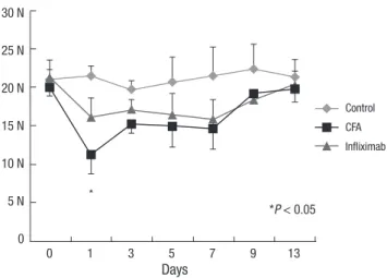
Control, CFA, and infliximab groups showed a similar base- line BF (20 to 21 N) at day 0. From day 1, a significant reduction in BF (nearly 50% of the baseline value) was observed in the CFA group and this reduction was statistically significant com- pared to that in the control group (Fig. 3) (P < 0.05). The reduc- tion in BF was maintained until day 7, and BF started to recover gradually from day 9.
In the infliximab group also, the reduction in BF was observed on day 1, and this reduction was maintained until day 7. How- ever, the degree of reduction was less remarkable compared to that in the CFA group (P < 0.05) and it did not demonstrate a statistically significant change compared to that in the control group (Fig. 3).
We analyzed the TMJ histology in the control, infliximab and CFA group. Normal, well preserved synovial lining was observ- ed in the control group. CFA group demonstrated severe inflam- Fig. 3. Changes in bite force from day 0 to day 13 in the control, complete Freund’s
adjuvant (CFA), and infliximab groups. *Statistically significant compared to that in the CFA group.
Days
0 1 3 5 7 9 13 30 N
25 N 20 N 15 N 10 N 5 N 0
*
*P < 0.05
Control CFA Infliximab
Fig. 4. Temeporomandibular joint of control (A), infliximab group (B), and CFA group (C) (H&E, × 40). The synovial membrane of control group (D) is smooth and lined by 1-2 sy- novial cells (H&E, × 400). In infliximab group (E), synovial membrane is lined by 2-3 cells (arrowhead) and shows moderate degree of chronic inflammation (H&E, × 400). The CFA group (F) shows severe inflammation and marked synovial hyperplasia which shows irregularly piled synovial cells (arrows) (H&E, × 400).
A
D
B
E
C
F
mation with marked synovial hyperplasia while infliximab group with moderate degree of chronic inflammation (Fig. 4A-F).
DISCUSSION
More than 20% of adults are affected by orofacial pain and some of the most difficult to treat forms of pain result from the TMJD.
Patients with TMJD or masticatory muscle inflammation report that chewing induces the highest levels of pain. Patients with TMJD experience significant and prolonged pain compared to normal individuals during opening and closing of the jaw while chewing for an extended period of time (8, 18, 19). Duration of chewing is also related to the development of pain, and Gavish et al. (20) reported that patients with TMJD can experience a significant increase in pain after chewing for 9 min. Therefore, the cardinal symptom of TMJD occurs while chewing, and we tried to demonstrate this phenomenon by using a high through- put, objective assay for BF measurement.
With respect to using the BF in mice to quantify the nocicep- tive behavior in response to TMJ inflammation, another inno- vative method has been reported recently which also shows a remarkable similiarity to the clinical features of TMJD. Dolan et al. (21) used the dolognawmeter to quantify the gnawing func- tion in three models of orofacial pain including TMJ inflamma- tion, masticatory myositis, and head and neck cancer. Their me- thod with the use of the dolognawmeter measures functional mechanical allodynia in the rodent of orofacial complex. How- ever, facial mechanical allodynia is not associated with chewing or mastication related pain, which is a dominant clinical feature of TMJD. Taken together, we believe that the direct measure- ment of BF provides a novel quantitive approach for quantify- ing TMJ pain in the mouse model.
Our data of BF measurement from 3 groups demonstrated a robust, reproducible, and valid method for quantifying TMJ pain in the mouse model. Baseline BF in the 3 groups measured at day 0 was recorded as 20-21 N. The control group also showed a consistent value ranging from 19-21 N from day 0 to day 13.
However, CFA injection into both TMJs dramatically attenuated the BF, which was observed in the CFA group. On day 1 in the CFA group, the BF was as low as 11 N which means a nearly 50%
reduction from the baseline value or compared to that in the control group. The maximum attenuation of BF occurred on day 1, and from day 3 to day 7, BF was maintained at 14-15 N.
From day 9, BF recovered to as much as 19 N. In contrast with the CFA group, the infliximab group maintained a fairly good BF after CFA injection into the TMJ. From day 1 to day 7, BF was maintained at 15-17 N, and this value was not statistically sig- nificant compared to that in the control group.
Wang et al. (7) assessed the nociceptive behavior of animals based on the head withdrawal test after injection of monosodium iodoacetate into the TMJ. The head withdrawal response was
significantly decreased from day 1 and continued to decrease until the first 3 weeks after monosodium iodoacetate injection.
There is some discrepancy between the period of reduction in BF in our results and the period of decrease in head withdrawal response. However, a direct comparison between these two re- sults has some limitations because we measured the BF and Wang et al. (7) assessed the hyperalgesia of skin around the TMJ using the von Frey filament. Chen et al. (11) also measured the attenuation in BF after injection of CFA into the TMJ, and they reported a prolonged and significant attenuation of BF over 9 days, which was similar to our result.
TNF-α is one of various cytokines that are mainly produced by mono-macrophages, NK cells, T cells, B cells, endothelial cells, fibroblasts, and osteoblasts. Local stress or inflammatory condition of the overlying synovial cells in the TMJ can produce proinflammatory cytokines such as IL-6 and TNF-α. The clini- cal features of TMJD include dysfunction and pain during mas- tication, which are basically the symptoms of inflammation (8).
In a study on TMJ synovial fluid analysis using ELISA, many cy- tokines such as TNF-α, IL-6, and IL-1 were found in the synovial fluid (8, 22, 23).
It is known that the effects of TNF-α neutralization are medi- ated by 2 receptors, TNF-RI and TNF-RII. Several studies dem- onstrated the expression of both TNF-RI and TNF-RII in rat dor- sal root ganglion (DRG) neurons (24-26), however, other stud- ies identified only TNF-RI in neurons, while localizing TNF-RII in non-neuronal cells in the DRG (27). Boettger et al. (12) dem- onstrated that systemic administration of the TNF-α neutraliz- ers etanercept and infliximab significantly attenuated inflam- mation-induced changes in locomotor and pain related behav- ior in rats with antigen induced arthritis. Chen et al. (28) also reported that neutralization of TNF-α with infliximab partially alleviated ovariectomy induced mechanical and thermal hy- peralgesia in rats, and they concluded that TNF-α plays an im- portant role in estrogen deficiency induced mechanical and thermal hyperalgesia.
It is postulated that the main neuronal target of TNF-α is a trigeminal ganglion, and Chen et al. (11) demonstrated that TMJ inflammation following CFA injection caused a significant increase in TRPV4 in the trigeminal ganglion according to West- ern blotting, and it reached a peak at day 3 after induction of in- flammation. They also reported that the increase in TRPV4 in the trigeminal ganglion showed a remarkable coincidence with the time course of BF attenuation.
The present study demonstrated that the reduction in BF caus- ed by injection of CFA into the TMJ could be partially alleviated by the administration of TNF-α neutralizing drug, infliximab.
Our experimental result supports the hypothesis that treatment with infliximab not only influences the inflammatory process, but also exerts direct antinociceptive effects. However, inflix- imab only partially improved BF reduction when compared
with control group. Therefore, increased expression of TNF-α may be just one mechanism of BF reduction, other factors may also be contributed.
Infliximab is a chimeric anti-TNF monoclonal antibody that is composed of human IgG1 kappa (constant region) and a mu- rine Fv (variable) region. Infliximab has been shown to bind with high affinity to both soluble and membrane bound TNF and is capable of neutralizing TNF in vitro and in vivo (16). We administered infliximab systemically via the intraperitoneal route; however, Ohtani et al. (29) demonstrated that the local intra-articular injection of anti-rabbit TNF-α monoclonal anti- body in a monoarthritis model was also effective in controlling local inflammation and degenerative joint changes.
Our study has some limitations. We did not check the proin- flammatory cytokines or chemokines in the blood or tissue. The study was not carried out regarding pain pathway such as noci- ceptors or neurotransmitters.
In conclusion, infliximab, a TNF-α neutralizing drug, could partially alleviate the reduction in BF caused by the injection of CFA into the TMJ.
DISCLOSURE
The authors have no conflicts of interest to disclose.
AUTHOR CONTRIBUTION
Conception and coordination of the study: Kim SH, Son CN, Hong JH. Design of ethical issues: Cho HC, Jung SW, Hur JA, Baek WK, Jung HR. Acquisition of data: Lee HJ. Data review:
Kim SH, Son CN, Baek WK, Hong JH. Statistical analysis: Son CN, Lee HJ, Hong JH. Manuscript preparation: Kim SH, Son CN, Cho HC, Jung SW, Hur JA, Jung HR. Manuscript approval:
all authors.
ORCID
Sang-Hyon Kim http://orcid.org/0000-0002-8030-7939 Chang-Nam Son http://orcid.org/0000-0002-1722-2190 Hyo-Jung Lee http://orcid.org/0000-0003-3126-0086 Ho-Chan Cho http://orcid.org/0000-0002-3727-000X Sung-Won Jung http://orcid.org/0000-0002-2300-742X Ji An Hur http://orcid.org/0000-0003-3219-8368 Won-Ki Baek http://orcid.org/0000-0001-9123-4096 Hye Ra Jung http://orcid.org/0000-0002-1477-6606 Ji Hee Hong http://orcid.org/0000-0003-3292-3822 REFERENCES
1. Turman JE Jr. The development of mastication in rodents: from neurons to behaviors. Arch Oral Biol 2007; 52: 313-6.
2. Yamada Y, Yamamura K, Inoue M. Coordination of cranial motoneu- rons during mastication. Respir Physiol Neurobiol 2005; 147: 177-89.
3. Westberg KG, Kolta A. The trigeminal circuits responsible for chewing.
Int Rev Neurobiol 2011; 97: 77-98.
4. Cadden SW, Orchardson R. Mastication and swallowing: 2. control.
Dent Update 2009; 36: 390-2, 4-6, 8.
5. Marquezin MC, Kobayashi FY, Montes AB, Gavião MB, Castelo PM. As- sessment of masticatory performance, bite force, orthodontic treatment need and orofacial dysfunction in children and adolescents. Arch Oral Biol 2013; 58: 286-92.
6. Kogawa EM, Calderon PS, Lauris JR, Araujo CR, Conti PC. Evaluation of maximal bite force in temporomandibular disorders patients. J Oral Rehabil 2006; 33: 559-65.
7. Wang XD, Kou XX, He DQ, Zeng MM, Meng Z, Bi RY, Liu Y, Zhang JN, Gan YH, Zhou YH. Progression of cartilage degradation, bone resorp- tion and pain in rat temporomandibular joint osteoarthritis induced by injection of iodoacetate. PLoS One 2012; 7: e45036.
8. Lee JK, Cho YS, Song SI. Relationship of synovial tumor necrosis factor alpha and interleukin 6 to temporomandibular disorder. J Oral Maxillo- fac Surg 2010; 68: 1064-8.
9. Nicoll SB, Hee CK, Davis MB, Winkelstein BA. A rat model of temporo- mandibular joint pain with histopathologic modifications. J Orofac Pain 2010; 24: 298-304.
10. Wu YW, Bi YP, Kou XX, Xu W, Ma LQ, Wang KW, Gan YH, Ma XC. 17-Be- ta-estradiol enhanced allodynia of inflammatory temporomandibular joint through upregulation of hippocampal TRPV1 in ovariectomized rats. J Neurosci 2010; 30: 8710-9.
11. Chen Y, Williams SH, McNulty AL, Hong JH, Lee SH, Rothfusz NE, Parekh PK, Moore C, Gereau RW 4th, Taylor AB, et al. Temporomandibular joint pain: a critical role for Trpv4 in the trigeminal ganglion. Pain 2013; 154:
1295-304.
12. Boettger MK, Hensellek S, Richter F, Gajda M, Stöckigt R, von Banchet GS, Bräuer R, Schaible HG. Antinociceptive effects of tumor necrosis fac- tor alpha neutralization in a rat model of antigen-induced arthritis: evi- dence of a neuronal target. Arthritis Rheum 2008; 58: 2368-78.
13. Gruber-Schoffnegger D, Drdla-Schutting R, Hönigsperger C, Wunder- baldinger G, Gassner M, Sandkühler J. Induction of thermal hyperalge- sia and synaptic long-term potentiation in the spinal cord lamina I by TNF-α and IL-1β is mediated by glial cells. J Neurosci 2013; 33: 6540-51.
14. Feldmann M, Maini RN. Anti-TNF alpha therapy of rheumatoid arthri- tis: what have we learned? Annu Rev Immunol 2001; 19: 163-96.
15. Sanmarti R, Ruiz-Esquide V, Hernández MV. Rheumatoid arthritis: a clinical overview of new diagnostic and treatment approaches. Curr Top Med Chem 2013; 13: 698-704.
16. Chen YF, Jobanputra P, Barton P, Jowett S, Bryan S, Clark W, Fry-Smith A, Burls A. A systematic review of the effectiveness of adalimumab, etaner- cept and infliximab for the treatment of rheumatoid arthritis in adults and an economic evaluation of their cost-effectiveness. Health Technol Assess 2006; 10: iii-iv, xi-xiii, 1-229.
17. Williams SH, Peiffer E, Ford S. Gape and bite force in the rodents Ony- chomys leucogaster and Peromyscus maniculatus: does jaw-muscle anat- omy predict performance? J Morphol 2009; 270: 1338-47.
18. Reiter S, Goldsmith C, Emodi-Perlman A, Friedman-Rubin P, Winocur E. Masticatory muscle disorders diagnostic criteria: the American Acad- emy of Orofacial Pain versus the research diagnostic criteria/temporo-
mandibular disorders (RDC/TMD). J Oral Rehabil 2012; 39: 941-7.
19. Winocur E, Gavish A, Finkelshtein T, Halachmi M, Gazit E. Oral habits among adolescent girls and their association with symptoms of temporo- mandibular disorders. J Oral Rehabil 2001; 28: 624-9.
20. Gavish A, Winocur E, Menashe S, Halachmi M, Eli I, Gazit E. Experimen- tal chewing in myofascial pain patients. J Orofac Pain 2002; 16: 22-8.
21. Dolan JC, Lam DK, Achdjian SH, Schmidt BL. The dolognawmeter: a novel instrument and assay to quantify nociception in rodent models of orofacial pain. J Neurosci Methods 2010; 187: 207-15.
22. Kubota E, Imamura H, Kubota T, Shibata T, Murakami K. Interleukin 1 beta and stromelysin (MMP3) activity of synovial fluid as possible mark- ers of osteoarthritis in the temporomandibular joint. J Oral Maxillofac Surg 1997; 55: 20-7; discussion 7-8.
23. Fu K, Ma X, Zhang Z, Pang X, Chen W. Interleukin-6 in synovial fluid and HLA-DR expression in synovium from patients with temporoman- dibular disorders. J Orofac Pain 1995; 9: 131-7.
24. Hensellek S, Brell P, Schaible HG, Bräuer R, Segond von Banchet G. The cytokine TNFalpha increases the proportion of DRG neurones expressing the TRPV1 receptor via the TNFR1 receptor and ERK activation. Mol Cell
Neurosci 2007; 36: 381-91.
25. Schäfers M, Geis C, Brors D, Yaksh TL, Sommer C. Anterograde trans- port of tumor necrosis factor-alpha in the intact and injured rat sciatic nerve. J Neurosci 2002; 22: 536-45.
26. Schäfers M, Lee DH, Brors D, Yaksh TL, Sorkin LS. Increased sensitivity of injured and adjacent uninjured rat primary sensory neurons to exog- enous tumor necrosis factor-alpha after spinal nerve ligation. J Neurosci 2003; 23: 3028-38.
27. Xu JT, Xin WJ, Zang Y, Wu CY, Liu XG. The role of tumor necrosis factor- alpha in the neuropathic pain induced by Lumbar 5 ventral root tran- section in rat. Pain 2006; 123: 306-21.
28. Chen BL, Li YQ, Xie DH, He QL, Yang XX. Blocking TNF-α with inflix- imab alleviates ovariectomy induced mechanical and thermal hyperal- gesia in rats. Neurol Sci 2012; 33: 527-33.
29. Ohtani T, Habu M, Khanal A, Yoshioka I, Matsukawa A, Tominaga K.
Local effects of intra-articular injection of anti-rabbit tumor necrosis fac- tor alpha monoclonal antibody in antigen-induced arthritis of the rab- bit temporomandibular joint. J Oral Pathol Med 2012; 41: 96-105.