• 검색 결과가 없습니다.

Technique of Coronary Transfer for TGA with Single Coronary Artery

N/A
N/A
Protected

Academic year: 2021

Share "Technique of Coronary Transfer for TGA with Single Coronary Artery"

Copied!
4
0
0

로드 중.... (전체 텍스트 보기)

전체 글

(1)

ISSN: 2233-601X (Print) ISSN: 2093-6516 (Online)

− 529 −

Department of Thoracic and Cardiovascular Surgery, Samsung Medical Center, Sungkyunkwan University School of Medicine Received: June 3, 2014, Revised: August 19, 2014, Accepted: September 3, 2014, Published online: December 5, 2014

Corresponding author: Tae-Gook Jun, Department of Thoracic and Cardiovascular Surgery, Samsung Medical Center, Sungkyunkwan University School of Medicine, 81 Irwon-ro, Gangnam-gu, Seoul 135-710, Korea

(Tel) 82-2-3410-3484 (Fax) 82-2-3410-0489 (E-mail) tg.jun@samsung.com

C

The Korean Society for Thoracic and Cardiovascular Surgery. 2014. All right reserved.

CC
This is an open access article distributed under the terms of the Creative Commons Attribution Non-Commercial License (http://creative- commons.org/licenses/by-nc/3.0) which permits unrestricted non-commercial use, distribution, and reproduction in any medium, provided the original work is properly cited.

Technique of Coronary Transfer for TGA with Single Coronary Artery

Tae Ho Kim, M.D., Jae Jun Jung, M.D., Yong Han Kim, M.D., Ji-Hyuk Yang, M.D., Ph.D., Tae-Gook Jun, M.D., Ph.D.

An eight-day-old neonate was diagnosed with dextro-transposition of the great arteries, atrial septal defect, patent ductus arteriosus, and a single sinus origin of the coronary arteries. The single coronary artery originated from the left sinus (sinus 2), had a proximal left circumflex arterial branch, and passed anteriorly to the right side of the aorta, further branching into the right coronary and left anterior descending arteries. We successfully performed an arterial switch operation and coronary transfer by tube graft reconstruction with autologous aortic tissue to treat the dextro-transposition of the great arteries and atrial septal defect with a single-sinus origin of the coronary arteries.

Key words: 1. Congenital heart disease (CHD) 2. Great vessel anomalies 3. Transposition

4. Coronary vessels

CASE REPORT

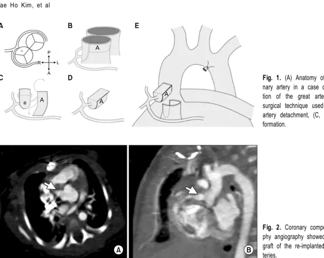
A male neonate weighing 3.9 kg who exhibited whole body cyanosis and desaturation upon birth was diagnosed with dextro-transposition of the great arteries, atrial septal de- fect, and patent ductus arteriosus associated with a single cor- onary artery pattern. The Leiden classification is often used in clinical contexts to describe coronary patterns. In this clas- sification, the right hand of an observer standing within the noncoronary sinus points to sinus 1 and the left hand points to sinus 2. In this patient, a single coronary artery originated from the left sinus (sinus 2), had a proximal left circumflex arterial branch, and passed anteriorly to the right side of the aorta, further branching into the right coronary and left ante- rior descending arteries. The left circumflex artery passed

posteriorly to the pulmonary artery, and the left anterior de- scending branch passed anteriorly to the aorta (Fig. 1A).

Surgery was performed eight days after birth with the neo- nate weighing 3.8 kg. After cardioplegic arrest with antegrade cold blood cardioplegia under moderate hypothermic car- diopulmonary bypass, the ascending aorta was transected and the coronary ostia were visualized. A single coronary artery originated from the left sinus, giving rise to a proximal left circumflex arterial branch that passed posterior to the pulmo- nary artery, then branched into the right coronary artery, and finally branched into the left anterior descending artery, pass- ing anteriorly to the aorta.

After the first dose of cardioplegia, the root catheter was removed and the ascending aorta was transected distally. The pulmonary artery was transected and brought anteriorly to the

Korean J Thorac Cardiovasc Surg 2014;47:529-532 □ Case Report □

http://dx.doi.org/10.5090/kjtcs.2014.47.6.529

(2)

Tae Ho Kim, et al

− 530 −

Fig. 1. (A) Anatomy of single coro- nary artery in a case of d-transposi- tion of the great arteries and (B) surgical technique used for coronary artery detachment, (C, D) tube graft formation.

Fig. 2. Coronary computed tomogra- phy angiography showed normal tube graft of the re-implanted coronary ar- teries.

aorta using the LeCompte maneuver. The distal ascending aorta was then anastomosed to the proximal pulmonary artery. Marking sutures were placed on the left sinus. A car- dioplegic catheter was then placed at the coronary sinus of the aorta (the neopulmonary artery), and a second dose of cardioplegia was infused while obliterating the aorta with forceps.

Next, coronary translocation was performed. The single coronary artery was removed from the aortic root along with a button from the coronary os. The coronary button was formed in a long U-shape instead of the usual round shape (Fig. 1B).

An arteriotomy was performed in the pulmonary artery (neoaorta), which was aligned with the opposite side of the left sinus and extended with Potts scissors. Coronary button reimplantation was performed by tube reconstruction of the coronary os with an aortic autograft patch (Fig. 1C, D). Then, the defect resulting from the resection of the proximal aorta

(neopulmonary artery) to create the button was reconstructed using a U-shaped patch of glutaraldehyde-fixed autologous pericardium.

All other steps of the procedure were routine, including the LeCompte maneuver, transatrial closure of the atrial septal defect, and division of the patent ductus arteriosus. After an aortic cross-clamping time of 120 minutes and a car- diopulmonary bypass time of 167 minutes, the child was weaned from cardiopulmonary bypass with low-dose ino- tropes, and the chest was primarily closed. The postoperative course was uneventful.

The child was extubated on postoperative day five, trans-

ferred to the ward on postoperative day 10, and discharged

home on postoperative day 15 in good clinical condition. His

left ventricular function was normal on a two-dimensional

echocardiogram. Coronary computed tomography angiography

showed a normal tube graft of the reimplanted coronary ar-

teries (Fig. 2).

(3)

Technique of Coronary Transfer

− 531 − DISCUSSION

The arterial switch operation has become the surgery of choice for correcting transposition of the great arteries [1,2].

Due to variations in the pattern of coronary arteries asso- ciated with the transposition of the great arteries and transfer of the coronary arteries during the arterial switch operation, much attention has been paid to the relationship between original patterns of coronary arteries and surgical outcomes [3,4]. Therefore, the coronary artery transfer technique is the most important step in attaining successful outcomes in arte- rial switch operations correcting transposition of the great arteries. Many techniques have been described, but each has its own advantages and disadvantages [5-8].

It is debatable whether autologous pericardium is a suitable material to use as a flap or tunnel in the coronary artery transfer, because the pericardium might cause long-term calci- fication, contraction, or thrombosis. Recently, a technique us- ing the patients’ own arterial wall, from either the aorta or the pulmonary artery, has become standard. No postoperative torsion or obstruction has been identified, and no thrombosis was found at a long-term follow-up. The introduction of this technique reduced the frequency of complications and pre- vented coronary artery torsion and deformation. In addition, adapting arterial vessel walls from either the aorta or the pul- monary artery into lateral walls or roofs has the benefit of al- lowing growth and recovery at the sites where the coronary artery was cut [9].

Our patient had a single ostium coronary artery originating from the rightward posterior sinus. The circumflex coronary artery arose from the single coronary artery, extending from the rightward posterior-facing sinus and passing behind the pulmonary artery. Next, the right coronary artery branched, and finally, the left anterior descending artery passed in front of the aorta. This pattern is extremely rare. In this situation, transfer of the coronary artery at a higher position than usual helps avoid kinking [10]. Some authors have even suggested placing the coronary button at or above the neoaortic anasto- mosis [11].

In this patient, resecting the coronary button in the usual round shape would have led to the possibility of the circum- flex artery kinking and creating tension on the right coronary

and left anterior descending arteries. Tube graft reconstruction was performed to avoid kinking or tension on the coronary arteries as well as to prevent postoperative myocardial ischemia. When performing tube graft reconstruction, using autologous pericardium is an option, although an aortic auto- graft was used in the present case. Autologous arterial tissue has the benefit of eliminating the risk of overstretching while preserving growth potential [12].

Tube graft reconstruction is a useful surgical technique for coronary translocation in patients with transposition of the great arteries associated with abnormal coronary artery varia- tion. Likewise, the use of an autologous aortic patch in tube reconstruction led to satisfactory results.

CONFLICT OF INTEREST

No potential conflict of interest relevant to this article was reported.

REFERENCES

1. Bove EL, Beekman RH, Snider AR, et al. Arterial repair for transposition of the great arteries and large ventricular septal defect in early infancy. Circulation 1988;78(5 Pt 2):III26-31.

2. Jatene AD, Fontes VF, Paulista PP, et al. Successful anatom- ic correction of transposition of the great vessels: a prelimi- nary report. Arq Bras Cardiol 1975;28:461-64.

3. Wernovsky G, Sanders SP. Coronary artery anatomy and transposition of the great arteries. Coron Artery Dis 1993;4:

148-57.

4. Planche C, Lacour-Gayet F, Serraf A. Arterial switch.

Pediatr Cardiol 1998;19:297-307.

5. Yacoub MH, Radley-Smith R. Anatomy of the coronary ar- teries in transposition of the great arteries and methods for their transfer in anatomical correction. Thorax 1978;33:

418-24.

6. Planche C, Bruniaux J, Lacour-Gayet F, et al. Switch oper- ation for transposition of the great arteries in neonates: a study of 120 patients. J Thorac Cardiovasc Surg 1988;96:

354-63.

7. Aubert J, Pannetier A, Couvelly JP, Unal D, Rouault F, Delarue A. Transposition of the great arteries: new techni- que for anatomical correction. Br Heart J 1978;40:204-8.

8. Parry AJ, Thurm M, Hanley FL. The use of ‘pericardial hoods’ for maintaining exact coronary artery geometry in the arterial switch operation with complex coronary anatomy.

Eur J Cardiothorac Surg 1999;15:159-64.

(4)

Tae Ho Kim, et al

− 532 − 9. Zheng JH, Xu ZW, Liu JF, Su ZK, Ding WX. Arterial

switch operation with coronary arteries from a single sinus in infants. J Card Surg 2008;23:606-10.

10. Quaegebeur JM, Rohmer J, Ottenkamp J, et al. The arterial switch operation: an eight-year experience. J Thorac Cardiovasc Surg 1986;92(3 Pt 1):361-84.

11. Lacour-Gayet F, Anderson RH. A uniform surgical technique

for transfer of both simple and complex patterns of the cor- onary arteries during the arterial switch procedure. Cardiol Young 2005;15 Suppl 1:93-101.

12. Mace L, Vanhuyse F, Jellimann JM, et al. Arterial switch

operation with a single coronary artery: the autograft

concept. Ann Thorac Surg 2009;87:1967-8.

수치

Fig. 1. (A) Anatomy of single coro- coro-nary artery in a case of  d-transposi-tion of the great arteries and (B)  surgical technique used for coronary  artery detachment, (C, D) tube graft  formation.

참조

관련 문서

Long-term outcomes of short dental implants supporting single crowns in posterior region: a clinical retrospective study of 5-10 years.. Osteotome sinus

Therefore, cTnT is a recommended biomarker for use in the detection of myocardial infarction (MI) and in acute coronary syndromes.[8] Indeed, several authors

Utility of T1-and T2-weighted high-resolution vessel wall imaging for the diagnosis and follow up of isolated posterior inferior cerebellar artery dissection with

- the right side : to move deoxygenated blood that is loaded with carbon dioxide from the body to the lungs - the left side : to receive oxygenated blood that has.. had most

The average position of the posterior superior alveolar artery, the wall thickness of the lateral wall, and the average volume of the maxillary sinus will

Pilot Confirm runway 09 left for landing. ATC gives you further instructions to turn right heading 310. On the radar display, there was an area to avoid which requires a

void makeTree(Object root, Object left, Object right): Creates a binary tree with root as the root element, left as the left subtree, right as the right

To read single character: getchar() function To read a string with space: