The Effect of Bonding Resin on Bond Strength of Dual-Cure Resin Cements
Duck-Su Kim, Sang-Hyuk Park, Gi-Woon Choi, Kyung-Kyu Choi*
Department of Conservative Dentistry, Division of Dentistry, Graduate of Kyung Hee University
The objective of this study is to evaluate the effect of an additional application of bonding resin on the bond strength of resin luting cements in both the light-cure (LC) and self-cure (SC) modes by means of the μTBS tests.
Three combinations of One-Step Plus with Choice, Single Bond with Rely X ARC, and One-Up Bond F with Bistite II were used. D/E resin and Pre-Bond resin were used for the additional appli- cation. Twelve experimental groups were made. Three mandibular 3rd molars were used in each group. Indirect composite blocks were cemented on the tooth surface. 1 × 1 ㎟ dentin-composite beam for μTBS testing were made and tested.
When total-etching dentin adhesives were used, an additional application of the bonding resin increased the bond strength (P < 0.05). However, this additional application didn’t influence the bond strength of self-etching dentin adhesives (P > 0.05).
In conclusion, the results suggest that an additional application of the bonding resin increases bond strength and enhances quality of bonding when using total-etching dentin adhesives. [J Kor Acad Cons Dent 32(5):426-436, 2007]
Key words : Dentin permeability, Resin-cement, Indirect composite, Nanoleakage, Bonding Resin, Bond Strength
- Received 2007.3.23., revised 2007.4.30., accepted 2007.6.26.-
Ⅰ. INTRODUCTION
Early resin cements were self-curing types. But, the self-curing resin cements take several minutes to be cured, and air voids included during the mixing of the base and the catalyst act as a defect
in the tooth-restoration bonded interface1). To compensate for the slow curing time of self-curing resin cements, new resin cements that combine light-curing with self-curing were developed. They were called dual-curing resin cements.
Dual-curing resin cements are used widely nowadays. Most of the clinicians use dual-curing resin cements for the bonding of all-ceramics, metal or metal-free inlays, veneers, crowns, indi- rect composite restorations, and endodontic fiber posts2-4). But dual-curing resin cements also have a problem, which is the polymerization contraction stress. Immediate light-curing of resin cements ABSTRACT
* Corresponding Author: Kyung-Kyu Choi Professor of Division of Dentistry, Graduate school of KyungHee University
1, Hoegi Dong, Dongdaemun Gu, Seoul, Korea, 130-702 Tel: 82-2-958-9337
E-mail:[email protected]
causes high polymerization contraction stress5). Because of the polymerization contraction stress, the interfacial and/or marginal defects are always present beneath the bonded indirect restora- tions6).
When clinicians select resin cements, the use of the dentin adhesive system is essential. Among these dentin adhesive systems, some total-etching single-bottle adhesives and single-step self-etch- ing adhesives are popular because of the conve- nience of use and less time-consuming procedure, but several problems such as low bond strength, increased marginal leakage, and durability of bonding can effect the longevity of restoration.
The bonding procedure of total-etching single- bottle adhesives includes priming with bonding simultaneously. To simplify the application step, an introduction of hydrophilic and acidic resin monomers are inevitable. For this reason, some total-etching single-bottle adhesives may not pro- vide the appropriate tooth structure for accept- able bonding when they are used with self-curing composites7). In detail, uncured acidic resin monomers within oxygen inhibition layer of total- etching single bottle adhesives react with the ini- tiator components of the chemical-cured compos- ites so that an acid-base reaction occurs before the radical polymerization reaction8). It has been known that the acidic resin monomers retard the polymerization of the self/dual-curing composites that are initiated by the means of peroxide-amine type binary redox catalysts9). When total-etching single-bottle adhesives are used, the volatile adhesive solvent evaporates quickly, and a con- tinuous transudation of dentinal fluid through the open dentinal tubules before the polymerization of the adhesives may result in the entrapment of water-filled blisters along the adhesive interface.
Therefore these water-filled blisters become defects in the adhesive layer which may cause bonding failure and post-operative hypersensitivi- ty10).
On the other hand, single-step self-etching adhesives combine etching, priming, and bonding into a single-step procedure. The acidity of single- step self-etching adhesives is usually high in
nature by the virtue of their self-etching capabili- ties. There is not only a redox reaction of the chemical components10), but there also was a report that single-step self-etching adhesives may act as a semi-permeable membrane to the denti- nal tubule11).
The purpose of this study is to evaluate the effect of an additional application of bonding resin contained in conventional three-step total-etching adhesives to both total-etch single-bottle and sin- gle-step self-etching adhesives on the bond strength of the dual-curing resin cements in both dual-curing and self-curing modes by the means of the μTBS test. In addition to the bond strength test, the TEM analysis for bonded interface was performed to support the results.
Ⅱ. MATERIALS AND METHODS 1. Materials
Three-combinations of One-Step Plus with Choice (Bisco Inc., Schaumburg, IL, USA), Single Bond with Rely X ARC (3M Dental Products, St.
Paul, MN, USA), and One-Up Bond F with Bistite II DC (Tokuyama Corp., Tokyo, Japan) were used. For the additional application, D/E bonding resin and Pre-bond resin (Bisco Inc., Schaumburg, IL, USA) were used. Pre-bond resin was used in the self-curing mode of dual-curing resin cements by mixing it with D/E bonding resin. Materials used in this study are listed in Table 1.
Thirty-six freshly extracted caries- and restora- tion-free human third molars were used in this study. Three teeth were selected randomly for each experimental group. Table 2 shows all the experimental groups.
2. Methods
⑴ Tooth preparation
The occlusal enamel and the superficial dentin of each tooth were removed with a high-speed diamond bur under a water coolant, and the exposed fresh dentin surface was polished with
wet 320-grit silicon carbide abrasive papers to create standard smear layers.
⑵ Restoration fabrication
6 × 6 × 3 ㎣ volume heat- and light-activated composite resin block (Tescera, Bisco Inc., Schaumburg, IL, USA) were made by using Teflon molds. The molds containing the uncured composites were placed inside an composite inlay processing chamber (Nitro-Therma-Lite; Bisco Inc.) and light-activated under a pressurized nitrogen atmosphere maintained at 551.6 ㎪ (i.e 80psi) for one complete cycle at 125℃ for 10 min- utes. The surface of the indirect composite block that would be bonded to the tooth surface was polished with wet silicon carbide abrasive papers
from 180- to 600-grit serially, then sandblasted with 20-50 ㎛ alumina oxide for 10 seconds under 30 psi.
⑶ Bonding procedure
Each dentin adhesive was applied to the pol- ished dentin according to the manufacturer’s instruction. Bonding resin of each dentin adhesive was applied to the sandblasted surface of the indirect composite block and light-cured according to the manufacturer’s instruction. For the groups that D/E bonding resin was used, bonding resin was applied and light-cured for 20 seconds on the previously applied adhesive layer. Pre-Bond resin was used after mixing it with D/E bonding resin in self-cure mode. Each dual-curing resin cement Table 1.Materials used in this study
Materials Type Component and Composition Batch No.
One-Step Plus Total-etch Biphenyl dimethacrylate, Hydroxyethyl methacrylate, 300012311
adhesive Acetone, Glass Frit
Choice Dual-curing Adhesive Paste: Strontium glass, amorphous silica, 0000011679 (Bisco, USA) cement Bis-GMA, UDMA, photoinitiator
Dual-cure catalyst paste: Amorphous silica, 0000007490 Bis-GMA, TEGDMA, benzoyl peroxide
Single Bond Total-etch Bis-GMA, HEMA, Bisphenol A OER
adhesive glycerolate dimethacrylate, Water Rely X ARC Diurethane dimethacrylate, Polyalkenoic acid
(3M, USA) Dual-curing copolymer,Ethanol, Water BHBH
cement Bis-GMA, TEGDMA, dimethacrylate polymer, zirconia filler, silica
One-Up Bond F Self-etch Bis-GMA, UDMA, HEMA, Bis-GMA, TEGDMA, 23074510902
adhesive benzoyl peroxide, BHT
Bistite II DC Dual-curing Primer 1A: Phosphoric acid monomer, acetone, water 118
(Tokuyama, cement Primer 1B: Initiator, alcohol, water 212
Japan) Primer 2: HEMA, acetone, water, 3133
Dual-cured resin cement
Paste A and B: MAC-10, methacrylic monomers, 84B-08R initiators, silica-zirconia filler, Bis-MPEPP, NPGDMA,
camphoroquinone, initiator
D/E bonding Bonding resin Bis-GMA, UDMA, HEMA 200003847
resin, in conventinoal Bis-GMA, TEGDMA, benzoyl peroxide, BHT 200005428 Pre-bond resin 3-step
(Bisco, USA) adhesive
was mixed and applied to the treated surface of the indirect composite block and it was then placed on the coated tooth surface. Then, 500g load was applied for 30 seconds along the long axis of the tooth. Excess resin cement was removed carefully. Either light-curing or self-cur- ing was performed on each experimental group.
Every tooth-composite bonded specimen was stored in distilled water at 37℃ for 24 hours.
⑷ Micro-tensile bond strength (μTBS) test Each tooth-composite bonded specimen was embedded in epoxy resin and sectioned occluso- gingivally into 1.0 ㎜ thick serial slabs using a Low-speed saw (Isomet, Buehler Ltd,Lake Bluff, IL, USA). These sectioned slabs were further sec- tioned into 1.0 × 1.0 ㎟ cross-section composite- dentin beams, according to the technique for the
‘non-trimming’version of the μTBS test12). The number of the composite-dentin beams of each experimental group varied from 23 to 38. Each composite-dentin beam was mounted on the μTBS testing zig with cyanoacrylate adhesive (Zapit;
Dental Ventures of America, USA) and the μTBS test was performed with an universal testing machine (EZ-Test, Shimadzu, Japan) at a crosshead speed of 1.0 ㎜/min.
⑸ Transmission electron microscope (TEM) analysis
The TEM analysis was performed in some experimental groups. Two teeth were selected for each experimental group. The occlusal enamel and dentin was removed with a high-speed dia- mond bur. The dentin surface was polished with wet 320-grit silicon carbide abrasive papers.
Dentin adhesives and corresponding dual-curing resin cements were applied to according manufac- turer’s instructions. To prevent any fracture of the diamond knife when using the ultramicro- tome, the indirect composite block was not luted.
Each specimen was stored in distilled water at 37
℃ for 24 hours and the 1.0 ㎜ thick slab was sec- tioned with a Low-speed saw. The two central slabs of each block were selected and so four slabs were examined for each group. Two-coats of nail varnish were applied, leaving 1.0 ㎜ from resin cement-dentin interface and each slab was stored in distilled water for 10 minutes. Each slab was immersed in 50 wt% ammoniacal silver nitrate solution for 24 hours in a dark room and washed with distilled water. Then each slab was stored in a developing solution for 8 hours under a fluores- cent light and washed with distilled water. Each slab was trimmed to 6 ㎜ width and fixed in Table 2.Classification of Bonding Protocol
Dentin Adhesive Curing Mode of The Use of Number of Group Code Resin Luting Cement Resin Cement Bonding Resin Spicemen
One-Step Plus Light-cure No 32 OC-LN
Yes 36 OC-LY
Choice Self-cure No 34 OC-SN
(Bisco, USA) Yes 36 OC-SY
Single Bond Light-cure No 31 SR-LN
Yes 38 SR-LY
Rely X ARC Self-cure No 35 SR-SN
(3M, USA) Yes 29 SR-SY
One-Up Bond F
Light-cure No 35 OB-LN
Yes 35 OB-LY
Bistite II DC
Self-cure No 28 OB-SN
(Tokuyama, Japan) Yes 30 OB-SY
a Karnovsky’s solution (2.5% glutaraldehyde and 2% paraformaldehyde in 0.1 M sodium phosphate buffer, pH 7.3) for 8 hours, then washed 3 times each for 10 minutes with 0.1 M sodium phosphate buffer solution (pH 7.4). Each specimen was post-fixed in a 1% osmium tetroxide solution for 2 hours and washed 3 times each for 10 minutes.
The post-fixed specimen was dehydrated by 50%, 70%, 80%, 90%, and 100% (2 times) serially for 10 minutes and embedded in epoxy resin along the composite-dentin interface.
After screening a 200 ㎚ semithin section of each specimen by using the Ultramicrotome equipped a diamond knife, a 90 ㎚ ultrathin section was made and collected on a 100-mesh formvar-coated copper grid. TEM analysis was performed with a 50 ㎸ Transmission Electron Microscope (JEM- 1010, JEOL Ltd., Tokyo, Japan) without an additional staining procedure.
⑹ Statistical analysis
The data was analyzed using One-way ANOVA and Duncan’s multiple comparison tests were performed at a 95% significance level.
Ⅲ. RESULTS
1. Micro-tensile bond strength (μTBS)
The μTBS of 12 experimental groups are shown in Table 3. As shown in Fig. 1, OC-LY and OC- SY showed higher μTBS values than that of OC- LN and OC-SN respectively. But, there was a significant difference in the light-curing mode only (P < 0.05). The μTBS of SR-LY and SR-SY
were higher than that of SR-LN and SR-SN respectively. There was also a significance in the light-curing mode only (P < 0.05). OB-LY and OB-SY showed slightly higher μTBS values than that of OB-LN and OB-SN respectively.
OC-LN showed the highest μTBS value among OC-LN, SR-LN, and OB-LN significantly (P <
0.05). When D/E bonding resin was applied addi- tionally, the μTBS of OC-LY and SR-LY were higher than that of OB-LY significantly (P <
0.05). In the self-curing mode, OC-SY, SR-SY and OB-SY showed higher μTBS values than OC- SN, SR-SN, and OB-SN respectively, but there was no significant difference (P > 0.05).
2. TEM analysis (Evaluation of nanoleakage)
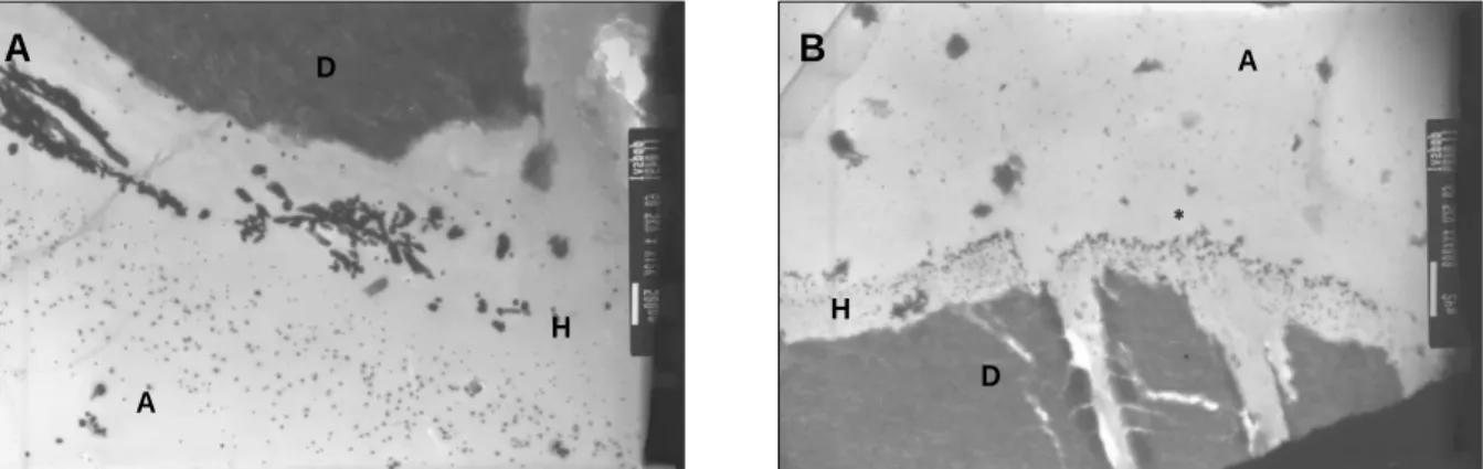
The TEM micrographs of OC-LN are shown in Figure 1. The hydrophilic adhesive primer infil- trated into the demineralized dentinal tubule thoroughly. Nanoleakage appeared at the top of the hybrid layer and within the adhesive layer.
The nanoleakage within the hybrid layer was con- centrated locally. These concentration of nanolea- kage are called the ‘water-tree’structure. The direction of ‘water-tree’goes from the dentinal tubule toward adhesive layer. On the other hand, the nanoleakage at the top of the hybrid layer was found along the interface of the hybrid layer and the adhesive layer. The thickness of the adhesive layer was about 8-10 ㎛ (Figure 1-B).
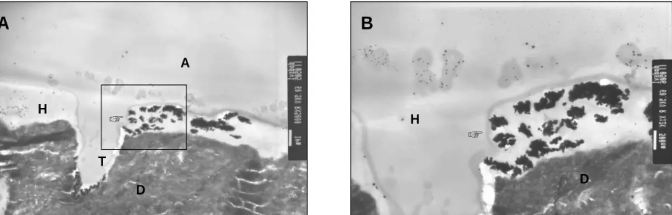
The TEM micrographs of OC-LY are shown in Figure 2. Although the ‘water-tree’structure could still be identified in the adhesive layer, the nanoleakage was reduced significantly and a
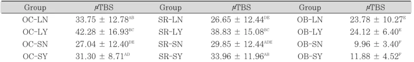
Table 3.Micro-tensile bond strength of all experimental groups (㎫ ± SD)
Group μTBS Group μTBS Group μTBS
OC-LN 33.75 ± 12.78AB SR-LN 26.65 ± 12.44DE OB-LN 23.78 ± 10.27E OC-LY 42.28 ± 16.93BC SR-LY 38.83 ± 15.08BC OB-LY 24.12 ± 6.40E OC-SN 27.04 ± 12.40DE SR-SN 29.85 ± 12.44ADE OB-SN 9.96 ± 3.40F OC-SY 31.30 ± 8.71AD SR-SY 33.96 ± 11.96AB OB-SY 11.88 ± 4.52F
* Same superscript means no statistically difference
Figure 2. TEM micrographs of OC-LY. A. Homogeneous adhesive layer appeared. Nanoleakage was not identified almost. Thickness of adhesive layer was about 14 - 16 ㎛. D: dentin, H: hybrid layer, A:
adhesive layer, R: resin cement, pointer: nanoleakage formed in hybrid layer. B. Magnification of box area in Figure 2-A. Structure of ‘water tree’was identified. T: dentinal tubule, pointer: cluster of nanoleakge.
Figure 1. TEM micrographs of OC-LN. A. Nanoleakage appeared both at the top of hybrid layer and within adhesive layer. ‘water-tree’structure identified within hybrid layer(pointer). D: dentin, H: hybrid layer, A: adhesive layer, pointer: nanoleakage toward adhesive layer. B. Nanoleakage occurred at the top of hybrid layer. The thickness of adhesive layer was estimated about 8 - 10 ㎛. T: dentinal tubule, R: resin cement, pointer: nanoleakage formed at the top of hybrid layer.
Figure 3. TEM micrographs of SR-LN. A. High magnification of hybrid layer. ‘Water-tree’structure was observed (pointer). At the top of hybrid layer, diffused nanoleakage was identified. D: dentin, H: hybrid layer, A: adhesive layer, Pointer: nanoleakage formed in hybrid layer. B. Nanoleakage formed at the top of hybrid layer (asterisk).
A B
A B
A B
A
H
H
T
D A
R
D
D
H
D
A
H
A
H
D
**
A
A
T
H D
R
☞
☞ ☞
☞
☞
Figure 4. TEM micrographs of SR-LY. A. Localized concentration of nanoleakage(‘water-tree’). in hybrid layer(pointer) D: dentin, H: hybrid layer, A: adhesive layer, T: dentinal tubule, pointer: nanoleakage formed in hybrid layer. B. magnification of box area in Figure 2-A. ‘Water-tree’structure formed in hybrid layer(pointer). Diffused nanoleakage at the top of the hybrid layer.
Figure 5. TEM micrographs of OB-LN. A. low magnification of resin cement-dentin interface. Nanoleakage appeared in adhesive-dentin interface. Globular filler was observed in adhesive layer (typical in One-Up Bond F). Representative ‘water-tree’structure appeared in adhesive layer (pointer) A: adhesive layer, D:
dentin, pointer: nanoleakage from dentin toward adhesive layer. B. Thin hybrid layer was observed (arrow). Unusual nanoleakage pattern appeared inside dentin (pointer). C. High magnification of ‘water- tree’structure.
A
A
B
A A
D D
T D
H H
D A
D
C B
☞ ☞
☞
☞
☞
homogeneous adhesive layer appeared. The thick- ness of the adhesive layer was about 14 - 16 ㎛ (Figure 2-A). In Figure 2-B, the localized concen- tration of the nanoleakage is observed in the hybrid layer.
The TEM micrographs of SR-LN were similar to OC-LN. The localized concentration of the nanoleakage appeared within the hybrid layer.
Moreover, the direction of the nanoleakage within the hybrid layer was same as that of OC-LN (Figure 3-A). Also, dispersed nanoleakage was observed in the adhesive layer (Figure 3-B).
The TEM micrographs of SR-LY are shown in Figure 4. The TEM micrographs of SR-LY were similar to that of OC-LY. The localized concentra- tion of the nanoleakage appeared in the hybrid layer. Dispersed nanoleakage was observed in the adhesive layer. But, a relatively homogenous adhesive layer was identified except for some localized concentration of the nanoleakage.
The TEM micrographs of OB-LN are shown in Figure 5. The thickness of the hybrid layer was very thin and globular filler particles were observed in the adhesive layer. The ‘Water-tree’
structure appeared in the adhesive layer and it runs along the dentinal tubules toward the adhe- sive layer. But an unusual nanoleakage inside the dentin was identified (Figure 5-B).
Ⅳ. DISCUSSION
This study demonstrated that the adhesive per- meability that was caused by the total-etching single-bottle dentin adhesive was reduced by the means of an additional application of the bonding resin contained in a conventional three-step dentin adhesive. Due to the more hydrophobic bonding resin, the permeability of the adhesive layer was reduced13). Also, the μTBS values of OC- LY and SR-LY increased significantly compared to OC-LN and SR-LN respectively.
Among the experimental groups which combined One-Step Plus with Choice, OC-LY showed higher μTBS value than OC-LN. In the self-curing mode, the μTBS value of OC-SY was higher than that of
OC-SN. In the TEM micrographs of OC-LN (Figure 1), the reticular type of ammoniacal sil- ver grain due to transudation of deep dentinal fluid away from interface between superficial dentin and hybrid layer after volatile acetone sol- vent in One-Step Plus evaporated quickly was observed clearly14). The spotted type of ammonia- cal silver grains appeared along the interface of the hybrid layer and also within the adhesive lay- er15). On the other hand, the TEM micrographs of OC-LY (Figure 2) showed reduced nanoleakage and thickened adhesive layers. Although the reticular type of ammoniacal silver grain could be easily identified, the spotted type of ammoniacal silver grain was found rarely. It was thought that the additional application of the more hydrophobic bonding resin contributed to the enhancement of the adhesive layer. These ultramorphological changes in the adhesive layer might be attributed to increase of the bond strength.
When Rely X ARC was combined with Single Bond, an additional application of the bonding resin increased the μTBS of the tooth-resin cement both in the light-curing and self-curing mode. In the TEM micrographs, SR-LN and SR- LY showed similar appearances as OC-LN and OC-LY respectively. But in the TEM micrographs of SR-LN (Figure 3), the reticular type of ammo- niacal silver grain was not observed as well as it was in OC-LN. The reason was thought to be due to the fact that the solvent of Single Bond is mostly water, while the solvent of One-Step Plus is acetone. It was postulated that since the water solvent in Single Bond is less volatile than the acetone in One-Step Plus, the transudation of dentinal fluids may be low16). In addition, the spotted type of ammoniacal silver grain was found mainly at the top of the hybrid layer. In SR-LY (Figure 4), the spotted type of ammoniacal silver grain was disappeared mostly. This reduction of the nanoleakage may have affected the tooth- resin cement bonding so that the μTBS value of SR-LY was higher than SR-LN.
In case of One-Up Bond F with Bistite II DC, the additional application of the bonding resin did
not have any influence on the μTBS of the tooth- resin cement both in the light-curing and the self- curing mode. In the TEM micrographs of OB-LN (Figure 5), the etching pattern of single-step self- etching adhesives was irregular and the resin tags were difficult to identify17). An agglomeration of adhesive fillers was also easily observed in the adhesive layer. The reticular type and the spotted type of ammoniacal silver grain were same as that of OC-LN and SR-LN. But, some nanoleak- age inside the dentin was found. The nanoleakage in the adhesive layer was thought to be usual.
Perhaps, the reason for this specific phenomenon was in the tooth used in the TEM specimen preparation. Further study should be performed for a closer examination. Although the μTBS val- ues of OB-LY and OB-SY did not increase as compared with OB-LN and OB-SN respectively, the durability of the bonding is expected to be affected due to the reduced nanoleakage18). Further study is necessary for the durability of bonding.
The self-curing composites have a slower rate of polymerization compared to the light-curing com- posites. The tendency of the acid-base reaction between the tertiary amines and the acidic resin monomers is higher in the self-curing composites than that of the dual-curing composites. But, according to the results, an additional application of the bonding resin would be clinically reliable for the self-curing composites. On the other hand, the groups that combined One-Up Bond F with Bistite II DC showed low μTBS values. In addition to the permeability of single-step self-etching adhesives, the acidic resin monomers play a major roll in reducing the bond strength19). In this study, the reduction rate in the μTBS value between OB-LN and OB-SN was about 60%, while it was about 20% in the groups that com- bined One-Step Plus with Choice and Single Bond with Rely X ARC.
According to this study, an additional applica- tion of the bonding resin to total-etching single- bottle adhesives is useful clinically. One point of concern is that such an additional application will
affect the fitness of the indirect restorations adversely. In the TEM micrographs, the increased film thickness after an additional application of bonding resin was about 4 - 6 ㎛. But, considering that the cement spaces in indirect ceramics or composite restorations are in the range of 50-100
㎛, a slight increase in the adhesive layer would not be a serious problem20,21). In a clinical situa- tion, the dentin adhesives tend to pool in the cor- ner of the cavity. Thus, clinicians have to pay attention to the pooling of the dentin adhesives.
Further studies to improve the defects of the dentin adhesive is necessary.
Ⅴ. CONCLUSION
This study was designated to evaluate the intrinsic permeability of both total-etching single- bottle adhesives and one-step self-etching adhe- sives and to correct their defects by an additional application of bonding resin contained in conven- tional three-step total-etching adhesives. The results followed as:
1. Both One-Step Plus with Choice and Single Bond with Rely X ARC which had an addi- tional application of in the light-curing mode showed significantly higher bond strengths than that of those not applied (P < 0.05).
2. For the groups that combined Bistite II with One-Up Bond F, there was no significant dif- ference in the groups with or whthout an additional application of the bonding resin in both light- and self-curing modes (P > 0.05) . 3. In the light-curing mode, the group which
combined Choice with One-Step plus without an additional application of the bonding resin showed a significant result than the groups that combined Rely X ARC with Single-Bond and Bistite II with One-Up Bond F (P <
0.05).
4. In self-curing mode, an additional application of the bonding resin did not have a significant effect on the bond strength of all experimen- tal groups (P > 0.05).
REFERENCE
1. Lutz F, Setcos JC, Philips RW, Roulet JF. Dental restorative resins: Type and characteristics. Dent Clin North Am 27:697-712, 1983.
2. Peumans M, Van Meerbeck B, Lambrechts P, Vanherle G. Porcelain veneers: a review of literature. J Dent 28:163-177, 2000.
3. Michelle CA, Abbariki M, Orr JF. The influence of lut- ing cement on the probabilities of survival and modes of failure of cast full-coverage crowns. Dent Mater 16:198-206, 2000.
4. Mannocci F, Innocenti M, Ferrari M, Watson TF.
Confocal and scanning electron microscopic study of teeth restored with fiber posts, metal posts, and com- posite resins. J Endod 25:789-794, 1999.
5. Braga RR, Ferracane JL, Condon JR. Polymerization contraction stress in dual-cure cements and its effect on interfacial integrity of bonded inlays. J Dent 30:333-340, 2002.
6. Peutzfeldt A, Asmussen E. A comparison of accuracy in seating and gap formation for three inlay/onlay tech- niques. Oper Dent 15:129-135, 1990.
7. Swift EJ Jr, Perdigao J, Combe EC, Simpson CH 3rd, Nunef MF. Effects of restorative and adhesive curing methods on dentin bond strengths. Am J Dent 14:137- 140, 2001.
8. Sanares AME, King NM, Itthagarun A, Tay FR, Pashley DH. Adverse surface interactions between one- bottle light-cured adhesives and chemical-cured com- posites. Dent Mater 17:542-556, 2001.
9. Tay FR, Pashley DH, Yiu CKY, Sanares AME, Wei SW. Factors contributing to the incompatibility between simplified-step adhesives and self-cured or dual-cured composites. Part I. Single-step self-etch adhesive. J Adhes Dent 5:27-40, 2003.
10. Tay FR, Pashley DH. Have Dentin Adhesives Becom Too Hydrophilic? J Can Dent Assoc 69:726-731, 2003.
11. Tay FR, Pashley DH, Suh BI, Carvalho RM,
Itthagarun A. Single-step adhesives are permeable membranes. J Dent 30:371-382, 2002.
12. Pashley DH, Carvalho RM, Sano H, Nakajima M, Yoshiyama M, Shono Y, Fernandes CA, Tay FR. The Microtensile Bond Test: A Review. J Adhes Dent 1:299-309, 1999.
13. Carvalho RM, Pegoraro TA, Tay FR, Pegoraro LF, Silva NRFA, Pashley DH. Adhesive permeability affects coupling of resin cements that utilize self-etch- ing primers to dentine. J Dent 32:55-65, 2004.
14. Pashley DH, Pashley EL, Carvalho RM, Tay FR. The effects of dentin permeability on restorative dentistry, Dent Clin North Am 46:211-245, 2002.
15. Tay FR, Pashley DH, Yiu C, Cheong C, Hashimoto M, Itou K, Yoshiyama M, King NM. Nanoleakage type and potential implications: evidence from unfilled and filled adhesives with the same resin composition. Am J Dent 17:182-190, 2004.
16. Reis A, Loguercio AD, Carvalho RM, Grande RM.
Durability of resin dentin interface: effects of surface moisture and adhesive solvent component. Dent Mater 20:669-676, 2004.
17. Tay FR, Pashley DH, Yoshiyama M. Two Modes of Nanoleakage Expression in Single-step Adhesives. J Dent Res 81:472-476, 2002.
18. Choi KK, Condon JR, Ferracane JL. The Effects of Adhesive Thickness on Polymerization Contraction Stress of Composite. J Dent Res 79:812-817, 2000.
19. Suh BI, Feng L, Pashley DH, Tay FR. Factors con- tributing to the incompatibility between simplified-step adhesives and chemically-cured or dual-cured compos- ites. Part III. Effect of acidic resin monomers. J Adhes Dent 5:267-282, 2003.
20. Audenino G, Bresciano ME, Bassi F, Carossa S. In vit- ro evaluation of fit of adhesively luted ceramic inlays.
Int J of Pros 12:342-347, 1999.
21. Molin MK, Karlsson SL, Kristiansen MS. Influence of film thickness on joint bend strength of a ceramic/resin composite joint. Dent Mater 12:245-249, 1996.
접착레진의 부가도포가 레진 시멘트의 결합강도에 미치는 영향에 대한 연구
김덕수∙박상혁∙최기운∙최경규*
경희대학교 대학원 치의학과 치과보존학교실
본 연구에서는 접착레진의 부가적인 도포가 레진 시멘트의 결합강도에 미치는 영향을 연구하였다.
One-Step Plus와 Choice, Single Bond와 Rely X ARC, One-Up Bond F와 Bistite II DC를 사용하였고 접 착레진으로 D/E Bonding resin과 Pre-Bond Resin을 선택하였다. 적용 및 광중합 유무에 따라 12개의 군을 설 정하였다.
제 3대구치의 건전한 상아질 면에 간접 복합레진 수복물을 제작하여 접착을 시행하고 1 × 1 ㎟의 시편을 만들어 미세인장강도를 측정하였다. 또한 투과전자현미경으로 접착계면을 관찰하였다. 그리하여 다음과 같은 결론을 얻을 수 있었다.
1. Single Bond와 Rely X ARC, 그리고 One-Step Plus와 Choice를 조합하고 광중합을 시행한 군에서, 접착 레진을 부가적으로 도포할 경우 미세인장강도가 증가하였다.
2. One-Up Bond F와 Bistite II DC를 조합한 군에서 접착레진의 부가적인 도포에 의한 미세인장강도의 차이 는 나타나지 않았다.
3. 광중합을 시행한 군들 중, One-Step Plus와 Choice를 조합한 군이 다른 군보다 높은 미세인장결합강도를 보 였다.
4. 자가중합만을 시행한 군간에는 접착레진의 부가적인 도포에 의한 차이가 나타나지 않았다.
투과전자현미경 관찰을 시행하여 광중합을 시행하고 접착레진을 부가적으로 도포한 실험군에서 미세누출이 감소 하고 접착층의 두께가 증가한 것을 확인할 수 있었다.
본 연구결과 완전산부식 상아질 접착제와 이중중합 레진 시멘트를 사용할 경우 부가적인 접착레진의 도포가 임상 적으로 유용할 수 있다는 사실을 확인할 수 있었다.
주요어: 상아질 투과성, 레진시멘트, 간접 복합 레진, 미세누출, 접착레진, 결합강도 국문초록