68 https://e-jcvi.org
One of the complications of percutaneous coronary intervention is stent dislodgement that can lead to dire consequences either in the form of surgery or death.1) Most of the time, dislodgment of the stent is identified in fluoroscopy. In some cases, it is missed and can be identified in cardiac computed tomography.2) We report a 42-year-old male, who presented with two hours history of typical chest pain and profuse sweating. On examination pulse was 54 bpm with blood pressure of 110/80 mmHg and clear chest on auscultation. Blood tests were normal except for the Troponin I which was elevated. 12 lead electrocardiogram showed ST segment elevation in II, II, aVF with reciprocals in lead I and aVL suggestive of acute inferior wall myocardial infarction. Coronary angiography showed totally occluded proximal right coronary artery (RCA) and no significant lesion in left coronary artery. The patient underwent primary percutaneous coronary intervention of RCA. During the procedure he developed catheter-induced dissection for which 3.5 × 30 mm resolute stent was deployed.
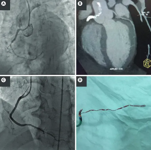
The actual lesion was distal to the stent. Another stent of 3.5 × 33 mm resolute stent was tried to cross through the stent and was presumed that stent has been deployed distally. On careful examination stent images in right coronary artery was not visualized. A search for stent dislodgement was done under fluoroscopy. After failure to detect the stent, patient shifted to CT room for CT image acquisition, which showed portion of stent in the RCA with its unfolded stents flipping in the aortic root. The stent was retrieved with a snare and 3.5 × 33 mm resolute stent was deployed distally. Coronary angiographic imaging after deployment of stent showed TIMI 3 flow with no dissection distal to stent (Figure 1). The hospital course was uneventful and patient was discharged with good condition.
J Cardiovasc Imaging. 2019 Jan;27(1):68-69 https://doi.org/10.4250/jcvi.2019.27.e2 pISSN 2586-7210·eISSN 2586-7296
Images in Cardiovascular Disease
Received: Sep 24, 2018 Revised: Oct 16, 2018 Accepted: Nov 20, 2018 Address for Correspondence:
Tariq Ashraf, FCPS
Department of Cardiology, National Institute of Cardiovascular Diseases, Rafiqui (H.J.) Shaheed Road, Karachi 75510, Pakistan.
E-mail: [email protected] Copyright © 2019 Korean Society of Echocardiography
This is an Open Access article distributed under the terms of the Creative Commons Attribution Non-Commercial License (https://
creativecommons.org/licenses/by-nc/4.0/) which permits unrestricted non-commercial use, distribution, and reproduction in any medium, provided the original work is properly cited.
ORCID iDs Tariq Ashraf
https://orcid.org/0000-0002-6680-1017 Parveen Akhter
https://orcid.org/0000-0002-4764-4757 Abdul Samad Achakzai
https://orcid.org/0000-0002-0248-506X Naveedullah Khan
https://orcid.org/0000-0001-7965-7555 Tahir Saghir
https://orcid.org/0000-0002-3148-8964 Syed Nadeem Hassan Rizvi
https://orcid.org/0000-0002-1901-3413 Kanwal Fatima Aamir
https://orcid.org/0000-0003-4904-3653
Tariq Ashraf , FCPS, Parveen Akhter , FCPS, Abdul Samad Achakzai , FCPS, Naveedullah Khan , FCPS, Tahir Saghir , FCPS,
Syed Nadeem Hassan Rizvi , MRCP, and Kanwal Fatima Aamir , FCPS
Department of Cardiology, National Institute of Cardiovascular Diseases, Karachi, Pakistan
Cardiac CT in Detecting a Dislodged Stent in Right Coronary Artery after Primary Percutaneous Coronary
Intervention
Conflict of Interest
The authors have no financial conflicts of interest.
REFERENCES
1. Ayça B, Okuyan E, Şahin İ, Dinçkal MH. Dislodgement of coronary stent due to rupture of stent balloon.
Turk Kardiyol Dern Ars 2015;43:93-4.
PUBMED | CROSSREF
2. Torre-Amione G, de la Pena-Almaguer E, Quintanilla J, et al. The use of cardiac CT as a roadmap for resolving coronary stent dislodgement. Arch Cardiol Mex 2018;88:318-9.
PUBMED | CROSSREF
69 https://e-jcvi.org https://doi.org/10.4250/jcvi.2019.27.e2
CT to Detect Dislodged Stent in RCA after Primary PCI
A B
D C
Figure 1. Coronary images by conventional angiogram and by CT images. (A) Coronary angiography showing stent in right coronary artery (RCA) with lesion distal to stent. (B) Dislodged stent extending from ostium of RCA to aortic root. (C) Coronary angiography after snaring and stenting of RCA. (D) Stent after retrieval.