119 A 47-year-old woman without a significant medical his-
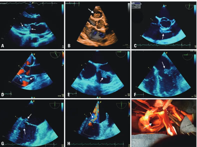
tory, including no history of intravenous drug abuse, no body piercings, and no tattoos, was referred with a 6-day history of high fever and arthralgia. Blood cultures were all positive for Staphylococcus aureus Meti-S. Transthoracic echocardiography revealed: 1) an irregular thickening of the posterior mitral valve root with mobile extensions projecting toward both the left ventricle and the left atrium (Fig. 1A, Supplementary movie 1); 2) a huge mobile vegetation rising from the upper portion of the right side of the interventricular septum (Fig.
1B, Supplementary movie 2). These findings were confirmed by transesophageal echocardiography (Fig. 1C-G, Supplemen- tary movie 3 and 4), which also disclosed a mild-to-moderate mitral regurgitation (Fig. 1H). A diagnosis of multisite infective endocarditis with right sided mural involvement was made. No point of entry was detected. The patient got quickly worse and was referred for emergency cardiac surgery.1) Intraoperative findings confirmed the presence of a bulky 3 cm-long vegetation attached to the right-sided interventricu- lar septal endocardial surface that reached the pulmonary valve orifice (Fig. 1I, Supplementary movie 5). On the left side of the heart, the surgeon noted the presence of a large vegeta- tion extending to the posterior free wall endocardium that was damaging the root of the mitral posterior leaflet, and destroy- ing several chordae tendineae and the top of the posterior papil- lary muscle. The surgical procedure consisted of a conservative posterior mitral valve repair and, on the right side, of a single septal vegectomy. The patient was discharged home 38 days
pISSN 1975-4612/ eISSN 2005-9655 Copyright © 2015 Korean Society of Echocardiography www.kse-jcu.org http://dx.doi.org/10.4250/jcu.2015.23.2.119
after surgery, in stable clinical condition.
Supplementary movie legends
Movie 1. Real-time two-dimensional transthoracic echocar- diography (parasternal long-axis view) showing an irregularly shaped vegetation rising from the root of the posterior mitral valve leaflet, and a mobile vegetation attached to the right side of the interventricular septum.
Movie 2. Real-time three-dimensional transthoracic echo- cardiography (parasternal long-axis view) showing the same left and right-sided endocarditic lesions as in Movie 1.
Movie 3. Real-time two-dimensional transesophageal echo- cardiography from a mid-esophageal right ventricular outflow view (transducer angle 77°) showing a large mural vegetation rising from the right side of the interventricular septum and projecting toward the pulmonary valve.
Movie 4. The same right-sided interventricular septal veg- etation as in Movie 3, displayed by real-time two-dimensional transesophageal echocardiography from a mid-esophageal right chambers view, transducer angle 0°.
Movie 5. Intraoperative surgeon’s view (right infundibulot- omy ventricular free wall approach) of the vegetation attached to the right side of the interventricular septum.
Reference
1. Yao F, Han L, Xu ZY, Zou LJ, Huang SD, Wang ZN, Lu FL, Yao YL.
Surgical treatment of multivalvular endocarditis: twenty-one-year single center experience. J Thorac Cardiovasc Surg 2009;137:1475-80.
• Received: November 29, 2014 • Revised: December 28, 2014 • Accepted: May 19, 2015
• Address for Correspondence: Dominique de Zuttere, Department of Clinical Physiology, Franco-Britannique Hospital, 4 rue Kléber, Levallois-Perret Cedex 92309, France Tel: +33-1-47-59-55-01, Fax: +33-1-47-59-59-75, E-mail: [email protected]
• This is an Open Access article distributed under the terms of the Creative Commons Attribution Non-Commercial License (http://creativecommons.org/licenses/by-nc/3.0) which permits unrestricted non-commercial use, distribution, and reproduction in any medium, provided the original work is properly cited.
Simultaneous Bilateral Infective
Endocarditis with Right Ventricular Mural Involvement
Dominique de Zuttere, MD1, Hervé Lardoux, MD1, Paulo Rocha, MD1,
Sylvie Plassart, MD2, Julie Sana-Sillard, MD2, and Jean-Michel Grinda, MD3
1Department of Clinical Physiology, Franco-Britannique Hospital, Levallois-Perret, France
2Intensive Care Unit, Franco-Britannique Hospital, Levallois-Perret, France
3CERIC, Ambroise-Paré Clinic, Neuilly-sur-Seine, France
KEY WORDS: Echocardiography · Endocarditis · Infective endocarditis · Cardiac surgery.
IMAGES IN CARDIOVASCULAR ULTRASOUND J Cardiovasc Ultrasound 2015;23(2):119-120
Journal of Cardiovascular Ultrasound 23 | June 2015
120
Fig. 1. Transthoracic (two-dimensional and three-dimensional) and two-dimensional transesophageal echocardiography display of the endocarditic lesions, and surgical view of the right-sided vegetation. A: Two-dimensional transthoracic echocardiography (parasternal long-axis view) showing an irregularly shaped vegetation rising from the root of the posterior mitral valve leaflet (arrows). B: Real-time three-dimensional transthoracic echocardiography (parasternal long-axis view) showing a huge vegetation attached to upper portion of the right side of the interventricular septum.
The same vegetation displayed by two-dimensional transesophageal echocardiography (C: mid-esophageal right ventricular outflow view, transducer angle 77°; D: mid-esophageal long-axis right ventricular outflow view, transducer angle 119°; E: mid-esophageal right chambers view, transducer angle 0°). F and G: The left heart vegetative lesion displayed by two-dimensional transesophageal echocardiography (F: mid-esophageal four- chamber view, transducer angle 0°; G: mid-esophageal two-chamber, transducer angle 89°). H: Transesophageal two-dimensional color Doppler flow recording of the mitral regurgitation (mid-esophageal two-chamber, transducer angle 89°). I: Intraoperative surgeon’s view (right infundibulotomy ventricular free wall approach) of the vegetation attached to the right side of the interventricular septum.
A B C
D E F
G H I