Copyright © 2012, the Korean Surgical Society J Korean Surg Soc 2012;83:246-249
http://dx.doi.org/10.4174/jkss.2012.83.4.246
CASE REPORT
Journal of the Korean Surgical Society
JKSS
pISSN 2233-7903ㆍeISSN 2093-0488
Received March 27, 2012, Revised July 2, 2012, Accepted July 17, 2012
Correspondence to: Dong-Sik Kim
Division of Hepato-bilio-pancreas Surgery & Liver Transplantation, Department of Surgery, Korea University College of Medicine, 73 Inchon-ro, Seongbuk-gu, Seoul 136-705, Korea
Tel: +82-2-920-6620, Fax: +82-2-928-1631, E-mail: [email protected]
This case report was presented as a poster during the 12th Congress of the Asian Society of Transplantation (CAST) held in September 25-28, COEX, Seoul, Korea.
cc Journal of the Korean Surgical Society is an Open Access Journal. All articles are distributed under the terms of the Creative Commons Attribution Non-Commercial License (http://creativecommons.org/licenses/by-nc/3.0/) which permits unrestricted non-commercial use, distribution, and reproduction in any medium, provided the original work is properly cited.
Liver abscess developed after cadaveric liver
transplantation due to ligation of an accessory right hepatic artery of the donor graft
Young-Dong Yu, Dong-Sik Kim, Geon-Young Byun, Sung-Ock Suh
Division of Hepato-bilio-pancreas Surgery & Liver Transplantation, Department of Surgery, Korea University College of Medicine, Seoul, Korea
It is important that extrahepatic arteries are identified precisely at the time of graft procurement. We present a case where the accessory right hepatic artery of the liver was ligated leading to postoperative liver abscess formation in the liver graft. A for- ty-seven-year-old female patient diagnosed with liver cirrhosis underwent orthotopic cadaveric liver transplantation due to altered mentality. The donor graft showed a variant of the hepatic artery anatomy where an accessory right hepatic artery arose from the superior mesenteric artery. This artery was accidentally transected during procurement. Since the back bleed- ing test using perfusion fluid was good, the artery was ligated. Postoperative abdominal computed tomography scan re- vealed a 6 cm low attenuating lesion in the liver. The patient underwent conservative treatment. We believe that even small accessory arteries (1 to 2 mm) should be reconstructed whenever possible to avoid postoperative complications such as liver abscess.
Key Words: Liver abscess, Liver transplantation, Postoperative complications, Hepatic artery
INTRODUCTION
Liver transplantation is the treatment of choice for pa- tients with end-stage liver disease. During the last decade, improvements in immunosuppressive drugs, surgical techniques, and preservation fluids have achieved better short-term and long-term outcomes. It is well known that during donor hepatectomy, recognition of hepatic arterial
variations is mandatory for the safety of the graft and of the recipient. The absence of an adequate hepatic arterial supply usually results in graft loss or ischemic biliary injuries. Vascular complications after liver transplantation are associated with a poor outcome for both the graft and the patient. The most common vascular complication after liver transplantation is hepatic artery thrombosis, occur- ring in 2 to 12% of transplants [1].
Liver abscess developed after cadaveric liver transplantation
thesurgery.or.kr 247
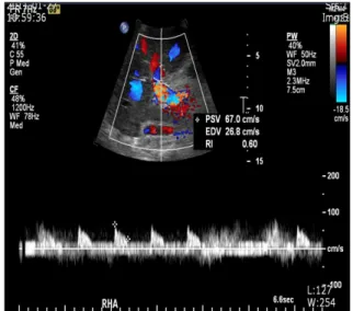
Fig. 1. Postoperative Doppler sonogram demonstrating normal pulsatile flow of the right hepatic artery with a resistive index of 0.6.
Fig. 2. (A) Postoperative computed tomography (CT) scan revealing a 6 cm multilobulating ill-defined heterogeneous low density lesion in the right posterior section of the liver. (B) The size of the lesion decreased on follow up CT scan.
The vascular anatomy of the liver is varied. The
"normal" anatomy i.e., the common hepaticartery origi- nating from the celiac trunk and branching close to the pa- renchyma into the left and right hepatic arteries is seen in
55 to 80% of the cases [2-5]. Under variant patterns, the lobes may receive blood supply from the superior mesen- teric artery (SMA), left gastric artery, aorta, or other viscer- al branches. These vessels may be accessory, occurring in addition to the normal arterial supply, or replaced, repre- senting the primary arterial supply to the lobe. It is im- portant that extrahepatic arteries are identified precisely at the time of graft procurement to avoid injuries that might compromise liver function. Thus, the presence of all arteries that are accessory or replaced must be demon- strated. Whether an individual vessel is accessory or re- placed is not always determined, because the intrahepatic branches are not dissected. In 42% of cases of variant hep- atic arterial anatomy a reconstruction was required before liver transplant [2-5]. We present a case where the ac- cessory right hepatic artery of the liver was ligated leading to postoperative liver abscess formation in the posterior section of the liver graft.
Young-Dong Yu, et al
248 thesurgery.or.kr
CASE REPORT
A forty-seven-year-old female patient diagnosed with cryptogenic liver cirrhosis underwent orthotopic cadav- eric liver transplantation due to altered mentality. Her model for end stage liver disease score was 33. During the operation, the donor graft showed a variant of the hepatic artery anatomy where anaccessory right hepatic artery arose from the SMA presumably supplying the posterior section of the liver graft. This artery was accidentally transected during procurement. At the back table, anasto- mosis of the accessory artery, the size of which was 1 mm, with the gastroduodenal artery (GDA) or splenic artery was not possible due to size discrepancy. Since the back bleeding test using organ preservation fluid (Custodiol HTK solution, Dr. Franz Köhler Chemie GmbH, Ben- sheim, Germany) was good, the artery was ligated.
Although the initial aspartateaminotransferase and alani- neaminotransferase levels were 2,871 IU/L and 1,539 IU/L, respectively, they began to decrease postoperatively. The postoperative doppler sonogram demonstrated normal wave form of the right and left hepatic arteries with a re- sistive index of 0.6 (Fig. 1). On the tenth postoperative day, an abdominal computed tomography (CT) scan among other studies was performed due to persistent fever and positive blood cultures for enterococci. The CT scan re- vealed a 6 cm multilobulating low attenuating lesion in the right posterior section of the liver (Fig. 2A). With the im- pression of liver abscess, the patient underwent con- servative treatment including antibiotic therapy using vancomycin and levofloxacin. On follow-up CT scans, the size of the lesion decreased without any draining proce- dure (Fig. 2B).
DISCUSSION
Michels' classic autopsy series of 200 dissections, pub- lished in 1966, defined the basic anatomic variations in hepatic arterial supply and has served as the benchmark for all subsequent contributions in this area. Variant pat- terns occurred in 45% of cases, and arteries could be de- fined as accessory or replaced because dissection was car-
ried into the liver substance. Michels' motivation was to maximize the database for surgeons performing proce- dures in and around the portahepatis, to avoid injury to vascular and ductal structures [2].
Complications associated with hepatic artery re- constructions are one of the major causes of graft loss and mortality after orthotopic liver transplantation (OLT) [6].
Hepatic artery complications after OLT include hepatic ar- tery thrombosis, hepatic artery stenosis, hepatic artery pseudoaneurysm, and hepatic artery fistula. The early complications of hepatic artery are usually caused by tech- nical problems. The late complications of hepatic artery are usually associated with hypercoagulable state, over transfusion of platelets and fresh frozen plasma during the surgery, severe rejection, and bileleakage [6]. The hepatic artery is relatively small (3 to 6 mm in diameter in adults) and has a very fragile intima that requires a careful atrau- matic manipulating technique during reconstruction [7].
The anatomical variations, diameter and length of hepatic artery, and injury to vessels including prolonged clamping of hepatic artery, kinking of a long artery, and hematoma of the artery wall from improper flushing afterclamping during operation, and the quality of recipient vessels and mismatch between donor and recipient arterial vessels should be carefully considered and managed preopera- tively and intraoperatively [6,7].
The detection of accessory hepatic arteries is an im- portant issue during liver harvesting procedures [3,8]. The most frequent is the presence of an accessory artery sup- plying the left hepatic lobe from the left gastric artery [2].
This variation is not by itself an indication for vascular re- construction [3,8]. What is important is the detection of an additional vessel supplying the right liver lobe from the SMA, which is also one of the most common anatomic var- iations of the donor hepatic artery in liver transplantation [2,3,8]. Most typically, this artery runs backward from the head of the pancreas to the rear portion of the hep- atoduodenal ligament. This topographic pattern mayof- ten be difficult to diagnose; it is frequently responsible for inadvertent damage to the arterial system during the har- vesting procedure [3,8]. Several methods for reconstruc- tion have been described [6-8]. In a report by Di Benedetto et al. [8], after preserving the SMA stump (with the right
Liver abscess developed after cadaveric liver transplantation
thesurgery.or.kr 249
hepatic artery branching from the SMA) originating from the aorta during donor procurement, they proposed a method in which the recipient common hepatic artery is anastomosed to the distal end of the donor SMA, and the proximal end of the donor SMA is anastomosed to the do- nor proper hepatic artery. Since the accessory right hepatic artery was inadvertently transected away from the origin of the SMA during procurement, in addition to size dis- crepancy between the accessory right hepatic artery and GDA or splenic artery, reconstruction using the above methods was not possible.
In a study by Ikegami et al. [9], during living-related liv- er transplantation (LRLT), where multiple graft arteries were present, both were anastomosed when the largest two were almost the same in diameter. When differences in diameter were noted among the graft arteries, the thick- est one was reconstructed first. When pulsatile blood flow- ed from the stumps that had not been anastomosed the re- maining arteries were not anastomosed. When pulsatile bleeding from the nonanastomosed stumps was not ob- served after rearterialization of the largest artery, all were anastomosed to the recipient hepatic arteries. They con- cluded that although several hepatic arteries may supply the potential allograft in LRLT, it is not always necessary to reconstruct all of them [9]. In the present case, back bleed- ing was tested using perfusion fluid. We believe that back bleeding might have been more accurately evaluated after reconstruction of the main hepatic artery.
However, Yanaga et al. [10] reported the clinical course of five patients with partial dearterialization of their hep- atic allografts. One patient died and three others suffered serious morbidity as a direct or indirect result of this complication. They concluded that partial dearterializa- tion of the liver allograft results in a serious and poten- tially life-threatening complication for which preservation of the complete hepatic arterial supply is important, even if this requires reconstruction of the aberrant vessels [10].
In addition, hepatic artery ligation can lead to liver in- farction and subsequent liver abscess [10].
In conclusion, although the liver abscess subsided in our patient, since whether an individual vessel is ac-
cessory or replaced is not always determined, we believe that even small accessory arteries (1 to 2 mm) should be re- constructed whenever possible even if good back bleeding exists to avoid postoperative complications such as liver abscess.
CONFLICTS OF INTEREST
No potential conflict of interest relevant to this article was reported.
REFERENCES
1. Tzakis AG, Gordon RD, Shaw BW Jr, Iwatsuki S, Starzl TE.
Clinical presentation of hepatic artery thrombosis after liv- er transplantation in the cyclosporine era. Transplantation 1985;40:667-71.
2. Michels NA. Newer anatomy of the liver and its variant blood supply and collateral circulation. Am J Surg 1966;
112:337-47.
3. Nelson TM, Pollak R, Herand OJ. Anatomic variants of the celiac, superior mesenteric, and inferior mesenteric ar- teries and their clinical relevance. Clin Anat 1988;1:75-91.
4. Hiatt JR, Gabbay J, Busuttil RW. Surgical anatomy of the hepatic arteries in 1000 cases. Ann Surg 1994;220:50-2.
5. Yang SH, Yin YH, Jang JY, Lee SE, Chung JW, Suh KS, et al.
Establishment of a guideline for the safe management of anatomical hepatic artery variations while performing ma- jor hepato-pancreatico-biliary surgery. J Korean Surg Soc 2009;76:100-8.
6. Proposito D, Loinaz Segurola C, Garcia Garcia I, Jimenez C, Gonzales Pinto I, Gomez Sanz R, et al. Role of anatomic variations and methods of hepatic artery reconstruction in the incidence of thrombosis following liver transplan- tation. Ann Ital Chir 2001;72:303-14.
7. Jones RM, Hardy KJ. The hepatic artery: a reminder of sur- gical anatomy. J R Coll Surg Edinb 2001;46:168-70.
8. Di Benedetto F, Cautero N, De Ruvo N, Masetti M, Montalti R, Gerunda GE, et al. A new reconstruction of the accessory donor right hepatic artery with interposition of the SMA in liver transplantation. Surgery 2006;140:835.
9. Ikegami T, Kawasaki S, Matsunami H, Hashikura Y, Nakazawa Y, Miyagawa S, et al. Should all hepatic arterial branches be reconstructed in living-related liver trans- plantation? Surgery 1996;119:431-6.
10. Yanaga K, Tzakis AG, Starzl TE. Partial dearterialization of the liver allograft. Transpl Int 1990;3:185-8.