ISSN 2234-3806 • eISSN 2234-3814
http://dx.doi.org/10.3343/alm.2015.35.2.226
Quantitative Measurement of Serum MicroRNA-21 Expression in Relation to Breast Cancer Metastasis in Chinese Females
Guinian Wang, M.D.1, Longzi Wang, M.D.2, Sijing Sun, M.S.3, Juan Wu, B.S.1, and Qinglu Wang, Ph.D.4
Departments of Laboratory Medicine1 and Surgical Medicine3, The First Hospital of Zibo City; Department of Pharmacy2, Zibo Vocational Institute; Key Laboratory of Biomedical Engineering4, Shandong Wanjie Medical College, Zibo, China
Background: Breast cancer is the most common type of cancer in females. Aberrant ex- pression of microRNA-21 (miR-21) has previously been reported in breast cancer tissue.
The aim of this study was to investigate expression levels of serum miR-21 in breast can- cer patients and evaluate its prognostic value in Chinese females.
Methods: Real-time quantitative (RQ)-PCR was used to analyze miR-21 expression in ar- chived serum, tumor tissue, and adjacent normal tissue from 549 participants (326 with breast cancer, 223 without breast cancer). We also analyzed associations between serum miR-21 expression and breast cancer subtypes and patient prognosis. Recurrence and survival were analyzed by using the multivariate Cox proportional hazards model.
Results: Expression of miR-21 was significantly higher in breast cancer tissues compared with normal adjacent breast tissues (P <0.001). The 2-ΔΔCt values for serum miR-21 in breast cancer patients versus healthy controls were 9.12±3.43 and 2.96±0.73, respec- tively. Multivariate Cox proportional hazards model suggested that serum miR-21 expres- sion was an independent poor prognostic factor for both recurrence (hazard ratio [HR]=
2.942; 95% confidence interval [CI]=1.420-8.325; P =0.008) and disease-free survival (HR=2.732; 95% CI=1.038-7.273, P =0.003) in breast cancer.
Conclusions: Increased serum miR-21 expression level was correlated with poor progno- sis of breast cancer patients, indicating that serum miR-21 may be a novel prognostic marker for recurrence and survival of breast cancer patients before resection.
Key Words: Biomarker, Breast cancer, Prognosis, Serum miR-21
Received: August 4, 2014 Revision received: October 1, 2014 Accepted: December 29, 2014 Corresponding author: Guinian Wang Department of Laboratory Medicine, The First Hospital of Zibo City, No.4, Emeishan East Road Boshan District Zibo, Shandong 255200, China
Tel: +86-533-4252214 Fax: +86-533-4253406 E-mail: [email protected]
© The Korean Society for Laboratory Medicine This is an Open Access article distributed under the terms of the Creative Commons Attribution Non-Commercial License (http://creativecom- mons.org/licenses/by-nc/3.0) which permits unrestricted non-commercial use, distribution, and reproduction in any medium, provided the original work is properly cited.
INTRODUCTION
In females, breast cancer (BC) is the most common type of can- cer and the second leading cause of death from cancer [1].
Though recent studies have focused on elucidating the molecu- lar mechanisms of BC, the exact mechanisms of BC initiation and progression remain unclear. Currently, conventional clinico- pathological factors are insufficient for accurate prediction of BC patient outcomes. Therefore, there is a need for development of novel markers that predict the course of BC and that can poten-
tially be used to develop treatment strategies.
MicroRNAs (miRNAs) are small (18-25 nucleotides long) non- coding RNA molecules that modulate the activity of specific messenger RNA (mRNA) targets involved in fundamental biolog- ical processes including development, apoptosis, proliferation, and differentiation. Following the discovery of lin-4 in Caenorhab- ditis elegans [2], thousands of miRNAs have been identified in a variety of organisms. Many studies have demonstrated that sev- eral miRNAs correlate with clinical and biological features of tu- mors, including tissue type, and are aberrantly expressed in vari-
ous types of cancer [3, 4]. Many recent studies have identified expression of microRNA-21 (miR-21) in the serum of BC pa- tients. Quantitative measurement of miR-21 expression has been previously reported in BC tissue [5]. These findings suggest that tumor-specific miRNAs in serum are derived not only from circu- lating blood cells but also from cancer cells. The miR-21 is a novel and promising biomarker for detection and diagnosis of biliary tract cancer [6, 7]. In particular, miR-21 has emerged as an oncogenic miRNA in BC, as it is the most consistently up- regulated miRNA in a wide range of cancers [8].
Here, we report that miR-21 plays a role in BC progression and has a prognostic value in a Chinese population. We investi- gated neoadjuvant chemotherapy treatment with either trastu- zumab or lapatinib in BC patients based on molecular factors such as estrogen receptor (ER) status, human epidermal growth factor receptor 2 (HER2) positivity, or triple negative status. To address these issues, our study quantitatively measured serum miR-21 expression and determined its relationship to BC prog- nosis, including recurrence and survival, after resection.
METHODS
1. Patients and samples
We examined 326 Chinese patients with invasive ductal carci- noma of the breast who underwent surgery between July 2009 and May 2014 at the First Hospital of Zibo City, China. Tissue and serum samples were collected from these 326 BC patients (154 patients in the trastuzumab arm and 172 patients in the lapatinib arm). For the validation study, serum samples were col- lected from 223 healthy volunteers. Healthy volunteers were confirmed to be free of malignant disease for >2 yr. Matched BC tumors and adjacent normal tissues were obtained from the First Hospital of Zibo City. Immediately after resection, tissue samples were frozen in liquid nitrogen and stored at -80°C until examined. Whole blood samples were collected in Serum Sepa- rator Tubes (Becton Dickinson, Franklin Lakes, NJ, USA) during routine sampling from 223 healthy volunteers and 326 BC pa- tients prior to treatment. Serum was harvested by centrifugation at 1,710 g for 10 min at 20°C after allowing blood to clot for 30 min at 35°C. Fresh serum samples were then aliquoted into ep- pendorf tubes and immediately frozen at -80°C until total RNA was isolated.
BC was staged according to 7th edition of the Cancer Staging Manual from the American Joint Committee on Cancer [9]. Re- currence was defined as a new tumor observed in the breast af- ter initial curative resection. Progression was defined as disease
with a higher tumor-node-metastasis stage at relapse. Diagno- ses were independently confirmed as primary BC for all patients using histopathological records (BC 1/2, early onset [BRCA1/2]
expression; ER/HER2 positivity; lymph node status; etc.). The Ethics Committee of the First Hospital of Zibo City approved this study, and written informed consent was obtained from all par- ticipants. For clinical results, miR-21 measurements were blinded. Detailed patient characteristics are listed in Table 1.
There were no differences in baseline characteristics between BC patients and normal subjects. Most of the patients with inva- sive ductal carcinoma of the breast had advanced-stage disease.
2. Isolation of total RNA and real-time quantitative (RQ)-PCR analysis
Total RNA was extracted from tumors, adjacent normal tissues, and serum samples using a commercial kit designed to isolate small molecular weight nucleic acids (miRNeasy Mini Kit; Qia- gen, Venlo, Limburg, Netherlands). We performed RQ-PCR us- ing the TaqMan miRNA Reverse Transcription Kit (Applied Bio- systems, Foster City, CA, USA), which utilized stem-loop reverse primers. Reactions were carried out in an ABI Prism 7000 PCR system (Applied Biosystems). U6 small nuclear (sn) RNA was used as an endogenous control. The PCR conditions included initial incubation at 50°C for 2 min and denaturing at 95°C for 10 min, followed by 40 cycles of 95°C for 15 sec and 60°C for 60 sec [10]. All samples were measured in duplicate.
To estimate miR-21 expression, an expression index (EI) was calculated using the formula: 1,000×2(-ΔCt), where ΔCt is the dif- ference in the threshold cycle (Ct) values between the target and the endogenous control used for analysis in the RQ-PCR results (ΔCt=CtmiR-21-CtU6 snRNA). The fold change in miR-21 ex- pression for each BC sample was calculated relative to the aver- Table 1. Baseline patient characteristics
N (%)*
Breast cancer (N=326) Healthy controls (N=223) Age (yr)
Median (range) 47 (27-65) 42 (25-56)
Clinical stage
I 109 (34)
II 151 (46)
III 66 (20)
Histological type
Ductal 326 (100)
*All subjects are females.
age expression in normal control samples. Fold changes were calculated based on the Ct values using the following formula:
RQ=2-ΔΔCt, where ΔΔCt=(CtmiR-21-CtU6 snRNA)tumor-(CtmiR-21-CtU6 snRNA) mean normal.
3. Statistical analysis
All statistical analyses were performed by using SPSS version 18.0 (SPSS Inc., Chicago, IL, USA). Values are presented as me- dians and ranges. Associations between the selected comparing groups were assessed by using Pearson’s chi-square tests. Sur- vival rates were calculated actuarially according to the Kaplan- Meier method, and survival curves were plotted; statistical dif- ferences were analyzed by using log-rank tests. The multivariate Cox proportional hazards model was used to assess the inde- pendent prognosis effect of serum miR-21 expression for risk of recurrence and disease-free survival. BC subtype and pathology factors included in the analysis were involved patient hormone positivity including HER2 positivity, triple negative status, and ER and progesterone receptor status. All tests comparing groups were 2-tailed, and P <0.05 was considered statistically significant.
RESULTS
1. miR-21 is significantly up-regulated in BC tissues
We analyzed the expression levels of miR-21 in paired BC tis- sues and normal adjacent breast tissues from 326 BC patients.Analysis by RQ-PCR revealed that miR-21 expression was
higher in BC tissues (median expression level=687.21; range=
98.95-4,981.31) than in normal adjacent breast tissues (median expression level=59.24; range=4.95-178.77; P <0.001).
2. Serum miR-21 expression in BC
Expression of miR-21 was detected in serum from BC patients, with expression levels that varied widely (EI values ranging from 25.4 to 9,230.2). The 2-ΔΔCt median expression of miR-21 was 0.67±0.15. We compared serum miR-21 expression between BC patients (n=326) and healthy controls (n=223). The 2-ΔΔCt values for BC patients versus healthy controls were 9.12±3.43 and 2.96 ±0.73, respectively. As shown in Fig. 1, association between serum miR-21 expression and tumor-node-metastasis stage were found to be up-regulated in BC patients (P =0.033).
3. Prognostic value of serum miR-21 expression in BC
We first performed survival analysis for all patients, and then for BC subtypes with differences in hormone positivity including ER status, HER2 positivity, or triple negative status (Table 2). The relationship between miR-21 expression and prognosis (recur- rence or survival) varied by disease stage. We investigated the serum levels of miR-21 in 326 BC patients before neoadjuvant chemotherapy treatment along with clinicopathological character- istics and pathological complete responses to trastuzumab or lapatinib-based neoadjuvant therapy. Our subgroup analysis re- vealed that recurrence was significantly different between pa- tients with high versus low miR-21 expression.To determine whether elevated serum miR-21 was a potential
Fig. 1. Association between serum microRNA-21 (miR-21) expres- sion and tumor-node-metastasis (TNM) stage. Significant: 2-sided Pearson’s chi-square test, P <0.05.
Abbreviations: miR-21, microRNA-21; TNM, tumor-node-metastasis.
90 80 70 60 50 40 30 20 10 0
I (N=109) II (N=157) III (N=66) TNM stage
P <0.05
Percentage (%)
miR-21 low expression miR-21 high expression
Fig. 2. Kaplan-Meier curves for recurrence-free survival in 326 breast cancer patients. Recurrence-free survival was significantly lower in patients with high serum miR-21 expression than in patients with low serum miR-21 expression.
1.0
0.8
0.6
0.4
0.2
0
0 10 20 30 40 50 60 Months after surgery
Low serum miR-21 expression (n=86)
High serum miR-21 expression (n=240) Log rank P =0.026
Recurrence-free survival
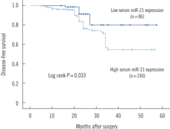
predictor of prognosis in BC patients, recurrence-free and dis- ease-free survival were analyzed, with the BC patients divided into subgroups according to serum miR-21 expression level. Ka- plan-Meier analysis demonstrated differences in the recurrence- free and disease-free survival curves in patients with high versus low expression of serum miR-21. As shown in Fig. 2, the high serum miR-21 expression group had a significantly shorter re- currence-free survival than in the low serum miR-21 expression group (P =0.026). Moreover, disease-free survival was signifi- cantly shorter in the high serum miR-21 expression group than in the low serum miR-21 expression group (P =0.033) (Fig. 3).
The multivariate Cox proportional hazards model indicated that serum miR-21 expression was an independent poor prog- nostic factor for both recurrence (HR=2.942; 95% CI=1.420- 8.325; P =0.008) and disease-free survival (HR=2.732; 95%
CI =1.038-7.273; P =0.003) in HER2-positive BC patients re- ceiving neoadjuvant therapy (Table 3).
DISCUSSION
In this study, we examined expression of miR-21 in tissues and serum from 326 BC patients and 223 healthy controls. We found that serum levels of miR-21 were significantly higher in BC pa- tients than in controls. This is in contrast to a study by Mar-Agu- ilar et al. [11] that found similar levels of miR-21 expression in BC patients and controls. Similarly, when Chan et al. [12] re- ported the 20 miRNAs that were most significantly differentially expressed in BC tumors, they found that only seven miRNAs
were overexpressed in both tumors and serum, and miR-21 was not identified as the most important diagnostic marker.
Also, miR-21 levels in serum showed a number of small differ- ences between BC patients who developed metastasis and BC patients who remain metastasis-free [12].
Notably, miR-21 is one of the most frequently studied onco- genic miRNAs (onco-miRNAs). Although a direct correlation between aberrant expression of miR-21 and BC has been previ- ously demonstrated, it is not clear whether suppression of miR- 21 alone will affect tumorigenesis [13]. In this report, we showed that miR-21expression was significantly higher in BC tissues Fig. 3. Kaplan-Meier curves for disease-free survival in 326 breast
cancer patients. Disease-free survival was significantly lower in pa- tients with high serum miR-21 expression than in patients with low serum miR-21 expression.
1.0
0.8
0.6
0.4
0.2
0
0 10 20 30 40 50 60 Months after surgery
Low serum miR-21 expression (n=86)
High serum miR-21 expression (n=240) Log rank P =0.033
Disease-free survival
Table 2. Associations between serum miR-21 expression and clini- copathological features
Variables
miR-21 expression level
P value High expression
(N=240, %) Low expression (N=86, %) Age (yr)
≥ 45 138 (71) 56 (29) 0.218
< 45 102 (77) 30 (23)
Tumor tissue (cm)
≥ 2 94 (75) 32 (25) 0.645
< 2 146 (73) 54 (27)
TNM stage
I 77 (71) 32 (29)
II 121 (80) 30 (20) 0.033*
III 42 (64) 24 (36)
BRCA1/2 status
Negative 235 (74) 83 (26) 0.471
Positive 5 (63) 3 (37)
ER status
Negative 75 (66) 38 (34) 0.031*
Positive 165 (77) 48 (23)
PR status
Negative 105 (70) 45 (30) 0.172
Positive 135 (77) 41 (23)
HER2 status
Negative 67 (66) 34 (34) 0.046*
Positive 173 (77) 52 (23)
Lymph node status
Invasion 177 (78) 51 (22) 0.012*
No invasion 63 (64) 35 (36)
*Significant: 2-sided Pearson’s chi-square test, P <0.05.
Abbreviations: miR-21, microRNA-21; BRCA1/2, breast cancer 1/2, early onset; ER, estrogen receptor; HER2, human epithelial growth factor recep- tor 2; PR, progesterone receptor; TNM, tumor-node-metastasis.
than in normal adjacent breast tissues (Table 1).
The miR-21 gene is located on chromosome 17q23.2, which is inside the common fragile site FRA17B. This region is fre- quently amplified in breast and lung cancer [14]. In BC, miR- 21 is an onco-miRNA. Elevated miR-21 expression may facili- tate tumor progression, and miR-21 expression may be up-reg- ulated by transforming growth factor-β [15]. This study provides a comprehensive investigation of the association between serum miR-21 and BC prognosis in Chinese females. We found an as- sociation between serum miR-21 expression and prognosis (re- currence and survival) that seemed to vary by disease stage.
Patients with high serum miR-21 expression had an increased risk of disease progression compared to patients with low serum miR-21 expression (Table 2).
There is a large amount of evidence indicating that miR-21 is associated with regulation of proliferation and differentiation during development. Also, it is possible to develop treatment strategies by targeting miR-21 [16]. Serum miRNA levels show a number of small differences in females who later develop can- cer versus those who remain cancer-free. Microarray analysis of miRNAs in the serum of BC patients has only recently been re- ported. Aberrantly expressed miR-21 in the blood of BC patients is occasionally reported, and expression of miR-21 and miR- 146a in plasma samples from BC patients might be useful for BC diagnosis in Indian females [17]. Our results show that ex- pression of serum miR-21 is up-regulated in BC patients com- pared to healthy controls in a population of Chinese females.
High expression of serum miR-21 may contribute to BC patho- genesis (P =0.033) (Fig. 1) and development of invasion and lymph node metastasis (P =0.012) (Table 2).
Although the impact of miR-21 on BC prognosis remains controversial, a few studies have reported promising associa- tions with miR-21 expression levels that could make this a novel potential biomarker for BC prognosis. Previously, Gao et al. [18]
reported that serum miR-21 was a more sensitive BC marker than cancer antigen (CA)15-3 or carcinoembryonic antigen; in particular, miR-21 was a potential tumor marker for the diagno- sis of early-stage BC. Also, miR-21 levels before and after che- motherapy demonstrated a detectable association with overall survival that was independent of anti-HER2 therapy. Investiga- tion of serum miR-21 has yielded encouraging data that sug- gests that the clinical relevance of miR-21 should be confirmed in a larger BC patient cohort [19]. When BC is suspected, clini- cians may check factors such as CA15-3, Ki-67, cytokeratin 8/18/19, BRCA1/2, and ER/HER2, which are still the main diag- nostic biomarkers [20, 21]. The BRCA1 and BRCA2 tumor sup- pressor genes are strong predictors of BC development; muta- tions in BRCA 1 and 2 are inherited in an autosomal dominant manner and play important roles in BC risk [22]. Reports have indicated that miR-21 functions as an oncogene and modulates tumorigenesis through regulation of genes such as bcl-2; there- fore, miR-21 may serve as a novel therapeutic target [23].
Though new technologies are emerging to improve the speci- ficity, stability, and efficiency of miRNA delivery and therapy, the final outcomes of this strategy remain uncertain [24]. Currently, clinicians do not have an effective molecular biology marker for BC. Expression profiles of circulating miRNAs may yield promis- ing biomarkers for diagnosis and assessment of the prognosis of cancer patients. Sensitive techniques allow the expression levels of many miRNAs to be determined, and this information can be Table 3. Prognosis of serum miR-21 for recurrence or disease-free survival in breast cancer
Features Recurrence-free survival Disease-free survival
HR 95% CI P value HR 95% CI P value
miR-21 expression 3.942 1.420-8.345 0.008* 2.932 1.038-7.273 0.003*
Age 0.951 0.836-4.571 0.746 1.147 0.726-3.859 0.289
Tumor size 1.135 0.783-6.901 0.461 0.973 0.674-5.730 0.307
Lymph node status 2.745 0.835-5.641 0.091 1.987 0.469-3.085 0.067
TNM stage 3.023 0.895-4.793 0.023* 2.075 0.906-5.673 0.031*
ER status 1.784 0.672-5.458 0.169 1.304 0.587-4.981 0.151
PR status 1.024 0.468-8.697 0.125 1.271 0.398-7.106 0.286
HER2 status 2.678 1.563-7.025 0.019* 1.948 0.976-6.597 0.025*
*Significant: 2-sided Pearson’s chi-square test, P <0.05.
Abbreviations: miR-21, microRNA-21; CI, confidence interval; ER, estrogen receptor; HER2, human epithelial growth factor receptor 2; HR, hazard ratio;
PR, progesterone receptor; TNM, tumor-node-metastasis.
used for diagnostic purposes [25]. The mechanism underlying miRNA-21 stability is still being investigated for BC detection [26].
The utility of miRNA profiles as potential diagnostic or prog- nostic markers for BC has been gaining interest [27]. Our study had some limitations, including small sample size and a limited ability to generalize our results since all our patients were Chi- nese females. Despite these limitations, our study provided initial data about the up-regulation of serum miR-21 in BC patients and suggested the diagnostic value of serum miR-21 in BC pa- tients with metastasis. Serum miR-21 may have clinical utility for monitoring and follow-up of BC patients with metastasis. Clearly, our results should be further validated by a prospective study in a multicenter clinical trial. The ideas in this study should be fur- ther explored by studies with larger sample numbers.
In conclusion, we found that serum miR-21 was up-regulated in BC patients, and increased miR-21 expression levels corre- lated with poor prognosis of BC patients. Our findings indicate that serum miR-21 may serve as a novel potential prognostic marker for recurrence and survival of BC patients before resection.
Authors’ Disclosures of Potential Conflicts of Interest
No potential conflicts of interest relevant to this article were re- ported.
Acknowledgments
This study was supported in part by the Research Program Foundation (No.2013) of the First Hospital of Zibo City, and the Natural Scientific Foundation of Shandong Province (grant No.
ZR2010CQ031).
REFERENCES
1. Jemal A, Bray F, Center MM, Ferlay J, Ward E, Forman D. Global cancer statistics. CA Cancer J Clin 2011;61:69-90.
2. Wightman B, Ha I, Ruvkun G. Posttranscriptional regulation of the het- erochronic gene lin-14 by lin-4 mediates temporal pattern formation in C. elegans. Cell 1993;75:855-62.
3. Michael MZ, O’Connor SM, van Holst Pellekaan NG, Young GP, James RJ. Reduced accumulation of specific microRNAs in colorectal neopla- sia. Mol Cancer Res 2003;1:882-91.
4. Calin GA, Sevignani C, Dumitru CD, Hyslop T, Noch E, yendamuri S, et al. Human microRNA genes are frequently located at fragile sites and genomic regions involved in cancers. Proc Natl Acad Sci USA 2004;
101:2999-3004.
5. Wang F, Zheng ZG, Guo J, Ding X. Correlation and quantitation of mi- croRNA aberrant expression in tissues and sera from patients with
breast tumor. Gynecol Oncol 2010:119:586-93.
6. Cortez MA, Bueso-Ramos C, Ferdin J, Lopez-Berestein G, Sood AK, Calin GA. MicroRNAs in body fluids--the mix of hormones and biomark- ers. Nat Rev Clin Oncol 2011;8:467-77.
7. Kishimoto T, Eguchi H, Nagano H, Kobayashi S, Akita H, Hama N, et al.
Plasma miR-21 is a novel diagnostic biomarker for biliary tract cancer.
Cancer Sci 2013;104:1626-31.
8. Zhao H, Shen J, Medico L, Wang D, Ambrosone CB, Liu S. A pilot study of circulating miRNAs as potential biomarkers of early stage breast can- cer. PLos One 2010;5:e13735.
9. Edge SB, Byrd DR, Carducci M, Compton CC, Fritz AG, Greene FL, et al.
eds. AJCC cancer staging manual. 7th ed. New York: Springer-Verlag, 2009.
10. Asaga S, Kuo C, Nguyen T, Terpenning M, Giuliano AE, Hoon DS. Direct serum assay for microRNA-21 concentrations in early and advanced breast cancer. Clin Chem 2011;57:84-91.
11. Mar-Aguilar F, Mendoza-Ramíreza JA, Malagón-Santiago I, Espino-Silva PK, Santuario-Facio SK, Ruiz-Flores P, et al. Serum circulating microR- NA profiling for identification of potential breast cancer biomarkers. Dis Markers 2013;34:163-9.
12. Chan M, Liaw CS, Ji SM, Tan HH, Wong CY, Thike AA, et al. Identifica- tion of circulating microRNA signatures for breast cancer detection. Clin Cancer Res 2013;19:4477-87.
13. Iorio MV, Ferracin M, Liu CG, Veronesse A, Spizzo R, Sabbioni S, et al.
MicroRNA gene expression deregulation in human breast cancer. Can- cer Res 2005; 65:7065-70.
14. Calin GA and Croce CM. Chromosomal rearrangements and microR- NAs: a new cancer link with clinical implications. J Clin Invest 2007;117:
2059-66.
15. Qian B, Katsaros D, Lu L, Preti M, Durango A, Arisio R, et al. High miR- 21 expression in breast cancer associated with poor disease-free sur- vival in early stage disease and high TGF-ß1. Breast Cancer Res Treat 2009;117:131-40.
16. Yan LX, Wu QN, Zhang Y, Li YY, Liao DZ, Hou JH, et al. Knockdown of miR-21 in human breast cancer cell lines inhibits proliferation, in vitro migration and in vivo tumor growth. Breast Cancer Res 2011;13:R2.
17. Kumar S, Keerthana R, Pazhanimuthu A, Perumal P. Overexpression of circulating miRNA-21 and miRNA-146a in plasma samples of breast cancer patients. Indian J Biochem Biophys 2013;50:210-4.
18. Gao J, Zhang Q, Xu J, Guo L, Li X. Clinical significance of serum miR- 21 in breast cancer compared with CA153 and CEA. Chin J Cancer Res 2013;25:743-8.
19. Müller V, Gade S, Steinbach B, Loibl S, von Minckwitz G, Untch M, et al.
Changes in serum levels of miR-21, miR-210, and miR-373 in HER2- positive breast cancer patients undergoing neoadjuvant therapy: a translational research project within the Geparquinto trial. Breast Cancer Res Treat 2014;147:61-8.
20. Tamaki K, Ishida T, Tamaki N, Kamada Y, Uehara K, Miyashita M, et al.
Analysis of clinically relevant values of Ki-67 labeling index in Japanese breast cancer patients. Breast Cancer 2014;21:325-33.
21. Leivonen SK, Mäkelä R, Ostling P, Kohonen P, Haapa-Paananen S, Kleivi K, et al. Protein lysate microarry analysis to identify microRNAs regulat- ing estrogen receptor signaling in braest cancer cell lines. Oncogene 2009;28:3926-36.
22. Fackenthal JD and Olopade Ol. Breast cancer risk associated with BR- CA1and BRCA2 in diverse populations. Nat Rev Cancer 2007;7:937-48.
23. Si ML, Zhu S, Wu H, Lu Z, Wu F, Mo YY. miR-21-mediated tumor growth.
Oncogene 2007;26:2799-803.
24. Li J, Shen L, Xiao XG, Fang L. MicroRNAs in breast cancer and breast cancer stem cells and their potential for breast cancer therapy. Chin
Med J 2013;126:2556-63.
25. Tsujiura M, Ichikawa D, Komatsu S, Shiozaki A, Takeshita H, Kosuga T, et al. Circulating microRNAs in plasma of patients with gastric cancers.
Br J Cancer 2010;102:1174-9.
26. Wu Q, Wang C, Lu Z, Guo L, Ge Q. Analysis of serum genome-wide mi- croRNAs for breast cancer detection. Clin Chim Acta 2012;413:1058-65.
27. Fu SW, Chen L, Man YG. miRNA biomarkers in breast cancer detection and management. J Cancer 2011;2:116-22.