Annals of Surgical Treatment and Research 277
pISSN 2288-6575 • eISSN 2288-6796 https://doi.org/10.4174/astr.2017.93.5.277 Annals of Surgical Treatment and Research
CASE REPORT
Cutaneous metastasis: a rare phenomenon of colorectal cancer
Dan Yang Wang, Feng Ye, Jian Jiang Lin, Xiao Xu1
Divisions of Colorectal Surgery, 1Hepatobiliary and Pancreatic Surgery, Department of Surgery, The First Affiliated Hospital, College of Medicine, Zhejiang University, Hangzhou, China
INTRODUCTION
Cutaneous metastases are discovered in only about 1.3%
of cases at the time of presentation of the primary tumor [1].
They may be the first manifestation of metastatic spread of an internal malignancy. Cutaneous metastases are com monly secondary to malignant melanoma, cancer of lung and breast.
The most common metastatic site of colorectal adenocarci noma is liver and lung. Meanwhile, cutaneous metastases also occur in 4%–6.5% of these metastatic cases, with abdominal skin being the most common site [2]. This often indicates a poor prognosis, but there is no standardized treatment so far.
CASE REPORT
A 76-year-old female was seen in March 2016, presenting with a 10-day history of hematochezia and change in bowel habit, including tenesmus, constipation, and defecating too
frequently. Digital rectal examination showed a huge mass, 6 cm from the anal margin, occupying the whole circle of the rectum. Abdominal CT, chest CT, pelvic MRI, and PET/CT demonstrated rectal tumor, multiple pelvic enlarged lymph nodes, no lung or liver metastasis, 2 suspicious nodules in the right adrenal gland and bottom of oral cavity respectively, with high fluorodeoxyglucose metabolism, clinical stage T4aN1Mx.
Colonoscopy failed due to the narrow intestine resulting from the biopsy-proven adenocarcinoma.
Her medical history included hypertension, chronic bron- chitis, and mild Alzheimer disease all of which were well con trolled pharmacologically. According to the guidelines, she should have received neoadjuvant chemotherapy. However, because of the incomplete intestinal obstruction, she under- went a low anterior resection of the rectum (Hartmann). Path- ologic findings showed poorly differentiated ulcerative adeno- carcinoma with lymph node metastasis, intravascular tumor thrombus, with no perineural invasion. Immunohistochemical Cutaneous metastases from colorectal cancer are extremely rare and generally appear several years after diagnosis or resection of the primary tumor. Although this phenomenon is uncommon, it is very important and often indicates a poor prognosis. We present a case of a 76-year-old female patient with multiple cutaneous metastatic nodules on the back, just 1 month after resection of rectal cancer. Unfortunately, the patient gave up the follow-up treatment due to her age and poor physical condition; she died 3 months later. In view of its rarity of occurrence and lack of experience in treatment, we reviewed the literature and report as follows.
[Ann Surg Treat Res 2017;93(5):277-280]
Key Words: Colorectal neoplasms, Neoplasm metastasis, Cutaneous
Reviewed January February March April May June July August September October November December
Received November 7, 2016, Revised January 26, 2017, Accepted February 21, 2017
Corresponding Author: Xiao Xu
Division of Hepatobiliary and Pancreatic Surgery, Department of Surgery, The First Affiliated Hospital, College of Medicine, Zhejiang University, 79, Qinchun Road, Hangzhou, Zhejiang Province 310003, China
Tel: +86-571-87236616, Fax: +86-571-87235128 E-mail: [email protected]
Copyright ⓒ 2017, the Korean Surgical Society
cc Annals of Surgical Treatment and Research is an Open Access Journal. All articles are distributed under the terms of the Creative Commons Attribution Non- Commercial License (http://creativecommons.org/licenses/by-nc/4.0/) which permits unrestricted non-commercial use, distribution, and reproduction in any medium, provided the original work is properly cited.
278
Annals of Surgical Treatment and Research 2017;93(5):277-280
results showed: MSH2(+), MSH6(+), MLH1(+), PMS2(+). The pathological stage was T4aN2Mx. The operation was successful, but postoperative complications like pulmo nary infection, hydrothorax, respiratory failure, and uri nary retention occurred. Fortunately, all of the complications were relieved by conservative treatment.
Curiously, in the postoperative period, we observed a rare pheno menon. One month after surgery, multiple subcutaneous nodules appeared on the patient’s back, hard, isolated, clear boundary, absence of tenderness, and 10–20 mm in size (Fig.
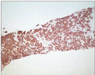
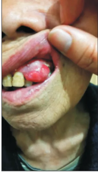
1). Core needle biopsy and histopathology confirmed it to be a metastatic poorly differentiated adenocarcinoma of colorectal origin positive for CK20, and negative for CDX2 and MUC2 (Figs. 2, 3). Meanwhile, one rapidly increasing gingiva nodule was observed as well (Fig. 4), regrettably, the patient refused biopsy. Therefore, we were not sure whether it was a metastatic lesion, but it was highly suspect according to the PET/CT result.
In addition, a small amount of atypical cells was found in the
pleural fluid. The patient gave up follow-up treatment due to her age and poor physical condition; she died 3 months after surgery, or, 2 months after the diagnosis of cutaneous meta- stasis.
DISCUSSION
Cutaneous metastases of cancer are rare, occurring in about 1.3% of cases at the time of presentation of the primary tumor, as previously mentioned. Patterns of cutaneous metastases vary among men and women. In men, melanoma, lung cancer, and colorectal cancer are the most common sites of cutaneous metastases. Breast cancer, colorectal cancer, and melanoma fre- quently metastasize to the skin in women [3]. These meta stases have also been associated with cancers of gastric, eso phageal,
Fig. 1. Multiple subcutaneous nodules on the patient’s back. Fig. 3. Immunohistochemical staining was positive for CK20 (×100).
A B
Fig. 2. Metastatic rectal adenocarcinoma involving skin: poorly differentiated adenocarcinoma, a few glands but largely com
posed of infiltrating nests of tumor cells (A: H&E, ×100; B: H&E, ×400).
Annals of Surgical Treatment and Research 279 prostatic, ovarian, hematologic, laryngeal, palatine-ton sillar,
pancreatic, parotid, thyroid, uterine origin, and miscella neous others.
Cutaneous metastases occur in about 3% of colorectal can- cers [4]. The most frequent sites of cutaneous metastasis from colorectal cancer are abdomen followed by extremities, peri- neum, head, neck, and penis. These metastases generally occur within the first 2 years after resection of the pri mary colorectal tumor and often present simultaneously with metastases to the liver, peritoneum, and lung. It is extremely rare that tumors metastasize to the back and gingiva (not confirmed) just 1 month after surgery, as in our case.
Cutaneous metastases may occur through lymphogenous spread, intravascular dissemination, direct extension of tu mor, and surgical implantation and spread along embryonal rem- nants such as the urachus.
In the majority of the metastases, the diagnosis was based on the morphologic appearances, histomorphology and immu- no histochemistry of the cutaneous lesion, in conjunction with comparison with the primary tumor morphology if it is avail- able. Cutaneous metastases can present in a variety of clinical manifestations, such as a rapidly growing painless der mal or subcutaneous nodules with intact overlying epidermis, or mimic inflammatory dermatosis. Ulceration, nodules, bullae, or fibrotic processes are the most common presentations of cutaneous metastases [2]. While most cancers present as soli- tary nodules, melanomas and carcinomas with an unknown pri mary frequently present as multiple nodules. Although meta stases cells may be more undifferentiated than the pri- mary tumor, careful microscopic examination usually reveals im portant clues. For example, colon cancer may be associated with mucinous cells. When tumors are poorly differentiated
or anaplastic, screening immunohistochemical studies such as CK7, CK20, CK19, and CDX2 can be very helpful [5].
Identi fication of cutaneous metastasis from an internal malig nancy indicates poor prognosis, as it usually reflects wide- spread disease. Survival after diagnosis of cutaneous metastasis ranges from 1 to 34 months [6]. Schoenlaub et al. [7] reported the median survival of cutaneous metastasis patients with colorectal primary tumours was 4.4 months. On the other hand, a retrospective study by Lookingbill et al. [1] showed a median sur vival of 18 months in patients with similar characteristics.
In our case, the patient died 3 months after surgery.
Treatment of metastatic carcinoma of the skin is limited and lacks any standardization strategy. Although management of metastatic colorectal cancer has been based on systemic chemo- therapy, surgical resection in selected patients with metastatic colorectal cancer offers the only possibility for long-term sur vival [8]. For isolated lesions, Nesseris et al. [6] suggested wide local excision and reconstruction. Nevertheless, another research paper proposed typical resection with a 1-cm margin of normal skin. Furthermore, in some areas of high cosmetic impor tance or when therapy is limited to palliation, excisions may be performed with very limited margins [5]. For patients with multiple cutaneous metastases or unresectable lesions, sys temic chemotherapy can be considered. However, there is no optimal chemotherapy regimen as of yet. Radiotherapy, poly chemotherapy, isolated limb perfusion, interferon alpha injec tions, cryotherapy, laser ablation, radiofrequency ablation, imiquimod 5% cream, and oncogene-targeted therapy were be also mentioned in some other studies [9,10].
In conclusion, cutaneous metastasis is a rare but important phenomenon, which should not be ignored, frequently indicates advanced disease and poor prognosis. Early diagnosis will be the key element in its effective management, which requires careful physical examination. Once any change in the skin is noted, further examination should be implemented. More effec tive treatment modalities need further exploration.
CONFLICTS OF INTEREST
No potential conflict of interest relevant to this article was reported.
ACKNOWLEDGEMENTS
This paper supported by Zhejiang Provincial Natural Science Foundation of China (grant number: LY13H030005).
Dan Yang Wang, et al: Cutaneous metastasis from colorectal cancer
Fig. 4. Rapidly increasing gingiva nodule.
280
Annals of Surgical Treatment and Research 2017;93(4):277-280
REFERENCES
1. Lookingbill DP, Spangler N, Helm KF.
Cutaneous metastases in patients with meta static carcinoma: a retrospective study of 4020 patients. J Am Acad Dermatol 1993;29(2 Pt 1):228-36.
2. Hashimi Y, Dholakia S. Facial cutaneous metastasis of colorectal adenocarcinoma.
BMJ Case Rep 2013;2013. pii: bcr2013009875.
http://doi.org/10.1136/bcr-2013-009875.
3. Cidon EU. Cutaneous metastases in 42 patients with cancer. Indian J Dermatol Venereol Leprol 2010;76:409-12.
4. Aravind B, Kumar R, Basnyat P. Cutaneous meta stases secondary to colorectal car- ci noma may not be as ominous as pre- viously thought: a case report and review
of the literature. BMJ Case Rep 2013;2013.
pii: bcr2013008556. https://doi.org/
10.1136/bcr-2013-008556.
5. Wong CY, Helm MA, Kalb RE, Helm TN, Zeitouni NC. The presentation, pathology, and current management strategies of cuta neous metastasis. N Am J Med Sci 2013;5:499-504.
6. Nesseris I, Tsamakis C, Gregoriou S, Ditsos I, Christofidou E, Rigopoulos D.
Cutaneous metastasis of colon adeno car- cinoma: case report and review of the lite- ra ture. An Bras Dermatol 2013;88(6 Suppl 1):56-8.
7. Schoenlaub P, Sarraux A, Grosshans E, Heid E, Cribier B. Survival after cutaneous
metastasis: a study of 200 cases. Ann Dermatol Venereol 2001;128:1310-5.
8. Oh SY, Kim DY, Suh KW. Oncologic outcomes following metastasectomy in colo rectal cancer patients developing dis- tant metastases after initial treatment.
Ann Surg Treat Res 2015;88:253-9.
9. Leong SP, Gershenwald JE, Soong SJ, Schadendorf D, Tarhini AA, Agarwala S, et al. Cutaneous melanoma: a model to study cancer metastasis. J Surg Oncol 2011;103:538-49.
10. Flaherty KT, Puzanov I, Kim KB, Ribas A, McArthur GA, Sosman JA, et al. Inhibition of mutated, activated BRAF in metastatic mela noma. N Engl J Med 2010;363:809-19.