Received January 31, 2018, Revised June 27, 2018, Accepted for publication July 24, 2018
Corresponding author: Jong Hee Lee, Department of Dermatology, Samsung Medical Center, Sungkyunkwan University School of Medicine, 81 Irwon-ro, Gangnam-gu, Seoul 06351, Korea. Tel: 82-2-3410-3549, Fax:
82-2-3410-3869, E-mail: bell711@hanmail.net ORCID: https://orcid.org/0000-0001-8536-1179
This is an Open Access article distributed under the terms of the Creative Commons Attribution Non-Commercial License (http://creativecommons.
org/licenses/by-nc/4.0) which permits unrestricted non-commercial use, distribution, and reproduction in any medium, provided the original work is properly cited.
Copyright © The Korean Dermatological Association and The Korean Society for Investigative Dermatology
Ann Dermatol Vol. 31, No. 1, 2019 https://doi.org/10.5021/ad.2019.31.1.6
ORIGINAL ARTICLE
The Important Factors Associated with Treatment Response in Laser Treatment of Facial Scars:
A Single-Institution Based Retrospective Study
Youngkyoung Lim1, Ji-Hye Park1, Dong-Youn Lee1, Na-Young Hwang2, Soohyun Ahn3, Jong Hee Lee1,4
1Department of Dermatology, Samsung Medical Center, Sungkyunkwan University School of Medicine, 2Statistics and Data Center, Samsung Medical Center, Seoul, 3Department of Mathematics, Ajou University, Suwon, 4Department of Medical Device Management &
Research, SAIHST, Sungkyunkwan University, Seoul, Korea
Background: There is increased interest in laser treatment of facial scars. Objective: To determine the factors associated with treatment response. Methods: We conducted an in- stitution-based retrospective study by including the patients treated with laser for facial scars from 2012 to 2015.
Treatment methods were determined with an algorithm ac- cording to individual scar characteristics. In each treatment session, either a 595-nm pulsed-dye laser or a non-ablative fractional laser was used, often in combination with a corti- costeroid injection. We evaluated treatment responses based on the number of treatment sessions required to reach the treatment endpoint. Data were analyzed using multi- nomial logistic regression analysis to examine the associa- tion between treatment response and various factors of the scar. Results: A total of eighty-four scars were analyzed. The onset of treatment (defined as the period between the injury and treatment initiation), used laser modality, and the loca- tion of the scar were all found to be significantly associated with treatment responses. Early implementation was more
likely to provide better treatment response. Scars on the per- ioral area were more likely to be associated with worse treat- ment response. Conclusion: The important factors for the treatment response in facial scars were the location of the scar and the timing of the initiation of treatment. Such in- formation can be used to predict treatment response and tai- lor the treatment plan to the patient, depending on scar characteristics. (Ann Dermatol 31(1) 6∼13, 2019)
-Keywords-
Face, Laser, Location, Scar
INTRODUCTION
Scarring is an inevitable result of the wound healing proc- ess1. Although scars are not representative of malignant le- sions, they can have significant physical and emotional impacts on human life2-5. Scarring can occur on any parts of the body, but facial scars can seriously impair a pa- tient’s quality of life5. Therefore, there has been a growing interest in preventing and minimizing facial scars in recent years. Various methods such as the use of silicone agents, intralesional steroid injection, 5-fluorouracil, laser therapy, radiation therapy, cryotherapy, bleomycin, and pressure therapy have been proposed6,7. In recent decades, laser therapy has been recognized as one of the most effective treatment methods for scars and many studies have pro- vided evidence for its benefit6,8-10. Different types of lasers such as pulsed-dye laser (PDL), non-ablative fractional la- ser (NAFL), and ablative fractional laser (AFL) have been used for different types of scars1,4,10-13.
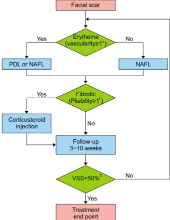
Fig. 1. The treatment algorithm: In one treatment session, either a 595-nm pulsed-dye laser (PDL) or a non-ablative fractional laser (NAFL) was used. Additionally, intralesional corticosteroid injection was employed in case of patients with pliability score
≥1. Patients were asked to return between 3 and 10 weeks subsequent to treatment to check the status of their scar and determine whether continuing treatment was needed or whether the scar reached its treatment endpoint. *Vascularity (normal=0, pink=1, red=2, purple=3); †Pliability (normal=0, supple=1, yielding=2, firm=3, banding=4, contracture=5); ‡The time point at which Vancouver scar scale (VSS) score was reduced by 50% was considered as the treatment endpoint.
Various factors, including patient characteristics and treat- ment-related factors, such as optimal treatment time and treatment modalities, can influence treatment outcomes.
However, standardized guidelines for facial scar treatment have yet to be established. This study was designed to evaluate the factors that may play an important role in scar treatment and offer an optimal guideline for facial scar treatment.
MATERIALS AND METHODS
Patients
We reviewed the clinical data of all the patients treated with laser therapy for facial scarring at the Dermatology Clinic of Samsung Medical Center (SMC) from March 2012 to April 2015. This study was performed by review- ing electronic medical records and clinical photographs of all the patients. The study was approved by the SMC Institutional Review Board (IRB File No. 2016-11-122).
A total of 245 facial scars of patients who did not have a previous keloid history were treated with lasers during the study period. If the time of wound occurrence was un- known, or the Vancouver scar scale (VSS) of each scar was not recorded before and after treatment, the scars were ex- cluded (38 scars) from the investigation. Patients who stopped receiving treatments before reaching their end- point, or those who did not return within three months of their last treatment were excluded from this study (81 scars). Therefore, all the included scars in this study had reached the treatment endpoint. We also excluded cases in which laser treatment sessions were added for other purposes (e.g., melasma treatment, removal of nevus or wart, etc.) during scar treatment (16 scars). Due to an out- break of Middle East Respiratory Syndrome, the out-pa- tient clinic in our center was closed for three months (from June to August) in 2015 and the treatment duration for 26 scars was extended; these scars were also excluded from this study. All patients included in this study were Asians with Fitzpatrick skin types III∼IV.
Treatment
All the treatment sessions were conducted by a single la- ser expert. The optimal laser modality was selected ac- cording to the state of individual scars and selection was automated with a treatment algorithm (Fig. 1). In one treat- ment session, either a 595-nm PDL (V-Beam; Candela Laser Corporation, Wayland, MA, USA) or an NAFL (Fraxel Re:Store DUAL; Solta Medical, Hayward, CA, USA) was used. We used the PDL with a fluence between 7∼9 J/cm2 with a 7-mm spot size and a pulse width of 1.5 milliseconds. The NAFL was set to a 15∼25 mJ energy
level with treatment level 8 and 4∼8 passes. An intrale- sional corticosteroid injection (2.5∼5.0 mg/ml of tri- amcinolone acetonide [TA]) was combined with the laser treatment in cases with a pliability score of 1 or greater.
Not more than 0.1∼0.5 ml of TA was injected per treat- ment session and was always used in conjunction with la- ser treatment. After the laser treatment, a cooling hydrogel was applied to all the patients for eight hours to calm down the treated sites, thus minimizing the risk of occur- rence of possible adverse events after treatment. Patients were instructed to use topical moisturizer three times a day for a week after the treatment and afterward twice a day until their next visit. They were also asked to avoid sun exposure and use a broad-spectrum sunscreen.
Patients were asked to follow-up between 3 and 10 weeks
to check the status of their scar and to determine whether the treatment needed to be continued or if the scar had reached its treatment endpoint.
End point and measurement
Scars were sub-classified as flat, atrophic, hypertrophic, or adhesive prior to treatment. Flat scars were defined as scars without textural irregularity compared with adjacent normal skin. Adhesive scars were defined as any scars (flat, atrophic or hypertrophic) with contractures to the skin underneath it. The treatment effect was evaluated by two independent dermatologists using the VSS14. Each subcategory of the VSS, including vascularity (normal=0, pink=1, red=2, purple=3), pigmentation (normal=0, hy- popigmentation=1, hyperpigmentation=2), pliability (nor- mal=0, supple=1, yielding=2, firm=3, banding=4, con- tracture=5), and height (normal=0, <2 mm=1, 2∼5 mm=
2, >5 mm=3) was measured prior to each treatment ses- sion and at every follow up visit. For atrophic or adhesive scars, the depth of the scar region, when compared with the surrounding tissue, was considered to be the height.
The time point at which a VSS reduction of 50% was ob- served was considered to be the treatment endpoint, which allowed for easier and more objective comparison.
The number of treatment sessions required to reach the treatment endpoint (NTSE) was evaluated. Treatment re- sponse was considered to be good when NTSE was 1 or 2, moderate when NTSE was 3 or 4, poor when NTSE was 5 or 6, and resistant when NTSE was 7 or higher.
Statistical analysis
Previous studies have suggested that the onset of treatment (OT), defined as the period between the injury and treat- ment initiation, is associated with scar prognosis5,15,16. We thought that OT might be one of the important factors in determining the treatment response. Therefore, we div- ided scars into three groups according to OT and inves- tigated the presence of significant differences in other variables. Simple descriptive statistics were used to de- scribe the overall study population by OT groups. Fisher’s exact test was used to compare the groups. The variables showing a significant difference in the comparison of OT groups were considered to be potential confounding variables. Next, considering the nature of the repeated measures and the fact that the treatment response is ordi- nal variable, generalized estimating equations for cumu- lative multinomial logistic regression analysis were performed. Further, a multivariable analysis was per- formed, considering the variables that were significant in the univariable analysis and the potential confounders from the comparison of the OT groups. Bonferroni cor-
rected p-values were calculated. All statistical analyses were performed using R version 3.3.0 (R Development Core Team, 2016). Statistical significance was set at p<
0.05.
RESULTS
Eighty-four scars of 64 patients were analyzed in this study. Most of the patients had a single facial scar (n=48), and 16 patients had two or more scars. Overall, the me- dian NTSE was 4 (range 1∼10) sessions. The median in- terval between treatment sessions was 6 (range 3∼10) weeks. The median treatment duration was 18.5 (range 3∼
65) weeks. The median initial VSS score was 5 (range 1∼
9), which reduced to 2 (range 0∼4) at the treatment endpoint.
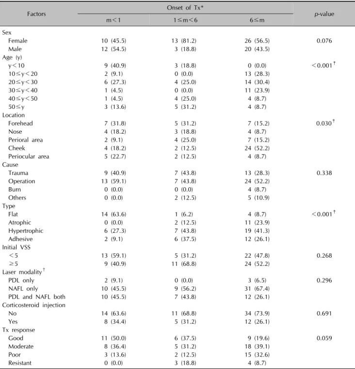
When scars were grouped by OT (m), the m<1, 1≤m<6, and m≥6 groups differed significantly from each other based on several other factors (Table 1). When the laser treatment started earlier, more scars were observed in the better treatment response group, but differences between the OT groups were not statistically significant (p=
0.0590). Scars of the m<1 group were more likely to be located on the forehead, while scars of the m>6 group were more likely to be located on the cheeks (p=0.030).
Flat scars were the most frequently occurring among the four scar types in the m<1 group (63.6%); however, hy- pertrophic scars were more frequent in the 1≤m<6 (43.8%) and m≥6 (41.3%) groups (p<0.001).
In univariable analyses, age, OT, and laser modalities (LM) were significantly associated with treatment response (Table 2). Subsequently, a multivariable analysis was per- formed considering the variables which were statistically significant in the univariable analysis and the potential confounders from the comparison of the OT groups. As a result, only three factors were found to be significantly as- sociated with treatment response; OT, LM, and the loca- tion of the scar. Among the OT groups, there was a sig- nificant difference in the treatment response of the scar treated earlier within a month and the scar treated after 6 months (p=0.0035). Considering scar locations, partic- ularly, scars on the perioral area and scars on the peri- ocular area showed a significant difference in treatment response (p=0.0324). The group of patients who were treated using both PDL and NAFL showed a significantly worse treatment response when compared to the group of patients treated using only PDL (p=0.0011).
DISCUSSION
This study was designed to identify the valuable factors for
Table 1. Different characteristics of facial scars according to treatment onset
Factors Onset of Tx*
p-value
m<1 1≤m<6 6≤m
Sex
Female 10 (45.5) 13 (81.2) 26 (56.5) 0.076
Male 12 (54.5) 3 (18.8) 20 (43.5)
Age (y)
y<10 9 (40.9) 3 (18.8) 0 (0.0) <0.001‡
10≤y<20 2 (9.1) 0 (0.0) 13 (28.3)
20≤y<30 6 (27.3) 4 (25.0) 14 (30.4)
30≤y<40 1 (4.5) 0 (0.0) 11 (23.9)
40≤y<50 1 (4.5) 4 (25.0) 4 (8.7)
50≤y 3 (13.6) 5 (31.2) 4 (8.7)
Location
Forehead 7 (31.8) 5 (31.2) 7 (15.2) 0.030‡
Nose 4 (18.2) 3 (18.8) 4 (8.7)
Perioral area 2 (9.1) 4 (25.0) 7 (15.2)
Cheek 4 (18.2) 2 (12.5) 24 (52.2)
Periocular area 5 (22.7) 2 (12.5) 4 (8.7)
Cause
Trauma 9 (40.9) 7 (43.8) 13 (28.3) 0.338
Operation 13 (59.1) 7 (43.8) 24 (52.2)
Burn 0 (0.0) 0 (0.0) 4 (8.7)
Others 0 (0.0) 2 (12.5) 5 (10.9)
Type
Flat 14 (63.6) 1 (6.2) 4 (8.7) <0.001‡
Atrophic 0 (0.0) 2 (12.5) 11 (23.9)
Hypertrophic 6 (27.3) 7 (43.8) 19 (41.3)
Adhesive 2 (9.1) 6 (37.5) 12 (26.1)
Initial VSS
<5 13 (59.1) 5 (31.2) 22 (47.8) 0.268
≥5 9 (40.9) 11 (68.8) 24 (52.2)
Laser modality†
PDL only 2 (9.1) 0 (0.0) 3 (6.5) 0.296
NAFL only 10 (45.5) 9 (56.2) 31 (67.4)
PDL and NAFL both 10 (45.5) 7 (43.8) 12 (26.1)
Corticosteroid injection
No 14 (63.6) 11 (68.8) 34 (73.9) 0.691
Yes 8 (34.4) 5 (31.2) 12 (26.1)
Tx response
Good 11 (50.0) 6 (37.5) 9 (19.6) 0.059
Moderate 8 (36.4) 5 (31.2) 18 (39.1)
Poor 3 (13.6) 2 (12.5) 15 (32.6)
Resistant 0 (0.0) 3 (18.8) 4 (8.7)
Values are presented as number (%). Percentages may not add up to 100 due to rounding. Tx: treatment, m: months, y: years, VSS:
Vancouver scar scale, PDL: 595-nm pulsed-dye laser, NAFL: non-ablative fractional laser. *The period between the injury and initiation of laser treatment. †Used laser modalities were selected via an algorithm according to the state of individual scars. ‡p<0.05.
predicting treatment response of laser treatment for facial scars. With the help of biostatistics specialists, we ana- lyzed possible influencing factors based on their treatment response, for which we determined values by the NTSE.
NTSE was defined as the number of treatments needed to achieve a VSS decrease of ≥50%, compared with the ini-
tial score. Therefore, the smaller NTSE values indicated the better treatment response and the larger NTSE values vice versa.
Among many treatment modalities, lasers have been suc- cessfully used for scar treatment, and among them, 585-nm or 595-nm PDL and NAFL have been consistently
Table 2. Univariable and multivariable analyses of the effects of various factors on treatment response Factors
Tx response (n=84) Univariableanalysis Multivariableanalysis Good
(n=26)
Moderate (n=31)
Poor (n=20)
Resistant
(n=7) p-value p-value
Sex
Female 13 (26.5) 20 (40.8) 12 (24.5) 4 (8.2) Ref. -
Male 13 (37.1) 11 (31.4) 8 (22.9) 3 (8.6) 0.4192
Age (y)
y<10 7 (58.3) 5 (41.7) 0 (0.0) 0 (0.0) Ref. Ref.
10≤y<20 3 (20.0) 2 (13.3) 8 (53.3) 2 (13.3) 0.0555 0.5306
20≤y<30 9 (37.5) 8 (33.3) 7 (29.2) 0 (0.0) 0.4719 0.7868
30≤y<40 2 (16.7) 8 (66.7) 0 (0.0) 2 (16.7) 0.0135‡ 0.2289
40≤y<50 1 (11.1) 4 (44.4) 2 (22.2) 2 (22.2) 0.1647 0.0908
50≤y 4 (33.3) 4 (33.3) 3 (25.0) 1 (8.3) 0.6548 0.2560
Location
Forehead 5 (26.3) 9 (47.4) 5 (26.3) 0 (0.0) 1 0.9783
Nose 4 (36.4) 4 (36.4) 2 (18.2) 1 (9.1) 0.0960 0.7947
Perioral area 2 (15.4) 2 (15.4) 6 (46.2) 3 (23.1) Ref. Ref.
Cheek 8 (26.7) 13 (43.3) 7 (23.3) 2 (6.7) 0.9076 0.3087
Periocular area 7 (63.6) 3 (27.3) 0 (0.0) 1 (9.1) 0.1707 0.0324‡
Cause
Trauma 12 (41.4) 8 (27.6) 8 (27.6) 1 (3.4) 0.9636 -
Operation 14 (31.8) 18 (40.9) 7 (15.9) 5 (11.4) Ref.
Burn 0 (0.0) 1 (25.0) 3 (75.0) 0 (0.0) 0.2085
Others 0 (0.0) 4 (57.1) 2 (28.6) 1 (14.3) 0.3210
Type
Flat 9 (47.4) 8 (42.1) 0 (0.0) 2 (10.5) 0.1281 0.8692
Atrophic 3 (23.1) 5 (38.5) 5 (38.5) 0 (0.0) 1 0.9336
Hypertrophic 6 (18.8) 9 (28.1) 13 (40.6) 4 (12.5) Ref. Ref.
Adhesive 8 (40.0) 9 (45.0) 2 (10.0) 1 (5.0) 1 0.4550
Onset of Tx (m)*
m<1 11 (50.0) 8 (36.4) 3 (13.6) 0 (0.0) Ref. Ref.
1≤m<6 6 (37.5) 5 (31.3) 2 (12.5) 3 (18.8) 0.6243 0.3576
6≤m 9 (19.6) 18 (39.1) 15 (32.6) 4 (8.7) 0.0028‡ 0.0035‡
Initial VSS
<5 17 (42.5) 11 (27.5) 9 (22.5) 3 (7.5) Ref. -
≥5 9 (20.5) 20 (45.5) 11 (25.0) 4 (9.1) 0.1479
Laser modality†
PDL only 3 (60.0) 2 (40.0) 0 (0.0) 0 (0.0) Ref. Ref.
NAFL only 22 (44.0) 18 (36.0) 9 (18.0) 1 (2.0) 0.5576 0.1439
PDL, NAFL both 1 (3.4) 11 (37.9) 11 (37.9) 6 (20.7) 0.0176‡ 0.0011‡
Corticosteroidinjection
No 22 (100.0) 0 (0.0) 0 (0.0) 0 (0.0) Ref. -
Yes 4 (6.5) 31 (50.0) 20 (32.3) 7 (11.3) 0.3618
Values are presented as number (%). Percentages may not add up to 100 due to rounding. Tx: treatment, y: years, m: months, VSS: Vancouver scar scale, PDL: 595-nm pulsed-dye laser, NAFL: non-ablative fractional laser, Ref.: reference. *The period between the injury and initiation of laser treatment. †Used laser modalities were selected via an algorithm according to the state of individual scars. ‡p<0.05.
reported to be effective and safe1,7,11,17,18. In this study, scars were treated with one of these two lasers based on a treatment algorithm. Corticosteroid injection (CSI) is the most popularly used treatment modality for scarring19,20. However, for facial scars, cosmetic concerns are a priority
and, therefore, possible adverse effects such as steroid-in- duced atrophy or CSI-related telangiectasia are not often acceptable to patients. For this reason, we limited the use of CSI to the lowest concentration possible and used it on- ly for scars with high pliability scores.
In the present study, scars showed a tendency of having better treatment response, when laser treatment was ini- tiated earlier following injury. The optimal timing for ini- tiating laser scar treatment has not yet been determined.
Traditionally, laser treatments are started after the wound had matured, which meant several months after trauma or surgery11,15. Some researchers have reported that laser treatment could be effectively started 2∼3 months after scar stabilization and when erythema had decreased at the surgical site21. However, earlier interventions have been emphasized in recent years to prevent hypertrophic scar formation15. Studies have shown that earlier interventions using PDL could be started without waiting for the wound to mature18. It has also been reported that early treatment with fractional lasers, such as AFL or NAFL, is benefi- cial15,16,22-24.
In general, the progression of a wound healing process comprises of the following phases; the inflammation phase for a few days, the proliferation phase for weeks, and the maturation phase for several months or years.
Previous studies have shown that hypertrophic scars gen- erally begin to develop 6 to 8 weeks after wound healing, grow rapidly for 3∼6 months, and then gradually regress after 6 months25,26. Lasers generate heat to cause in- flammation and increase the permeability of blood ves- sels, matrix metalloproteinase production, and degrada- tion of collagen fibers17,18. Moreover, targeted blood-ves- sel destruction induces hypoxia in the tissue, thereby re- ducing fibroblast proliferation and interfering with colla- gen deposition17,18. Thus, early implementation of laser treatment can shorten the acute inflammation stage, accel- erate scar maturation, and prevent excessive scar for- mation17. Consistently, the results of the current study sug- gested that early active intervention of scars may reduce the number of treatments and show better treatment re- sponse rather than the conventional wait-and-see or de- layed scar treatment until the natural course of scar maturation.
Interestingly, our results showed that the location of the scar was also significantly related to treatment outcome and especially scars on the perioral area had worse treat- ment response than scars on other areas. Particular areas of the body are more likely to develop hypertrophic scars, especially the front chest, shoulder, and the lower abdo- men7,27. This might be due to different degrees of mechan- ical tension at each region7,28. The perioral area is also an example of a highly movable area that is associated with larger mechanical tension due to the attachment of masti- catory or facial muscles. Therefore, scars occurring on the perioral area are expected to respond poorly to the treatment.
The superiority of certain laser modality over others in scar treatment has not been reported until now9,16,17. However, the present study showed the statistically sig- nificant association of LM with treatment response in the multivariable analysis, which could be due to the nature of our treatment algorithm in which only one type of laser can be used in a single treatment session. To be precise, if both PDL and NAFL had been used on a scar, it was evi- dent that the scar must have been treated at least twice.
However, in the PDL only group or the NAFL only group, patients treated with only one session of each laser treat- ment is included. Therefore, among the three groups of LM, in the group using both PDL and NAFL, NTSE could inevitably be predicted to be higher than the groups using only one modality among PDL and NAFL.
This study has a few limitations. First, because this study was the retrospective analysis of patients, comparative analysis with the untreated control groups was not carried out. Second, incomplete clinical data and follow-up loss limited the number of scars that we could include in the analyses. Third, the exclusion criteria eliminating the pa- tients who had stopped receiving treatments before reach- ing the endpoint of treatment might obtain biased results with better treatment response. Fourth, the use of different detailed treatment methods, depending on individual scar characteristics, was a serious analysis concern. However, all the scars were treated by one laser specialist following the same algorithm, and LM was controlled in our multi- variable analysis. Lastly, the study population was limited to Asian patients with Fitzpatrick skin type III or IV.
Despite these limitations, the discovery that OT is asso- ciated with treatment response is of major clinical significance. In the daily practice, patients with facial scars often prefer to get treated as soon as possible and to the maximum extent possible and do not wish to wait for the natural course of scar maturation. Our results provide in- direct evidence to the argument that early active laser in- tervention for scars yields better and faster outcomes com- pared to the conventional wait-and-see scar treatment that recommends delayed treatment until scar maturation.
Although we investigated the effects of several factors on the scar-treatment response, other factors may also affect the outcomes of scar treatment. In addition to the factors analyzed in the present study, anemia, ischemia, hor- mones, stress, smoking, alcohol consumption, obesity and body mass index, medications, and underlying conditions (immunocompromised, diabetes, malnutrition, etc.) are known to influence wound healing18,29,30. Therefore, in fu- ture studies, the above-mentioned factors should be considered.
In conclusion, the most important factors associated with
the treatment response in facial scars were the location of the scar and the timing of the start of treatment. The latter seemed to be the only modifiable factor for patients to achieve better treatment response among the factors we analyzed. Therefore, physicians should advise patients with a facial scar that early active intervention may help them in reducing the number of treatment sessions re- quired, save cost and time, and achieve more desirable clinical outcomes. In addition, our results revealed that scars on different locations may show a different response to the treatment. Especially, patients with scars on the per- ioral area might require more treatment sessions. Such in- formation can be used to predict treatment response and to tailor the treatment plan, depending on the character- istics of the scar. To ensure the generalizability of these re- sults, further research is required involving larger pop- ulations at multiple centers over a longer period of time.
CONFLICTS OF INTEREST
The authors have nothing to disclose.
ORCID
Youngkyoung Lim, https://orcid.org/0000-0002-6409-2704 Ji-Hye Park, https://orcid.org/0000-0002-6699-5202 Dong-Youn Lee, https://orcid.org/0000-0003-0765-9812 Na-Young Hwang, https://orcid.org/0000-0001-6282-5946 Soohyun Ahn, https://orcid.org/0000-0001-5016-5469 Jong Hee Lee, https://orcid.org/0000-0001-8536-1179
REFERENCES
1. Balaraman B, Geddes ER, Friedman PM. Best reconstructive techniques: improving the final scar. Dermatol Surg 2015;
41 Suppl 10:S265-S275.
2. Bock O, Schmid-Ott G, Malewski P, Mrowietz U. Quality of life of patients with keloid and hypertrophic scarring.
Arch Dermatol Res 2006;297:433-438.
3. Lawrence JW, Mason ST, Schomer K, Klein MB. Epidemiology and impact of scarring after burn injury: a systematic review of the literature. J Burn Care Res 2012;33:136-146.
4. van Drooge AM, Vrijman C, van der Veen W, Wolkerstorfer A. A randomized controlled pilot study on ablative fractional CO2 laser for consecutive patients presenting with various scar types. Dermatol Surg 2015;41:371-377.
5. Parry I, Sen S, Palmieri T, Greenhalgh D. Nonsurgical scar management of the face: does early versus late intervention affect outcome? J Burn Care Res 2013;34:569-575.
6. Gold MH, Berman B, Clementoni MT, Gauglitz GG, Nahai F, Murcia C. Updated international clinical recommendations on scar management: part 1--evaluating the evidence.
Dermatol Surg 2014;40:817-824.
7. Tziotzios C, Profyris C, Sterling J. Cutaneous scarring:
pathophysiology, molecular mechanisms, and scar reduction therapeutics part II. Strategies to reduce scar formation after dermatologic procedures. J Am Acad Dermatol 2012;66:13- 24; quiz 25-26.
8. Haedersdal M, Moreau KE, Beyer DM, Nymann P, Alsbjørn B. Fractional nonablative 1540 nm laser resurfacing for thermal burn scars: a randomized controlled trial. Lasers Surg Med 2009;41:189-195.
9. Vasily DB, Cerino ME, Ziselman EM, Zeina ST. Non-ablative fractional resurfacing of surgical and post-traumatic scars. J Drugs Dermatol 2009;8:998-1005.
10. Cervelli V, Gentile P, Spallone D, Nicoli F, Verardi S, Petrocelli M, et al. Ultrapulsed fractional CO2 laser for the treatment of post-traumatic and pathological scars. J Drugs Dermatol 2010;9:1328-1331.
11. Anderson RR, Donelan MB, Hivnor C, Greeson E, Ross EV, Shumaker PR, et al. Laser treatment of traumatic scars with an emphasis on ablative fractional laser resurfacing:
consensus report. JAMA Dermatol 2014;150:187-193.
12. Vrijman C, van Drooge AM, Limpens J, Bos JD, van der Veen JP, Spuls PI, et al. Laser and intense pulsed light therapy for the treatment of hypertrophic scars: a systematic review. Br J Dermatol 2011;165:934-942.
13. Verhaeghe E, Ongenae K, Bostoen J, Lambert J. Nonablative fractional laser resurfacing for the treatment of hypertrophic scars: a randomized controlled trial. Dermatol Surg 2013;
39:426-434.
14. Baryza MJ, Baryza GA. The Vancouver scar scale: an administration tool and its interrater reliability. J Burn Care Rehabil 1995;16:535-538.
15. Park KY, Oh IY, Seo SJ, Kang KH, Park SJ. Appropriate timing for thyroidectomy scar treatment using a 1,550-nm fractional erbium-glass laser. Dermatol Surg 2013;39:1827- 1834.
16. Jung JY, Jeong JJ, Roh HJ, Cho SH, Chung KY, Lee WJ, et al.
Early postoperative treatment of thyroidectomy scars using a fractional carbon dioxide laser. Dermatol Surg 2011;37:
217-223.
17. Jin R, Huang X, Li H, Yuan Y, Li B, Cheng C, et al. Laser therapy for prevention and treatment of pathologic excessive scars. Plast Reconstr Surg 2013;132:1747-1758.
18. Cooper JS, Lee BT. Treatment of facial scarring: lasers, filler, and nonoperative techniques. Facial Plast Surg 2009;25:
311-315.
19. Atiyeh BS. Nonsurgical management of hypertrophic scars:
evidence-based therapies, standard practices, and emerging methods. Aesthetic Plast Surg 2007;31:468-492; discussion 493-464.
20. Ledon JA, Savas J, Franca K, Chacon A, Nouri K.
Intralesional treatment for keloids and hypertrophic scars: a review. Dermatol Surg 2013;39:1745-1757.
21. Kunishige JH, Katz TM, Goldberg LH, Friedman PM.
Fractional photothermolysis for the treatment of surgical scars. Dermatol Surg 2010;36:538-541.
22. Choe JH, Park YL, Kim BJ, Kim MN, Rho NK, Park BS, et al.
Prevention of thyroidectomy scar using a new 1,550-nm
fractional erbium-glass laser. Dermatol Surg 2009;35:1199- 1205.
23. Kim HS, Lee JH, Park YM, Lee JY. Comparison of the effectiveness of nonablative fractional laser versus ablative fractional laser in thyroidectomy scar prevention: a pilot study. J Cosmet Laser Ther 2012;14:89-93.
24. Kim SG, Kim EY, Kim YJ, Lee SI. The efficacy and safety of ablative fractional resurfacing using a 2,940-Nm Er:YAG laser for traumatic scars in the early posttraumatic period.
Arch Plast Surg 2012;39:232-237.
25. Gold MH, McGuire M, Mustoe TA, Pusic A, Sachdev M, Waibel J, et al. Updated international clinical recommenda- tions on scar management: part 2--algorithms for scar prevention and treatment. Dermatol Surg 2014;40:825-831.
26. Chun Q, ZhiYong W, Fei S, XiQiao W. Dynamic biological
changes in fibroblasts during hypertrophic scar formation and regression. Int Wound J 2016;13:257-262.
27. Agha R, Ogawa R, Pietramaggiori G, Orgill DP. A review of the role of mechanical forces in cutaneous wound healing. J Surg Res 2011;171:700-708.
28. On HR, Lee SH, Lee YS, Chang HS, Park C, Roh MR.
Evaluating hypertrophic thyroidectomy scar outcomes after treatment with triamcinolone injections and copper bromide laser therapy. Lasers Surg Med 2015;47:479-484.
29. Guo S, Dipietro LA. Factors affecting wound healing. J Dent Res 2010;89:219-229.
30. Khalil H, Cullen M, Chambers H, Carroll M, Walker J.
Elements affecting wound healing time: an evidence based analysis. Wound Repair Regen 2015;23:550-556.