The Natural Course of Biopsy-Proven Isolated Microscopic Hematuria: A Single Center Experience of 350 Patients
The increasing interest in healthcare and health screening events is revealing additional cases of asymptomatic isolated microscopic hematuria (IMH). However, a consensus of the evaluation and explanation of the IMH prognosis is controversial among physicians. Here, we present the natural course of IMH together with the pathological diagnosis and features to provide supportive data when approaching patients with IMH. We retrospectively evaluated 350 patients with IMH who underwent a renal biopsy between 2002 and 2011, and the pathological diagnosis and chronic histopathological features (glomerulosclerosis, interstitial fibrosis, and tubular atrophy) were reviewed. Deterioration of renal function was examined during follow up. The patients with IMH were evaluated for a mean of 86 months. IgA nephropathy was the most common diagnosis in 164 patients (46.9%).
Chronic histopathological changes were observed in 166 (47.4%) but was not correlated with proteinuria or a decline in renal function. Ten patients developed proteinuria, and all of them had IgA nephropathy. Three patients progressed to chronic kidney disease with an estimated glomerular filtration rate < 60 mL/min/1.73 m2 but none progressed to end stage renal disease. In conclusion, IMH had a generally benign course during 7-years of observation, although IgA nephropathy should be monitored if it progresses to proteinuria.
Future prospective randomized studies may help conclude the long-term prognosis and lead to a consensus for managing IMH.
Keywords: Hematuria; Kidney; Biopsy; Proteinuria; Glomerulonephritis, IGA; Kidney Failure, Chronic
Hae Min Lee, Ji In Hyun, Ji-Won Min, Kyungsoo Lee, Yong Kyun Kim, Euy Jin Choi, and Ho Cheol Song Division of Nephrology, Department of Internal Medicine, Bucheon St. Mary’s Hospital, College of Medicine, The Catholic University of Korea, Bucheon, Korea
Received: 5 November 2015 Accepted: 23 February 2016 Address for Correspondence:
Ho Cheol Song, MD
Division of Nephrology, Department of Internal Medicine, Bucheon St. Mary’s Hospital, College of Medicine, The Catholic University of Korea, 327 Sosa-ro, Wonmi-gu, Bucheon 14647, Korea
E-mail: [email protected]
http://dx.doi.org/10.3346/jkms.2016.31.6.909 • J Korean Med Sci 2016; 31: 909-914
INTRODUCTION
Despite the high prevalence of microscopic hematuria in adults, a consensus on the extent of evaluation remains controversial (1-3). Even more, the exact definition of microscopic hematuria is unclear (4,5). The scarcity of long-term data related to disease progression of isolated microscopic hematuria (IMH) is related to the equivocal consensus (6). In contrast to the lack of interest and data, an increasing number of people are receiving health screening and are diagnosed with incidental asymptomatic mi- croscopic hematuria. IMH can be transient; however, our pre- vious study focused on persistently existing microscopic hema- turia (7).
Several studies have discussed IMH but most reports are ei- ther out of date or short-term retrospective studies, and patho- logical diagnostic data are extremely rare (5,6,8,9), which is re- lated to the controversy surrounding renal biopsy in patients with IMH. Renal biopsy is supported by some groups because the pathology can suggest a prognostic factor (1,10). However, some nephrologists do not recommend a biopsy because iden- tifying prognostic factors will not change the course of manage- ment (11,12).
These trends and debates lead us to design a study to present the long-term natural course of single-center patients with bi- opsy-proven isolated microscopic hematuria. The follow up fo- cused on whether patients progressed to chronic kidney disease (estimated glomerular filtration rate [eGFR] < 60 mL/min/1.73 m2 for > 3 months) or significantly high proteinuria (> 300 mg/
g urine protein to creatinine ratio [PCR]), as the presence of ei- ther marker suggests declining kidney function. Our study is unique because we focused only on patients with IMH. This was a 7-year retrospective, single-center study.
MATERIALS AND METHODS Clinical definitions
The terms concerning microscopic hematuria need to be clari- fied. We defined IMH as the presence of two or more red cells per high-power field on a microscopic examination and absence of any other clinical symptom or signs. The absence of protein- uria was confirmed by a repeated negative dipstick test or < 300 mg/g in spot urine PCR. Trace proteinuria was neglected from the subjects.
Nephrology
Estimated glomerular filtration rate
The Modification of Diet in Renal Disease (MDRD) equation:
186.3 × [serum creatinine (sCr) (mg/dL)]-1.154 × [age (years)]
-0.203 × (0.742 if female) was used to determine eGFR (13). Our laboratory does not use the isotope dilution mass spectrometry MDRD study equation due to global usage of the MDRD equa- tion (14).
Subjects
A retrospective study was designed for subjects who had under- gone renal biopsy from February 2002 to January 2011. Patients were meticulously evaluated for the presence of other second- ary causes of hematuria, such as malignancy or urolithiasis. Any patient with such a secondary cause was excluded from biopsy.
Renal biopsy for IMH was recommended by a single clinician to exclude any selection bias. The clinician recommended bi- opsy to every patient entering the clinic with IMH. Only those who agreed to the procedure received the biopsy. Also, patients were included in the study if they fulfilled the criteria listed be-
low (Fig. 1).
Consecutive presence of > 2 red blood cells/high power mi- croscopic field (test was repeated three times), sCr < 1.2 mg/dL, eGFR > 90 mL/min/1.73 m2, urine PCR < 300 mg/g (spot urine test was done only once), resting systolic blood pressure < 140 mmHg, resting diastolic blood pressure < 90 mmHg, sterile urinalysis except hematuria, absence of gross hematuria, and patients > 15 years old. All patients had undergone either ul- trasonography or computed tomography to exclude any sec- ondary causes. Patients with a chronic medical history or pres- ence of a family history suggestive of Alport’s syndrome were also excluded. Finally, 350 of the 1,508 patients (23%) were en- rolled for the analysis.
Renal biopsy
Renal biopsy was done under ultrasonographic guidance. An 18G core needle and automated biopsy gun were used to target the inferior pole of the left kidney. All biopsy specimens were examined by light microscopy, immunofluorescence, and elec-
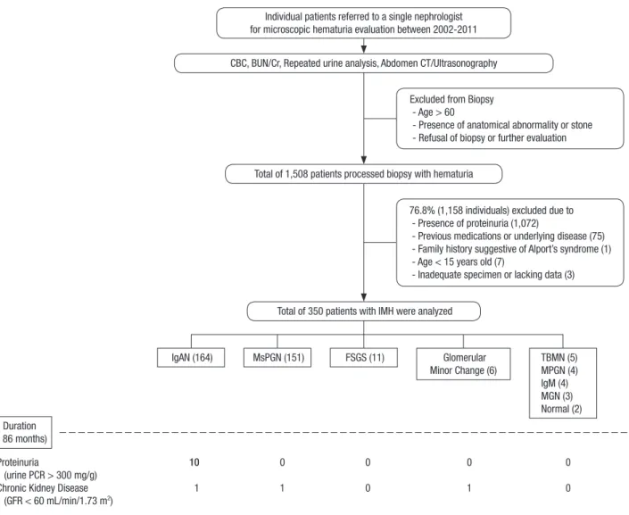
Fig. 1. Process of evaluation and results of isolated microscopic hematuria patients.
F-U Duration (mean 86 months)
Proteinuria 10 0 0 0 0
(urine PCR > 300 mg/g)
Chronic Kidney Disease 1 1 0 1 0
(GFR < 60 mL/min/1.73 m2)
Individual patients referred to a single nephrologist for microscopic hematuria evaluation between 2002-2011
Excluded from Biopsy - Age > 60
- Presence of anatomical abnormality or stone - Refusal of biopsy or further evaluation
76.8% (1,158 individuals) excluded due to - Presence of proteinuria (1,072)
- Previous medications or underlying disease (75) - Family history suggestive of Alport’s syndrome (1) - Age < 15 years old (7)
- Inadequate specimen or lacking data (3) CBC, BUN/Cr, Repeated urine analysis, Abdomen CT/Ultrasonography
Total of 1,508 patients processed biopsy with hematuria
Total of 350 patients with IMH were analyzed
IgAN (164) MsPGN (151) FSGS (11) Glomerular
Minor Change (6) TBMN (5) MPGN (4) IgM (4) MGN (3) Normal (2)
tron microscopy. Specimens of inadequate quality or quantity were excluded. The main focus of the biopsy results was the pa- thologic diagnosis and the presence of chronic histopathologi- cal features (tubular atrophy, interstitial fibrosis, and glomeru- losclerosis).
Statistical analysis
All data are expressed as mean ± standard deviation. Student’s t-test, the χ2 test, or Fisher’s exact test were used to compare the data and detect differences. A two-sided P value < 0.05 was con- sidered significant. All analyses were done using SPSS ver. 18.0 software (SPSS Inc., Chicago, IL, USA). A multivariate model (Cox proportional hazard model) was used to evaluate factors (age at biopsy, body mass index [BMI], initial blood pressure, initial proteinuria, baseline Cr, and presence of chronic histo- pathological changes in the biopsy leading to a progression to significant proteinuria).
Ethics statement
This study protocol was approved by the institutional review board of The Catholic University of Korea (No. HC15RIS0077).
The institutional review board waived the requirement for writ- ten consent from participants due to the retrospective design.
RESULTS
Baseline patient characteristics
The baseline clinical characteristics of the 350 consecutive pa- tients are listed in Table 1. Age ranged from 15 to 58 years and the mean was 40 ± 12 years. There were 78 males (22.3%) and 272 females (77.7%) included. The mean baseline urine PCR was 80 ± 30 mg/g with a mean follow up of 86 months. Mean body mass index was 23.1 ± 3.2 kg/m2. Mean systolic blood pres- sure was 104.5 ± 8.3 mmHg, and mean diastolic blood pressure was 66.2 ± 6.8 mmHg. Mean baseline sCr was 0.72 ± 0.14 mg/
dL with a mean eGFR of 101.38 ± 23.55 mL/min/1.73 m2.
Renal histology findings
The renal pathologic diagnoses of the 350 patients are shown in Table 2. The dominant pathologic diagnosis was IgA nephropa- thy (164/350, 46.9%), followed by idiopathic mesangial prolifer- ative glomerulonephritis (151, 43.1%). Eleven (3.1%) patients had focal segmented glomerulosclerosis, six (1.7%) had minor glomerular changes, five (1.4%) had thin basement membrane nephropathy, four (1.1%) had membranous proliferative glo- merulonephritis, four (1.1%) had IgM nephropathy, four (1.1%) had membranous glomerulonephritis, and two (0.6%) had nor- mal biopsy results.
The number of patients who presented with a chronic histo- pathological feature at baseline is listed in Table 3. Nearly half (166, 47.4%) of the patients had chronic pathological changes.
Segmental or global glomerulosclerosis was seen in 91 patients (26%). Tubular atrophy and interstitial fibrosis was shown in Table 1. Baseline clinical characteristics of isolated microscopic hematuria patients
Characteristics Isolated microscopic hematuria (n = 350)
Age, yr 40.26 ± 11.69
Female (n, %) 272 (77.7)
Body Mass Index, kg/m2 23.13 ± 3.17
Systolic Blood Pressure, mmHg 104.5 ± 8.3 Diastolic Blood Pressure, mmHg 66.2 ± 6.8
Serum Creatinine, mg/dL 0.72 ± 0.14
eGFR, mL/min/1.73 m2 101.38 ± 23.55
Urine Protein-Creatinine Ratio, mg/g 80 ± 30
Hemoglobin, g/dL 12.89 ± 1.23
Glucose, mg/dL 96.24 ± 11.32
Total Protein, g/dL 7.49 ± 0.48
Albumin, g/dL 4.49 ± 0.34
Total Cholesterol, mg/dL 175.20 ± 31.88
Triglyceride, mg/dL 117.82 ± 59.39
Duration of follow up, mon 85.81 ± 30.27
Values are mean ± SD.
Table 2. Renal pathologic diagnosis of isolated microscopic hematuria patients Renal biopsy results Total (n = 350) Percentage (%)
IgA nephropathy 164 46.9
Idiopathicmesangial proliferative GN 151 43.1
Focal segmental glomerulosclerosis 11 3.1
Glomerular minor changes 6 1.7
Thin basement membrane nephropathy 5 1.4
Membranous proliferative GN 4 1.1
IgM nephropathy 4 1.1
Membranous GN 3 0.9
Normal 2 0.6
GN, Glomerulonephritis.
Table 3. Baseline chronic histopathologic features of isolated microscopic hematuria patients
Lesions IgAN
(n = 164) IMPGN
(n = 151) FSGS
(n = 11) GMC
(n = 5) TBMN
(n = 5) MPGN
(n = 4) IgMN
(n = 4) MGN
(n = 3) Normal
(n = 2) Total IMH (n = 350)
Total chronicity No. (%) 75 (45.7) 70 (46.4) 11 (100) 4 (66.7) 3 (60) 1 (25) 0 2 (66.7) 0 166 (47.4)
G. sclerosis No. (%) 42 (26.5) 31 (20.5) 11 (100) 3 (50) 2 (40) 1 (25) 0 1 (33.3) 0 91 (26.0)
T. atrophy No. (%) 59 (36) 56 (37.1) 7 (63.6) 4 (66.7) 2 (40) 1 (25) 0 1 (33.3) 0 130 (37.1)
I. fibrosis No. (%) 59 (36) 50 (33.1) 7 (63.6) 4 (66.7) 2 (40) 1 (25) 0 1 (33.3) 0 124 (35.4)
IgAN, IgA nephropathy; IMPGN, idiopathic mesangial proliferative glomerulonephritis; FSGS, focal segmental glomerulosclerosis; GMC, glomerular minor changes; TBMN, thin basement membrane nephropathy; IgMN, IgM nephropathy; MGN, membranous glomerulonephritis; No., number of subjects; G. sclerosis, glomerulosclerosis; T. atrophy, tubu- lar atrophy; I. fibrosis, interstitial fibrosis.
130 (37.1%) and 124 (35.4%) patients, respectively.
Clinical outcomes
The clinical characteristics of the 350 patients and follow-up data are shown in Table 4. Follow-up duration was 12-148 months (mean, 85.81 ± 30.27 months). Systolic and diastolic blood pres- sure increased significantly (P < 0.001). However, mean blood pressure was within a normotensive level. sCr level increased significantly from a baseline of 0.72 ± 0.14 mg/dL to 0.76 ± 0.18 mg/dL at the end of follow up (P < 0.001). eGFR decreased from 101.4 ± 23.6 mL/min/1.73 m2 to 92.5 ± 18.2 mL/min/1.73 m2 (P < 0.001). The baseline urine PCR increased from a mean of 80 ± 30 mg/g to 100 ± 120 mg/g after a mean of 86 months (P = 0.003).
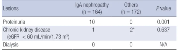
Ten patients developed significant proteinuria (> 300 mg/g in urine PCR) at the end of follow-up (Table 5). All 10 had been diagnosed with IgA nephropathy using the initial biopsy. The urine PCR increased in other patients but was always < 0.3. Three patients progressed to chronic kidney disease (CKD) with an eGFR < 60 mL/min/1.73 m2. A 19-year-old male who was diag- nosed with IgA nephropathy class II developed significant pro- teinuria after 17 months and his eGFR declined to < 60 mL/min/
1.73 m2 after 63 months. The eGFR of a 43-year-old female diag- nosed with idiopathic mesangial proliferative glomerulonephri- tis also declined to < 60 mL/min/1.73 m2 after 134 months. A 48-year-old woman with a minor glomerular lesion but chronic histopathological changes on biopsy progressed to an eGFR < 60 mL/min/1.73 m2 after 66 months.
Ten of 164 patients (6.1%) in the IgA group presented with significant proteinuria at the end of the follow up. A chi-square analysis of these patients showed IgA nephropathy compared to that in the other group (P = 0.001). A multivariate Cox pro- portional hazard model analysis was performed to evaluate in- dependent risk factors for the proteinuria and CKD, including age, sex, BMI, blood pressure, Cr level, urine protein-Cr ratio, biopsy results, and the presence of chronic changes on the bi- opsy. However, no significant risk factors leading to such events were detected.
DISCUSSION
This study is unique and the results are significant for clinicians.
First, we evaluated patients with IMH who had histopathologi- cal reports. Second, long-term data after renal biopsy are rare in patients with IMH. Our data include a maximum 12 years fol- low up with a mean follow up of 86 months (7-9,15). In addition, this was a large retrospective dataset of 350 patients with IMH from a single center. The single center and single physician de- creases the possibility of selection bias.
The pathological diagnoses were diverse among patients with IMH. IgA nephropathy was the most common followed by idio- pathic mesangial proliferative glomerulonephritis, with preva- lence rates of 46.9% and 43.1%, respectively. The result that IgA nephropathy was the most common pathology is compatible with previous reports (7-9,15). However, our data suggest a low prevalence (1.4%) of thin basement membrane disease in con- trast to other reports. These contrasting reports may be related to different prevalence rates of thin basement membrane dis- ease (TBMN) among geographic areas (16). TBMN is generally thought to be a benign renal disease. It has a very low risk of progressing to renal failure, whereas other chronic glomerulo- nephritis diseases, such as IgA nephropathy, generally result in decreased renal function during a long-term follow up. Such possibility supports the need for renal biopsy, which may lead to early aggressive management. Nevertheless, the necessity and risks of renal biopsy for these patients remain controversial.
The Oxford classification is a pathologic classification of IgA nephropathy with prognostic variables (17). The main six path- ological variables are mesangial cellularity score, segmental sclerosis, endocapillary hypercellularity, cellular/fibrocellular crescents, percentage of fibrosis/tubular atrophy, and the arte- riosclerosis score. Among these variables, chronic lesions, such as tubular atrophy, interstitial fibrosis, and glomerulosclerosis, have been reported to be the most meaningful histological pre- dictors (18,19). In our study, 45.7% (75/164) subjects with IgA nephropathy presented with one or more of these chronic le- sions. However, we were unable to find an association between the chronic changes and declining renal function. The same re- sult occurred with those who eventually progressed to signifi- cant proteinuria. These negative outcomes are thought to be re- Table 4. Clinical characteristics in the isolated microscopic hematuria patients during
the follow-up
Characteristics Baseline End of F/U P value*
Patients, No. (%) 350 (100) 350 (100) NS
SBP, mmHg 104 ± 8 117 ± 12 < 0.001
DBP, mmHg 66 ± 7 70 ± 9 < 0.001
Cr, mg/dL 0.72 ± 0.14 0.76 ± 0.18 < 0.001
eGFR, mL/min/1.73 m2 101.4 ± 23.6 92.5 ± 18.2 < 0.001
Urine PCR, mg/g 80 ± 30 100 ± 120 0.003
Follow up duration, mon - 85.81 ± 30.27 NS
SBP, systolic blood pressure; DBP, diastolic blood pressure; Cr, creatinine; eGFR, esti- mated glomerular filtration rate; PCR, protein to creatinine ratio.
NS: P > 0.05.
*P value were obtained by Student t-test.
Table 5. Occurrence of significant proteinuria or chronic kidney disease at the end of the follow-up
Lesions IgA nephropathy
(n = 164)
Others
(n = 172) P value
Proteinuria 10 0 0.001
Chronic kidney disease
(eGFR < 60 mL/min/1.73 m2) 1 2* 0.637
Dialysis 0 0 N/A
*The 2 subjects among the “Others” group was 1 membranous proliferative glomeru- lonephritis and 1 glomerular minor changes.
lated to the follow-up duration. Our study included patients who were evaluated for > 12 years; however, the mean dura- tion was 86 months, which is slightly more than 7 years. Previ- ous reports using the Oxford classification generally included
> 10 years of cohort data from patients with IgA nephropathy (11,12,17). We also evaluated whether such chronic changes was associated in patients with IMH rather than IgA, but no as- sociation was found.
Proteinuria at the initial diagnosis was a poor prognostic fac- tor in several studies (7,9). None of our patients with IMH had significant proteinuria at the initial evaluation. However, signif- icant proteinuria became evident in 10 patients with IgA ne- phropathy during the clinical course. Chow et al. (7) reported that each 0.1 g/day increase in baseline proteinuria is related to a two-fold relative risk of developing adverse renal events. We only had one patient who developed proteinuria in 18 months and progressed to CKD with an eGFR < 60 mL/min/1.73 m2 at the 63 month-follow-up. We expect that longer duration obser- vations would show a negative association between proteinuria and renal function. In addition, all of the patients who developed proteinuria had IgA nephropathy. Renal function decreased to an eGFR < 60 mL/min/1.73 m2 in three patients with IMH dur- ing the follow up, which is < 1% of all patients with IMH, and may be related to the relatively short duration of observations.
Several limitations in our study should be mentioned. The retrospective design lead to selection bias. To overcome this bias, we recruited subjects from a single physician at a single center. Another limitation was the lack of baseline data related to microalbuminuria. However, we provided spot urine pro- tein-Cr ratio and repeated urinalysis data before the biopsy to minimize the risk of pre-existing proteinuria.
In summary, we described the natural course and necessity for intervention to determine the pathology of patients with IMH.
Our results show that the prevalence of proteinuria increased during the 7 years follow up but none of the cases progressed to advanced CKD (eGFR < 30 mL/min/1.73 m2). Therefore, we conclude that IMH has a generally benign course despite the increased rate of proteinuria. Yet, IgA nephropathy should be observed when a rapid progression to proteinuria is detected.
DISCLOSURE
The authors have no potential conflicts of interest to disclose.
AUTHOR CONTRIBUTION
Research conception & design: Lee HM, Song HC. Data acqui- sition: Lee HM, Lee K, Hyun JI, Min JW. Data analysis and in- terpretation: Lee HM, Song HC. Statistical analysis: Lee HM.
Drafting of the manuscript: Lee HM, Song HC. Revision of the manuscript: Lee HM, Hyun JI, Min JW, Lee K, Song HC. Receiv-
ing grant: Song HC. Approval of final manuscript: all authors.
ORCID
Ho Cheol Song http://orcid.org/0000-0002-9849-8091 Hae Min Lee http://orcid.org/0000-0003-0131-1284 Ji In Hyun http://orcid.org/0000-0001-8207-5302 Ji-Won Min http://orcid.org/0000-0001-6295-8095 Kyungsoo Lee http://orcid.org/0000-0002-6781-0986 Yong Kyun Kim http://orcid.org/0000-0002-1871-3549 Euy Jin Choi http://orcid.org/0000-0002-3124-7829
REFERENCES
1. Fuiano G, Mazza G, Comi N, Caglioti A, De Nicola L, Iodice C, Andreucci M, Andreucci VE. Current indications for renal biopsy: a questionnaire- based survey. Am J Kidney Dis 2000; 35: 448-57.
2. Michael J, Jones NF, Davies DR, Tighe JR. Recurrent haematuria: role of renal biopsy and investigative morbidity. BMJ 1976; 1: 686-8.
3. Paone DB, Meyer LE. The effect of biopsy on therapy in renal disease. Arch Intern Med 1981; 141: 1039-41.
4. Cohen RA, Brown RS. Clinical practice. Microscopic hematuria. N Engl J Med 2003; 348: 2330-8.
5. Sutton JM. Evaluation of hematuria in adults. JAMA 1990; 263: 2475-80.
6. Vivante A, Afek A, Frenkel-Nir Y, Tzur D, Farfel A, Golan E, Chaiter Y, Sho- hat T, Skorecki K, Calderon-Margalit R. Persistent asymptomatic isolated microscopic hematuria in Israeli adolescents and young adults and risk for end-stage renal disease. JAMA 2011; 306: 729-36.
7. Chow KM, Kwan BC, Li PK, Szeto CC. Asymptomatic isolated microscop- ic haematuria: long-term follow-up. QJM 2004; 97: 739-45.
8. Shen P, He L, Jiang Y, Wang C, Chen M. Useful indicators for performing renal biopsy in adult patients with isolated microscopic haematuria. Int J Clin Pract 2007; 61: 789-94.
9. Yamagata K, Yamagata Y, Kobayashi M, Koyama A. A long-term follow- up study of asymptomatic hematuria and/or proteinuria in adults. Clin Nephrol 1996; 45: 281-8.
10. Richards NT, Darby S, Howie AJ, Adu D, Michael J. Knowledge of renal histology alters patient management in over 40% of cases. Nephrol Dial Transplant 1994; 9: 1255-9.
11. To KF, Choi PC, Szeto CC, Li PK, Tang NL, Leung CB, Wang AY, Ho KK, Wong TY, Lui SF, et al. Outcome of IgA nephropathy in adults graded by chronic histological lesions. Am J Kidney Dis 2000; 35: 392-400.
12. Walsh M, Sar A, Lee D, Yilmaz S, Benediktsson H, Manns B, Hemmelgarn B. Histopathologic features aid in predicting risk for progression of IgA nephropathy. Clin J Am Soc Nephrol 2010; 5: 425-30.
13. Levey AS, Bosch JP, Lewis JB, Greene T, Rogers N, Roth D; Modification of Diet in Renal Disease Study Group. A more accurate method to estimate glomerular filtration rate from serum creatinine: a new prediction equa- tion. Ann Intern Med 1999; 130: 461-70.
14. Lee CS, Cha RH, Lim YH, Kim H, Song KH, Gu N, Yu KS, Lim CS, Han JS, Kim S, et al. Ethnic coefficients for glomerular filtration rate estimation by the Modification of Diet in Renal Disease study equations in the Kore- an population. J Korean Med Sci 2010; 25: 1616-25.
15. Kim BS, Kim YK, Shin YS, Kim YO, Song HC, Kim YS, Choi EJ. Natural his-
tory and renal pathology in patients with isolated microscopic hematu- ria. Korean J Intern Med 2009; 24: 356-61.
16. Hall CL, Bradley R, Kerr A, Attoti R, Peat D. Clinical value of renal biopsy in patients with asymptomatic microscopic hematuria with and without low-grade proteinuria. Clin Nephrol 2004; 62: 267-72.
17. Working Group of the International IgA Nephropathy Network and the Renal Pathology Society, Roberts IS, Cook HT, Troyanov S, Alpers CE, Amore A, Barratt J, Berthoux F, Bonsib S, Bruijn JA, et al. The Oxford clas-
sification of IgA nephropathy: pathology definitions, correlations, and re- producibility. Kidney Int 2009; 76: 546-56.
18. Daniel L, Saingra Y, Giorgi R, Bouvier C, Pellissier JF, Berland Y. Tubular lesions determine prognosis of IgA nephropathy. Am J Kidney Dis 2000;
35: 13-20.
19. D’Amico G. Tubulointerstitium as predictor of progression of glomerular diseases. Nephron 1999; 83: 289-95.