This is an Open Access article distributed under the terms of the Creative Commons Attribution Non-Commercial License (http://creativecommons.org/licenses/by-nc/4.0/) which permits unrestricted non-commercial use, distribution, and reproduction in any medium, provided the original work is properly cited.
Copyright © 2016. Anatomy & Cell Biology
one such persistent jugulocephalic vein during our dissection classes and the aim of this case report is to discuss the clini- cal implications of this rare variation. The knowledge of this variation may be very useful to the radiologists, orthopedic surgeons, and plastic surgeons.
Case Report
During routine dissection classes for medical undergradu- ates, we observed a rare termination of cephalic vein of the right side in an adult male cadaver aged approximately 70 years. The variation was unilateral. The cephalic vein had a normal origin and course till it reached the delto-pectoral triangle. Upon reaching the delto-pectoral triangle, instead of piercing the clavipectoral fascia and terminating into the axil- lary vein, it ascended in front of the lateral part of the clavicle and terminated by opening into the external jugular vein.
It received a large muscular vein from sternocleidomastoid muscle just before its termination. It also gave a communi- cating vein at the delto-pectoral triangle, which pierced the clavipectoral fascia and opened into the axillary vein. Exter- nal jugular vein received the posterior external jugular vein and terminated into the subclavian vein. These variations are
Introduction
Cephalic vein is one of the superficial veins of the upper limb. It runs along the lateral border of the forearm and the arm, passes through the delto-pectoral groove, pierces the clavipectoral fascia at the delto-pectoral triangle and opens into the axillary vein. Variations of cephalic vein are rare. Its variations include its total absence, termination into subcla- vian vein, and abnormal communication with the external jugular vein [1-5]. In very rare cases, it crosses superficial to the clavicle and terminates into external jugular vein. This type of termination is called persistent jugulocephalic vein be- cause it is derived from an embryonic channel that connects the cephalic vein with the external jugular vein [6]. We found
Case Report
http://dx.doi.org/10.5115/acb.2016.49.3.210 pISSN 2093-3665 eISSN 2093-3673
Corresponding author:
Satheesha B. Nayak
Department of Anatomy, Melaka Manipal Medical College (Manipal Campus), Manipal University, Madhav Nagar, Manipal, Udupi District, Karnataka State 576 104, India
Tel: +91-820-2922519, Fax: +91-820-2571905, E-mail: [email protected]
A rare case of persistent jugulocephalic vein and its clinical implication
Prakashchandra Shetty, Satheesha B. Nayak, Rajesh Thangarajan, Melanie Rose D’Souza
Department of Anatomy, Melaka Manipal Medical College (Manipal Campus), Manipal University, Manipal, Inida
Abstract: Persistence of jugulocephalic vein is one of the extremely rare variations of the cephalic vein. Knowledge of such a variation is of utmost importance to orthopedic surgeons while treating the fractures of the clavicle, head and neck surgeons, during surgery of the lower part of neck, for cardiothoracic surgeons and radiologists during catheterization and cardiac device placement. We report the persistent jugulocephalic vein in an adult male cadaver, observed during the routine dissection classes. The right cephalic vein ascended upwards, superficial to the lateral part of the clavicle and terminated into the external jugular vein. It also gave a communicating branch to the axillary vein below the clavicle. We discuss the embryological and clinical importance of this rare variation.
Key words: External jugular vein, Jugulocephalic vein, Cephalic vein, Axillary vein, Subclavian vein Received June 22, 2016; Revised August 17, 2016; Accepted August 23, 2016
Jugulocephalic vein
http://dx.doi.org/10.5115/acb.2016.49.3.210
Anat Cell Biol 2016;49:210-212
211
www.acbjournal.org
shown in Figs. 1 and 2. The diameters of the veins observed were as follows: jugulocephalic vein, 0.3 cm; external jugular vein, 0.6 cm; axillary vein, 1 cm; cephalic vein, 0.5 cm; basilic vein, 0.4 cm; and communicating vein, 0.3 cm. The other veins in this case did not have any variations.
Discussion
Cephalic and external jugular veins are the two superficial veins which are used in many clinical and surgical proce- dures. A thorough knowledge of their variations can contrib- ute greatly to the success of any radiological or surgical pro- cedures. During embryonic development, the preaxial vein of the upper limb develops into cephalic vein and the postaxial vein develops into basilic vein. During early stages of develop- ment, the cephalic vein drains into a venous plexus of neck.
The external jugular vein develops from this plexus. Thus, in the early fetal life, cephalic vein is a tributary of external jugular vein. In a later stage of development, the cephalic vein establishes a connection with the axillary vein, after which it loses its connection with the external jugular vein. If it fails to lose connection with the external jugular vein, then it is called persistent jugulocephalic vein [3, 6-9]. Cephalic and external jugular veins are generally used for central venous catheterizations. Many device implanters prefer cephalic vein cut down for the placement of defibrillator leads instead of di- rectly puncturing the axillary or subclavian veins to avoid the risk of pneumothorax, subclavian crush, and other complica- tions. However, when there is a persistence of jugulocephalic vein, the catheterization may not be successful [10]. In a study
conducted by Loukas et al. [1], the cephalic vein terminated into subclavian vein after crossing anterior and superior to the clavicle in 0.2% of cases. In these cases, it gave a com- municating branch to the external jugular vein. Nayak and Soumya [11], have also reported a communication between cephalic and external jugular veins superficial to the clavicle.
But in that case, the cephalic vein terminated into the axil- lary vein by passing below the clavicle. In the current case, the cephalic vein terminated into external jugular vein by passing superficial to the clavicle and gave a communicating branch to the axillary vein. Supraclavicular course of cephalic vein is very rare and available reports indicate that it occurs only in 0.2% cases [12]. Cephalic vein with supraclavicular course must be avoided during pacemaker implantation due to the risk of skin erosion or lead fracture. Świętoń et al. [13]
suggest that supraclavicular course of cephalic vein might significantly affect the first time or repeated placement of car- diac implantable electronic device. According to Wysiadecki et al. [6] persistence of jugulocephalic vein increases the risk of complications during the clavicular fractures, cephalic vein catheterization, or head and neck surgery. Lin et al. [14]
recommend external jugular vein cut-down in cases of failure of cephalic vein cut-down procedures. Apart from the above said procedures, the persistence of jugulocephalic vein is of cosmetic concern also, and can be safely excised for the same reason [15].
Variations of cephalic vein are rare. Among its known vari- ations, persistence of jugulocephalic vein is extremely rare.
Knowledge of the same is quite useful to orthopedic surgeons, plastic surgeons, head and neck surgeons, cardiovascular sur- geons and more importantly, to the radiologists. Possibility of Fig. 1. Dissection of the upper part of the right arm and right side of
the neck showing JCV. A, anterior; COM, communication between cephalic vein and axillary vein; DLT, deltoid muscle; EJV, external jugular vein; I, inferior; JCV, jugulocephalic vein; MV, muscular vein from sternocleidomastoid; P, posterior; PM, pectoralis major muscle; S, superior; SCM, sternocleidomastoid muscle; TRP, trapezius muscle.
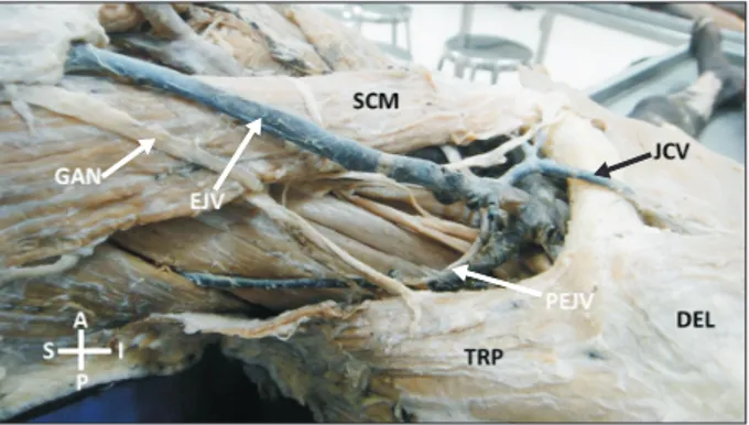
Fig. 2. Dissection of the upper part of the right arm and right side of the neck showing JCV. A, anterior; DEL, deltoid muscle; EJV, external jugular vein; GAN, great auricular nerve; I, inferior; JCV, jugulocephalic vein; P, posterior; PEJV, posterior external jugular vein;
S, superior; SCM, sternocleidomastoid muscle; TRP, trapezius muscle.
Anat Cell Biol 2016;49:210-212 Prakashchandra Shetty, et al
212
www.acbjournal.org http://dx.doi.org/10.5115/acb.2016.49.3.210
persistence of jugulocephalic vein has to be kept in mind dur- ing catheterizations, placement of cardiac devices and while treating the fractures of clavicle.
References
1. Loukas M, Myers CS, Wartmann CT, Tubbs RS, Judge T, Curry B, Jordan R. The clinical anatomy of the cephalic vein in the delto- pectoral triangle. Folia Morphol (Warsz) 2008;67:72-7.
2. Lee H, Lee SH, Kim SJ, Choi WI, Lee JH, Choi IJ. Variations of the cubital superficial vein investigated by using the intravenous illuminator. Anat Cell Biol 2015;48:62-5.
3. Standring S. Gray’s anatomy: the anatomical basis of clinical practice. 40th ed. Philadelphia, PA: Churchill Livingstone Else- vier; 2011. p.777-822, 899-906.
4. Snell RS. Clinical anatomy by regions. 8th ed. New Delhi: Wolter Kluwer; 2008.
5. Hollinshead WH. Anatomy for surgeons. Vol. 3. 2nd ed. New York: Harper and Row; 1966.
6. Wysiadecki G, Polguj M, Topol M. Persistent jugulocephalic vein:
case report including commentaries on distribution of valves, blood flow direction and embryology. Folia Morphol (Warsz) 2015 Sep 18 [Epub]. http://dx.doi.org/10.5603/FM.a2015.0084.
7. Thyng FW. The anatomy of a 17.8 mm human embryo. Am J Anat 1914;17:31-112.
8. Sadler TW, Langman J. Langman's medical embryology. 7th ed.
Baltimore, MD: Williams & Wilkins; 1995.
9. Moore KL, Persaud TV. The developing human: clinically ori- ented embryology. 6th ed. Philadelphia, PA: W. B. Saunders;
1998.
10. Lau EW, Liew R, Harris S. An unusual case of the cephalic vein with a supraclavicular course. Pacing Clin Electrophysiol 2007;
30:719-20.
11. Nayak BS, Soumya KV. Abnormal formation and communica- tion of external jugular vein. Int J Anat Var 2008;1:15-6.
12. De Maria E, Cappelli S. Cephalic vein with a supraclavicu- lar course: rare, but do not forget it exists! J Cardiovasc Med (Hagerstown) 2013 Aug 25 [Epub]. http://dx.doi.org/10.2459/
JCM.0b013e32836132d6.
13. Świętoń EB, Steckiewicz R, Grabowski M, Stolarz P. Selected clinical challenges of a supraclavicular cephalic vein (CV) in cardiac implantable electronic device (CIED) implantation. Folia Morphol (Warsz) 2015 Dec 29 [Epub]. http://dx.doi.org/10.5603/
FM.a2015.0125.
14. Lin CH, Yu JC, Lee YT, Wu HS. Conversion from cephalic vein to external jugular vein: success rate increased on totally im- plantable access ports with cut-down method. Surg Innov 2013;
20:566-9.
15. Brook WH, Smith CJ. Clinical presentation of a persistent jugu- locephalic vein. Clin Anat 1989;2:167-73.