Ansa Pancreatica-Type
Anatomic Variation of the Pancreatic Duct in Patients with Recurrent Acute
Pancreatitis and Chronic Localized Pancreatitis
급성 재발성 췌장염과 국소성 만성 췌장염 환자에서 발견된 고리형 췌관(Ansa Pancreatica) 유형의 췌관 변이
Jiyeon Ha, MD , Kyung Won Kim, MD* , Jin Hee Kim, MD, Seung Soo Lee, MD, Hyoung Jung Kim, MD, Jae Ho Byun, MD, Moon-Gyu Lee, MD
Department of Radiology and Research Institute of Radiology, University of Ulsan College of Medicine, Asan Medical Center, Seoul, Korea
Ansa pancreatic is a rare variation of pancreas duct. Ansa pancreatica is characterized by focal accessory duct atrophy and an additional curved duct linking main and accessory ducts replac- ing atrophied duct. Ansa pancreatica is considered as a predisposing factor of recurrent pan- creatitis. Pancreatitis can be localized in pancreas head and uncinate process, because pancre- as head and uncinate process might be drained through the additional hooked duct of ansa pancreatica. We reports three cases of localized chronic or recurrent pancreatitis cases with underlying ansa pancreatica type anatomic variation.
Index terms Pancreatic Duct; Anatomic Variation; Pancreatitis; Computed Tomography, X-Ray
INTRODUCTION
Many congenital anatomic variations of the pancreatic duct have been described in the literature, such as complete or incomplete pancreatic divisum, annular pancreas, and ansa pancreatica. Among various types of pancreas ductal anomalies, pancreas di- visum is the most frequent anatomic variation, whereas ansa pancreatica is a rare ana-
Received June 15, 2018 Revised August 2, 2018 Accepted August 15, 2018
*Corresponding author Kyung Won Kim, MD Department of Radiology and Research Institute of Radiology, University of Ulsan
College of Medicine, Asan Medical Center,
88 Olympic-ro 43-gil, Songpa-gu, Seoul 05505, Korea.
Tel 82-2-3010-4377 Fax 82-2-3010-2768
E-mail [email protected] This is an Open Access article distributed under the terms of the Creative Commons Attribu- tion Non-Commercial License (https://creativecommons.org/
licenses/by-nc/4.0) which permits unrestricted non-commercial use, distribution, and reproduc- tion in any medium, provided the original work is properly cited.
ORCID iDs Kyung Won Kim https://
orcid.org/0000-0002-1532-5970 Jiyeon Ha
https://
orcid.org/0000-0003-3496-4134
jksronline.org
366
tomic variation, with a reported prevalence of 1.1% (1, 2).
Ansa pancreatica is characterized by focal accessory duct atrophy and an additional curved duct linking main and accessory ducts replacing atrophied duct. Ansa pancreatica is consid- ered as a predisposing factor for recurrent pancreatitis (1). Adibelli et al. (3) reported that pa- tients with recurrent pancreatitis had a higher frequency of ansa pancreatica than the gener- al population (11.1% vs. 0.85%). The mechanisms of ansa pancreatica causing acute pancreatitis have not been clear yet. However, recent evidence consistently have suggested that the curved duct cause impaired pancreatic juice drainage, mainly from pancreas head and uncinate process, resulting in recurrent focal pancreatitis (1, 3).
There have only a few articles exploring the ansa pancreatica and recurrent/chronic pan- creatitis, thus ansa pancreatica is an under-reported disease entity (1, 3-8). As comprehensive evaluation of the pancreatic duct variation by imaging such as computed tomography (CT) or magnetic resonance cholangiopancreatography (MRCP) is extremely important in order to correctly guide the next management steps, radiologists should be aware of this rare disease entity. Thus, we intend to comprehensively analyze the imaging findings with three cases of recurrent pancreatitis or localized chronic pancreatitis in patients with ansa pancreatica.
CASE REPORT
We reported the following three cases of recurrent pancreatitis with underlying ansa pan- creatica ductal anomaly, as described in Fig. 1. Interestingly, the pancreatitis was localized in the pancreas head and uncinate process in all three cases.
CASE 1
A 24-year-old female patient presented with recurrent acute pancreatitis. The first episode of acute pancreatitis was 17 years ago and the most recent prior event was 3 years ago. At the time of hospital administration, serum amylase level was 145 U/L (normal range, 30-110 U/L) and lipase level was 136 U/L (normal range, 13-60 U/L), which were moderately higher than
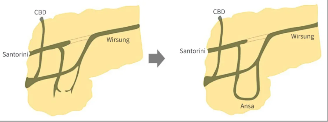
Fig. 1. Development of ansa pancreatica type anatomic variation. A curved additional duct is formed in ansa pancreatica replacing atrophied accessory duct during pancreatic duct development. The accessory duct arises from the main pancreatic duct and runs a hooked course anteriorly to the main duct ending in or around the minor papilla.
CBD = common bile duct
Santorini Santorini
Wirsung
Ansa
CBD CBD
Wirsung
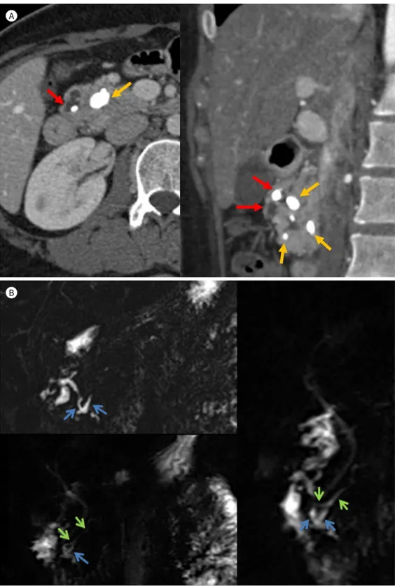
Fig. 2. A 24-year-old female patient with recurrent history of acute pancreatitis.
A, B. Axial scan and coronal (A) reconstructed image of contrast-enhanced CT reveal dilated duct of Santorini with impacted pancreaticolith within the dilated duct of Santorini (red arrows). In addition, multiple pancre- as parenchymal calcifications clustered in pancreatic head suggesting localized form of chronic pancreatitis (orange arrows). The coronal T2 weighted images of MRI and magnetic resonance cholangiopancreatogra- phy (B) show dilated duct of Santorini (blue arrows) with curved appearance and normal appearing duct of Wirshung (green arrows) suggesting ansa pancreatica.
A
B
jksronline.org
368
the upper normal limits.
Abdominal CT and MRCP were performed to evaluate recurrent pancreatitis and the un- derlying predisposing cause. Abdominal CT revealed clustered pancreatic parenchymal cal- cification in the pancreas head, suggesting localized chronic pancreatitis. Also several pan- creaticoliths were impacted within the dilated duct of Santorini continuing to the minor papillae (Fig. 2A). The MRCP revealed a normal appearing duct of Wirsung draining to major papillae, a dilated duct of Santorini, and another curved duct between the former two ducts, suggesting ansa pancreatica type anatomic variation (Fig. 2B).
CASE 2
A 62-year-old male patient presented with an incidental pancreas lesion on routine check- up. He denied any symptoms of abdominal discomfort at the time of visit. Indeed, he expe- rienced several episodes of acute abdominal pain during his whole life without knowing the exact cause of pain. The patient had rarely consumed alcohol before. At the time of visit, laboratory examination was not remarkable except for hyperlipidemia.
Abdominal CT showed a 2-cm size nodular lesion with multiple calcification at the pancre- as uncinate process, suggesting localized chronic pancreatitis (Fig. 3A). Pancreatic ductal dil- atation and pancreaticolith were not obvious on the CT scan. The patient underwent MRCP for further characterization of the pancreas lesion. MRCP showed normal appearance of the duct of Wirsung and another hooked duct arising from the flexion point of the pancreas duct draining to minor papillae, suggesting ansa pancreatica type ductal variation (Fig. 3B).
CASE 3
A 29-year-old male patient was referred from other hospital to evaluate the cause of recur- rent acute pancreatitis. He had not consumed alcohol before and he had no biliary stone disease. At the time of visit, laboratory examination was not remarkable. However, he had suffered from frequent episodes of abdominal pain and the most recent prior event was 2 months ago.
Abdominal CT revealed mild pancreas swelling localized in the pancreas head. Pancreas duct dilatation and panceaticolith were not obvious on the CT scan. Follow-up MRI and MRCP were performed 2 months after the CT scan. MRI showed improvement of pancreas head swelling. MRCP revealed arched duct linking the main pancreatic duct draining to the minor papillae, which was compatible with ansa pancreatica type anatomic variation.
DISCUSSION
Here, we reported three cases of recurrent pancreatitis localized in the pancreas head and uncinate process with underlying ansa pancreatica ductal anomaly. Ansa pancreatica is a rare type of pancreas ductal variation first reported by Dawson and Langman in 1961 (4). The reported prevalence of this type ductal variation is 1.1% (3). The ansa pancreatica is a ductal variation related with the developmental process of the pancreas.
The pancreatic duct is constituted of a dorsal duct (or a duct of Santorini) and a ventral
duct (or duct of Wirsung). The ventral duct or duct of Wirsung arises from the main pancre- atic duct, which empties through the major duodenal papillae. The dorsal duct or duct of Santorini forms the accessory pancreatic duct, draining to the minor duodenal papilla. These two ducts fuse in the pancreas head portion as a result of asymmetric duodenal rotation dur- ing gestational age 6 to 8 weeks. The ventral pancreatic bud rotates 180° counterclockwise, arriving at the dorsal pancreatic bud. During the development of the pancreatic duct, a vari- able degree of accessory duct atrophy occurs. Ansa pancreatica is characterized by focal ac- cessory duct atrophy around its junctions to the main pancreatic duct, which is replaced by Fig. 3. A 62-year-old male patient with calcified nodule in uncinate process.
A, B. The axial contrast-enhanced CT (A) reveals a small nodular lesion (circle) in the pancreas uncinate pro- cess suggesting localized chronic pancreatitis. The magnetic resonance cholangiopancreatography and axi- al T2 weighted images (B) reveal a hooked additional pancreatic duct arise from normal appearing main pancreatic duct (yellow arrows) draining to minor papillae (red arrows).
A
B
jksronline.org
370
an additional curved duct linking main and accessory ducts. This curved additional duct is formed from the proximal portion of the dorsal duct and inferior branches of both dorsal and ventral ducts. Thus, in ansa pancreatica, the accessory duct arises from the main pan- creatic duct and runs a hooked course anteriorly to the main duct ending in or around the minor papilla (5, 7, 9).
Ansa pancreatica type ductal anatomic variation might be a predisposing factor for recur- rent or chronic pancreatitis. Few observations reported coexistence of acute idiopathic pan- creatitis and underlying ansa pancreatica anatomic variation. But it is unclear whether the coexistence of theses two conditions is a simple coincidence or causal relationship (5-7). Ishii et al. (10) reported that approximately 7% of the patients with ansa pancreatica presented with acute pancreatitis. In ansa pancreatica ductal variation, the arched additional duct meets the main duct at on oblique angle, whereas the other tributaries of the main pancreat- ic duct join at a right angle. Based on this ductal variation, the pancreas area served by the additional arched duct has poor pancreatic juice drainage, resulting in recurrent pancreatitis (1). When condition predisposing pancreatitis such as heavy alcoholism or functional steno- sis of the sphincter of Oddi, this anatomic arrangement makes patients more vulnerable to the development of pancreatitis.
Summarizing, ansa pancreatica is a predisposing factor of recurrent pancreatitis localized in pancreas head and uncinate process, because drainage of pancreatic juice from the pan- creas head and uncinate process might be impaired due to the hooked duct linking main and accessory ducts. Through our experience and current consistent evidence, we would propose that if there is recurrent pancreatitis, especially localized in the pancreas head and uncinate process, underlying ductal anomaly such as ansa pancreatica should be considered in the di- agnosis and management of these patients.
Conflicts of Interest
The authors have no potential conflicts of interest to disclose.
Acknowledgments
This study was supported by a grant (No. 2017R1A2B3011475) from the National Research Founda- tion of Korea.
REFERENCES
1. Prasanna LC, Rajagopal KV, Thomas HR, Bhat KM. Accessory pancreatic duct patterns and their clinical im- plications. J Clin Diagn Res 2015;9:AC05-AC07
2. Dimitriou I, Katsourakis A, Nikolaidou E, Noussios G. The main anatomical variations of the pancreatic duct system: review of the literature and its importance in surgical practice. J Clin Med Res 2018;10:370-375 3. Adibelli ZH, Adatepe M, Imamoglu C, Esen OS, Erkan N, Yildirim M. Anatomic variations of the pancreatic
duct and their relevance with the Cambridge classification system: MRCP findings of 1158 consecutive pa- tients. Radiol Oncol 2016;50:370-377
4. Dawson W, Langman J. An anatomical-radiological study on the pancreatic duct pattern in man. Anat Rec 1961;139:59-68
5. Bhasin DK, Rana SS, Nanda M, Gupta R, Nagi B, Wig JD. Ansa pancreatica type of ductal anatomy in a pa- tient with idiopathic acute pancreatitis. JOP 2006;7:315-320
6. Kim HM, Park JY, Kim MJ. Ansa pancreatica: a case report of a type of ductal variation in a patient with idio- pathic acute recurrent pancreatitis. J Korean Soc Radiol 2010;63:83-86
7. Jarrar MS, Khenissi A, Ghrissi R, Hamila F, Letaief R. Ansa pancreatica: an anatomic variation and a rare cause of acute pancreatitis. Surg Radiol Anat 2013;35:745-748
8. Hayashi TY, Gonoi W, Yoshikawa T, Hayashi N, Ohtomo K. Ansa pancreatica as a predisposing factor for re- current acute pancreatitis. World J Gastroenterol 2016;22:8940-8948
9. Borghei P, Sokhandon F, Shirkhoda A, Morgan DE. Anomalies, anatomic variants, and sources of diagnos- tic pitfalls in pancreatic imaging. Radiology 2013;266:28-36
10. Ishii H, Arai K, Fukushima M, Maruoka Y, Hoshino M, Nakamura A, et al. Fusion variations of pancreatic ducts in patients with anomalous arrangement of pancreaticobiliary ductal system. J Hepatobiliary Pan- creat Surg 1998;5:327-332
급성 재발성 췌장염과 국소성 만성 췌장염 환자에서 발견된 고리형 췌관(Ansa Pancreatica) 유형의 췌관 변이
하지연 · 김경원* · 김진희 · 이승수 · 김형중 · 변재호 · 이문규
고리형 췌관(ansa pancreatica)은 드문 췌관 변이로 원인 미상의 급성 재발성 췌장염 또는 만성 췌장염의 선행요인이 될 수 있다는 가능성이 보고되어 있다. 저자 등은 췌장 두부에 국 한된 급성 재발성 췌장염 또는 췌장 두부에 국한된 만성 췌장염 환자에서 선행요인으로 고리 형 췌관이 발견된 증례 3예를 경험하였기에 문헌 고찰과 함께 보고한다.
울산대학교 의과대학 서울아산병원 영상의학과, 영상의학연구소