https://doi.org/10.14734/PN.2020.31.2.74 pISSN 2508-4887•eISSN 2508-4895
Chan-mi Lim, MD, Min-Jung Choi, MD, Hae-rin Jeon, MD, Sun-young Jung, MD, Suk-young Kim, MD, PhD Department of Obstetrics and Gynecology, Gachon University Gil Medical Center, Incheon, Korea
Objective: Chronic kidney disease (CKD) is a known risk factor of pregnancy that increases the rates of preterm birth, intrauterine growth restriction, and preeclampsia. And maternal CKD may worsen due to pregnancy itself. However, few studies have examined these problems in Korea.
Methods: The cases of 29 women with CKD who delivered singleton at our institute between 2006 and 2018 were retrospectively reviewed. Cases with major fetal malformation, intrauterine fetal death, a history of kidney transplantation, or insufficient information for the parameters were excluded. The subjects were divided into early-stage (stages 1 and 2) and late-stage (stages 3 to 5) groups according to the midterm glomerular filtration rate (GFR). Demographic profiles, renal func- tion, perinatal complications, and changes in GFR from pre-conception to postpartum were com- pared and analysed.
Results: The incidence of preeclampsia (28.6% vs. 75.0%; P=0.038), anemia (28.6% vs. 75.0%;
P=0.038), cesarean section (42.9% vs. 100.0%; P=0.009), small for gestational age (14.3% vs. 62.5%;
P=0.019) and the need for neonatal intensive care unit (38.1% vs. 87.5%, P=0.035) were significantly higher in the late-stage group than those in the early-stage group. Gestational age at delivery (38.1 vs. 35.4 weeks, P=0.021) and birth weight (2,970 vs. 2,200 g, P=0.006) were significantly lower in the late-stage group than those in the early-stage group. GFR decreased significantly after delivery compared to pre-conception in both group (P=0.028, both).
Conclusion: Late-stage CKD is a poor prognostic factor for pregnancy outcomes. Pregnancy deterio- rates renal function even in early-stage. Close monitoring and management are required for pregnant women with any stage of CKD.
Key Words: Pregnancy, Renal insufficiency, chronic, Glomerular filtration rate, Maternal health, Preg- nancy outcome
Introduction
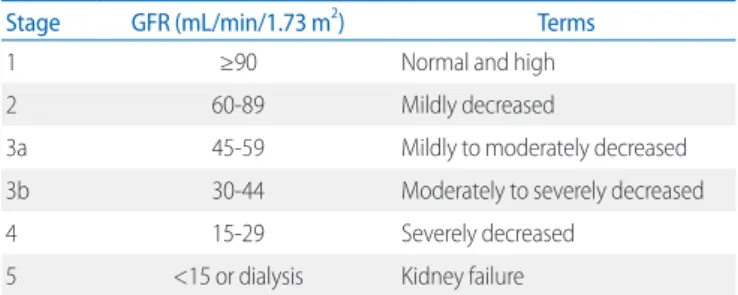
Chronic kidney disease (CKD) is defined as an abnormal renal structure or function that persists for more than 3 months. Specifically, patients are diagnosed as having CKD when a decreased glomerular filtration rate (GFR; <60 mL/min/1.73 m2) or one or more markers of kidney damage (albuminuria, urine sediment abnormalities, electrolyte and other abnormalities due to tubular disorders, abnormalities detected by histology, structural abnor
malities detected by imaging, and history of kidney transplantation) are present for more than 3 months. CKD is stratified into five stages based on GFR level (mL/min/1.73 m2) as Table 1.1
The prevalence of CKD is 0.16% in women of childbearing age.27 Earlystage CKD (stage 1 and 2) affects 3.2% of pregnant women,6 while 1 of 750 pregnant women has late
Received: 16 August 2019 Revised: 4 October 2019 Accepted: 24 January 2020 Correspondence to Suk-young Kim, MD, PhD Department of Obstetrics and Gynecology, Gachon University Gil Medical Center, 21 Namdong-daero 774beon-gil, Namdong-gu, Incheon 21565, Korea
Tel: +82-32-460-3254 Fax: +82-32-460-3290 E-mail: [email protected] Copyright© 2020 by The Korean Society of Perinatology
This is an Open Access article distributed under the terms of the Creative Com- mons Attribution Non-Commercial License (http://creativecommons.org/
license/by-nc/4.0/), which permits unrestricted non-commercial use, distribution, and reproduction in any
Maternal and Perinatal Outcomes in Preg
nancy by Chronic Kidney Disease Stage
Based on Midterm Glomerular Filtration
Rate
neonatal intensive care unit (NICU) admission, preeclampsia, and cesarean section (C/S).812 According to a metaanalysis, the combined risk of gestational hypertension, preeclampsia, eclampsia, and maternal mortality was 2 to 10 times higher than that of the control group.8 The risk increases with worsening renal function, especially in latestage CKD.911,1320 Conversely, pregnancy alone may deteriorate maternal renal function.16,20 An estimated 5075% of pregnant women with latestage CKD experienced disease progression.2126
Although many studies have shown adverse effects of CKD on pregnancy outcomes and the aggravation of CKD in pregnancy, only a few studies in Korea have examined that issue. Therefore, we performed this retrospective study to evaluate whether latestage CKD is a higher risk factor of adverse pregnancy outcomes than earlystage CKD and to what extent. We also analyzed the effect of pregnancy on early and latestage CKD progression in Korea.
Methods
This study was conducted in a tertiary medical center, Gachon University Gil Medical Center in Incheon, Korea. Women who were diagnosed as having CKD before or during pregnancy by a nephrologist were referred to the Department of Obstetrics and Gynecology of our center. Among them, patients delivered singleton baby at our institute between January 2006 and De
cember 2018 were enrolled in this study. Seven patients were excluded from the study because of insufficient information for analysis (three patients), loss of followup (one patient), intrauterine fetal death (one patient) and a history of kidney transplantation before pregnancy (two patients). Finally, 29
cases were included in our study. Their medical records were obtained and retrospectively analyzed. This study had approval of Institutional Review Boards (IRB No. GAIRB2019210).
Their GFRs were calculated using the CKDEpidemiology Collaboration equation. The patients were categorized in five stages based on their GFR in the midterm of pregnancy because some patients were diagnosed as having CKD during pregnancy.
The participants were divided into the earlystage (stages 1 and 2) and latestage (stages 3 to 5) groups.
The baseline parameters of patients were obtained, includ ing age, body mass index, preexisting hypertension or diabetes mellitus, obstetrical history, gestational age (GA) at delivery, GA at first referral to the Department of Obstetrics and Gyne
cology, type of renal disease, CKD duration, dialysis, serum creatinine level, and hemoglobin level. To assess CKD progres
sion during pregnancy, we used the GFRs based on the serum creatinine level before conception, in the midterm (the second trimester; between 14 weeks and 28 weeks), within 2 weeks before delivery, and within 6 weeks after delivery. When the serum creatinine level in the second trimester was not available, the level at the GA proximate to second trimester was used.
Obstetrical outcomes were set as C/S, preterm birth (GA <37 weeks or <34 weeks), anemia and preeclmapsia. Anemia was defined when the lowest hemoglobin level in pregnant period was below 11.0 g/dL (first or third trimester) or 10.5 g/dL (second trimester).27 And preeclampsia was diagnosed when the baseline blood pressure rises newly or worsens with aggravated proteinuria, thrombocytopenia, elevated liver enzyme levels, cerebral symptoms or pulmonary edema.28 Perinatal outcomes were birth weight, small for GA (SGA) which was defined in case that the birth weight was below the 10th percentile for the GA, IUGR defined when estimated fetal weight was below 10th percentile for the GA with oligohydramnios or abnormal Doppler findings in antenatal sonography, neonatal death, low 5min Apgar score (<7), and NICU admission.
Descriptive statistics were performed to summarize the sub
jects’ characteristics. Continuous data are shown as median and range and analyzed using the MannWhitney U test. GA converted to a decimal scale was used. Categorical data are presented as percentages, and the frequencies across cate
gories were assessed using Fisher’s exact test. To ascertain CKD progression during pregnancy, changes in GFR from pre
Table 1. Classification of Chronic Kidney Disease by Stage 1 to 5 according to GFR
Stage GFR (mL/min/1.73 m2) Terms
1 ≥90 Normal and high
2 60-89 Mildly decreased
3a 45-59 Mildly to moderately decreased
3b 30-44 Moderately to severely decreased
4 15-29 Severely decreased
5 <15 or dialysis Kidney failure
Abbreviation: GFR, glomerular filtration rate.
median hemoglobin level was 12.2 g/dL in the earlystage group and 10.5 g/dL in the latestage group (P=0.028).
Regarding pregnancy outcomes, 42.9% of patients delivered by C/S in the earlystage group versus 100% in the latestage group (Table 4). The most common indication of C/S in the earlystage group was prior C/S (four cases), which was fol
lowed by breech presentation, severe IUGR, preeclampsia with uncontrolled blood pressure, fetal anomaly and maternal request (one case each). Eight patients in the latestage group delivered by C/S due to prior C/S (four cases), fetal distress (two cases), preeclampsia with uncontrolled blood pressure (one case), and aggravation of CKD (one case). The rates of preterm birth before GA 37 weeks (33.3% vs. 62.5%) and before 34 weeks (9.5% vs. 12.5%) did not differ significantly between the two groups. The incidence of preeclampsia was 28.6% in the earlystage group and 75.0% in the latestage group (odds ratio, 7.5; P=0.038). There were no cases of eclampsia. The rate of anemia was significantly higher in the latestage group than that in the earlystage group (odds ratio, 7.5; P=0.038). The median birth weights were 2,970 g and 2,200 g in the early and latestage groups, respectively, which showed significantly low in latestage (P=0.006). The incidence of SGA was significantly conception to postdelivery were assessed using the Wilcoxon
signed rank test. The statistically analyses were performed using SPSS version 22.0 (IBM Corp., New York, NY, USA), and values of P<0.05 were considered statistically significant.
Results
The clinical profiles of 29 patients enrolled in the study are shown in Table 2. The median GA at delivery was 37.3 weeks;
there was a significant difference in GA at delivery between the earlystage and latestage groups (38.1 vs. 35.4 weeks, P=0.021). Eleven patients (37.9%) had hypertension and six patients (20.7%) had diabetes mellitus. The prevalence of pre
existing hypertension was significantly high in the latestage group (19.0% vs. 87.5%, P=0.001). Regarding renal disease type, glomerulonephritis was the most common in the early
stage group (76.2%), while glomerulonephritis (37.5%) and di
abetic nephropathy (37.5%) both were the most common type in the latestage group. The serum creatinine levels and the cal
culated GFRs differed significantly between the early and late
stage groups regardless of the measured time (Table 3). The
Table 2. Clinical Characteristics of Women with CKD
Characteristic Overall (n=29) Early-stage (n=21) Late-stage (n=8) P-value
Age (years) 31.0 (24.0-47.0) 31.0 (24.0-37.0) 31.5 (25.0-47.0) 0.487
Body mass index (kg/m2) 21.7 (18.8-28.3) 21.5 (18.8-28.3) 22.1 (21.3-24.9) 0.301
Nulliparity 15 (51.7) 12 (57.1) 3 (37.5) 0.427
Abortion history 9 (31.0) 6 (28.6) 3 (37.5) 0.675
GA at delivery (weeks) 37.3 (29.1-40.7) 38.1 (30.4-40.7) 35.4 (29.1-37.4) 0.021
GA at referral (weeks) 9.0 (4.0-36.0) 8.0 (4.0-36.0) 12.0 (7.0-29.0) 0.793
Hypertension 11 (37.9) 4 (19.0) 7 (87.5) 0.001
Diabetes mellitus 6 (20.7) 3 (14.3) 3 (37.5)
CKD duration (years) 4.0 (0.0-17.0) 2.0 (0.0-10.0) 4.5 (1.0-17.0) 0.093
Dialysis 4 (13.8) 0 (0.0) 4 (50.0)
Type of renal disease
Glomerulonephritis 19 (65.5) 16 (76.2) 3 (37.5) -
Diabetic nephropathy 5 (17.2) 2 (9.5) 3 (37.5) -
Hypertensive nephropathy 1 (3.4) 0 (0.0) 1 (12.5) -
SLE 2 (6.9) 2 (9.5) 0 (0.0) -
Unknown 2 (6.9) 1 (4.5) 1 (12.5) -
Values are presented as median (range) or number (%). Analyzed by Mann-Whitney U test and Fisher’s exact test.
Abbreviations: CKD, chronic kidney disease; GA, gestational age; SLE, systemic lupus erythematosus.
valve regurgitation with no other complication. He was born by C/S due to antenatal detection of those anomalies and expected need for neonatal resuscitation, however his 5min Apgar score was 10 and the anomalies were mild.
To assess changes in kidney function regarding CKD stage, the median GFR was compared among measurement points (Table 5). In earlystage CKD, the midterm GFR increased compared to the preconception GFR, decreased again before delivery, and decreased further postpartum. On the other hand, the median high in latestage group (P=0.019), whereas, the incidence of
IUGR did not differ significantly between the two groups (P=
0.068). There were no cases of neonatal death in either group.
The NICU admission rate was 38.1% in the earlystage group versus 87.5% in the latestage group (odds ratio, 11.4; P=0.035).
There was one baby in the earlystage group who was born at GA 34.7 weeks and 1,500 g in weight had a low 5min Apgar score below 7 points. Another baby in the earlystage group had anomalies of mild pulmonary artery stenosis and tricuspid
Table 3. Laboratory Characteristics of Women with CKD
Overall (n=29) Early-stage (n=21) Late-stage (n=8) P-value
Serum creatinine (mg/dL)
Pre-conception 0.8 (0.4-11.6) 0.7 (0.4-1.5) 3.0 (1.4-11.6) <0.001
Midterm 0.7 (0.4-10.2) 0.6 (0.4-1.2) 4.0 (1.3-10.2) <0.001
2 weeks before delivery 0.9 (0.3-9.4) 0.7 (0.3-2.0) 4.2 (1.6-9.4) <0.001
6 weeks after delivery 0.9 (0.4-11.8) 0.8 (0.4-1.9) 5.0 (1.6-11.8) <0.001
GFR (mL/min/1.73 m2)
Pre-conception 99.0 (4.0-136.0) 115.0 (48.0-136.0) 23.5 (4.0-49.0) <0.001
Midterm 116.5 (4.0-142.0) 122.0 (61.0-142.0) 16.0 (4.0-54.0) <0.001
2 weeks before delivery 86.0 (5.0-149.0) 116.0 (33.0-149.0) 14.0 (5.0-42.0) <0.001
6 weeks after delivery 85.0 (4.0-133.0) 100.0 (35.0-133.0) 11.5 (4.0-42.0) <0.001
Hemoglobin (g/dL) 11.3 (7.1-15.2) 12.2 (7.1-15.2) 10.5 (8.6-11.6) 0.028
Values are presented as median (range). Analyzed by Mann-Whitney U test.
Abbreviations: CKD, chronic kidney disease; GFR, glomerular filtration rate.
Table 4. Comparison of Pregnancy Outcomes by CKD Stage
Obstetric and perinatal outcomes Overall (n=29) Early-stage (n=21) Late-stage (n=8) OR (95% CI) P-value Obstetric outcomes
Cesarean section 17 (58.6) 9 (42.9) 8 (100.0) - 0.009
Preterm birth (<37 weeks) 12 (41.4) 7 (33.3) 5 (62.5) 3.33 (0.61-18.15) 0.218
Preterm birth (<34 weeks) 3 (10.3) 2 (9.5) 1 (12.5) 1.36 (0.11-17.42) 1.000
Preeclampsia 12 (41.4) 6 (28.6) 6 (75.0) 7.50 (1.17-48.15) 0.038
Anemia 12 (41.4) 6 (28.6) 6 (75.0) 7.50 (1.17-48.15) 0.038
Perinatal outcomes
Birth weight (g) 2,658 (820-4,275) 2,970 (1,470-4,275) 2,200 (820-2,545) - 0.006
SGA 8 (27.6) 3 (14.3) 5 (62.5) 10.00 (1.52-65.68) 0.019
IUGR 7 (24.1) 3 (14.3) 4 (50.0) 6.00 (0.95-38.08) 0.068
Anomaly 1 (3.4) 1 (4.8) 0 (0.0) - 1.000
Neonatal death 0 (0.0) 0 (0.0) 0 (0.0) - -
5-min Apgar score <7 1 (3.4) 1 (4.8) 0 (0.0) - 1.000
Admission in NICU 15 (51.7) 8 (38.1) 7 (87.5) 11.38 (1.27-110.42) 0.035
Values are presented as median (range) or number (%). Analyzed by Mann-Whitney U test and Fisher’s exact test.
Abbreviations: CKD, chronic kidney disease; OR, odds ratio; CI, confidence interval; SGA, small for gestational age; IUGR, intrauterine growth restriction; NICU, neonatal intensive care unit.
GFR declined steadily from preconception to postpartum in the latestage CKD. There was significant decrease in GFR pre
conception (115.0 mL/min/1.73 m2) versus postpartum (100.0 mL/min/1.73 m2) in earlystage group (P=0.028). In latestage group, the change of GFR between preconception (23.5 mL/
min/1.73 m2) and postpartum (11.5 mL/min/1.73 m2) was also significant (P=0.028).
Discussion
Delivery by C/S occurs more frequently in women with CKD than in those with a normal pregnancy.11,12 Piccoli et al.11 reported a higher C/S rate in patients with CKD versus controls (54.8% vs.
27.2%, respectively). This result is similar to that in our patients with CKD (56.7%). In our study, the rate of C/S in the latestage group was significantly higher than that in the earlystage group (100% vs. 42.9%, respectively), which is thought to be due to the more unfavourable maternal and fetal condition of late
stage CKD including preeclampsia, fetal distress, and worsening renal function. A cohort study reported a 9.1% incidence of preeclampsia in earlystage CKD, which is significantly higher than that in the normal population.29 Our study showed a higher prevalence of 28.6% in patients with earlystage CKD and a much higher rate of 75.0% in the latestage group (P=0.038).
This significant difference in the incidence of preeclampsia between early and latestage CKD is consistent with preceding data.16,20,23 An erythropoietin deficiency in CKD patients induces anemia, which is associated with fetal growth restriction, espe
cially in the latestage.26 The incidence of anemia was signifi
cantly higher in pati ents with latestage CKD (28.6% vs. 75.0%) as reported by preceding studies.20,30 Several studies demon
strated a diverse prevalence of preterm birth of 22.444% in CKD pa tients versus 4.9% in the normal population.9,19,29 In our study, there was a 41.4% preterm birth rate in overall CKD
patients, which is consistent with other studies. More than 75%
of patients with stage 3 CKD and almost all patients with stage 45 CKD gave birth to a premature baby.8,13,26,31 However, a study in India reported a comparable incidence of preterm birth rate in both stages (45.4% in earlystage, 56.5% in latestage).20 Similar to the result of an Indian study, there was no significant difference in the preterm birth rate between two stages in our study, whereas the significant gap of the median GA at delivery (38.1 vs. 35.4 weeks, P=0.021). This is thought be because of the early intervention to unfavourable fetal and maternal condi
tion in latestage CKD. The median birth weight was 2,970 g in the earlystage group and 2,200 g in the latestage group, which showed a significant difference (P=0.006). The result is probably due to earlier GA at delivery with latestage CKD.
The adverse effect of CKD stage on SGA remains disputable with various results of preceding studies.9,20,30 In this study, the incidence of SGA was 14.3% in the earlystage group and 62.5%
in the latestage group, which showed significantly difference (P=0.019). However, the incidence of IUGR had no significant difference (P=0.068). Piccoli et al.26 reported that more than 40% of babies born to women with latestage CKD required NICU admission. In our study, the NICU admission rate in the latestage group was 87.5%, which was significantly higher than 38.1% of rate in earlystage group (P=0.035).
The risk of CKD progression differed among several studies (2275%) and remains controversial.20,2326 According to the results of preceding studies, there was a significant decrease in GFR 6 weeks after delivery versus before conception at all CKD stages.16,20 A study by Davidson et al.30 reported a signi
ficant difference in GFR between before conception and 6 weeks and 12 months postpartum only in the latestage group.
On the other hand, a metaanalysis showed no association between pregnancy and deterioration of renal function.18 In our study, comparison of preconception and 6 weeks postpartum revealed a significant decrease in GFR in both stage group Table 5. Changes in GFR Over Time in Early- and Late-stage CKD (mL/min/1.73 m2)
Stage Pre-conception Midterm 2 weeks before delivery 6 weeks after delivery P-value*
Early 115.0 (48.0-136.0) 122.0 (61.0-142.0) 116.0 (33.0-149.0) 100.0 (35.0-133.0) 0.028
Late 23.5 (4.0-49.0) 16.0 (4.0-54.0) 14.0 (5.0-42.0) 11.5 (4.0-42.0) 0.028
Values are presented as median (range). Analyzed by Wilcoxon signed rank test.
Abbreviations: GFR, glomerular filtration rate; CKD, chronic kidney disease.
*Pre-conception vs. 6 weeks after delivery.
(P=0.028, both). The changing pattern of GFR in the early
stage resembled the physiological change in renal blood flow and hyperfiltration in pregnancy peaking in midterm, while the GFR value decreased steadily over time in the latestage.
Furthermore, the decline in GFR was faster and a greater pro
portion of patients progressed to a higher CKD stage in the late
stage group.16,20 Four out of 16 patients in stage 1 CKD, three out of five patients in stage 2, one out of two patients in stage 3, and two out of two patients in stage 4 progressed to the next stage 6 weeks after delivery in our study, supporting that higher initial stage is adverse factor of CKD aggravation.
This is the largest study to assess pregnancy and renal outcomes in pregnant women with CDK in Korea by comparing those with early and latestage disease. However, its size is too small to evaluate other factors known to affect adverse preg nancy outcomes and deterioration of renal function such as CKD pathology, proteinuria, hypertension, and required therapies.6,11,3236 Another limitation is that our study was retro
spec tive, so missing data could not be controlled for. Further multivariate analyses with larger populations are needed to more clearly verify the outcomes. The new attempt to set mid
term GFR as the baseline value can be a limitation, but it can be a good basis for identifying new useful predictors.
In conclusion, latestage CKD raised the risk of many adverse maternal and perinatal outcomes compared with earlystage CKD. Renal function deterioration induced by pregnancy was influenced regardless of CKD stage. Therefore, women with CKD, even in earlystage, must plan for pregnancy, and when they conceive, adequate and close multidisciplinary monitoring should be performed by obstetricians, nephrologists, and neonatologists to ensure better pregnancy and renal outcome.
Conflict of interest
No potential conflict of interest relevant to this article was reported.
References
1) Inker LA, Astor BC, Fox CH, Isakova T, Lash JP, Peralta CA, et al. KDOQI US
commentary on the 2012 KDIGO clinical practice guideline for the evaluation and management of CKD. Am J Kidney Dis 2014;63:713-35.
2) Williams D, Davison J. Chronic kidney disease in pregnancy. BMJ 2008;
336:211-5.
3) Hou S. Historical perspective of pregnancy in chronic kidney disease.
Adv Chronic Kidney Dis 2007;14:116-8.
4) Fischer MJ. Chronic kidney disease and pregnancy: maternal and fetal outcomes. Adv Chronic Kidney Dis 2007;14:132-45.
5) Vidaeff AC, Yeomans ER, Ramin SM. Pregnancy in women with renal disease. Part I: general principles. Am J Perinatol 2008;25:385-97.
6) Munkhaugen J, Lydersen S, Romundstad PR, Widerøe TE, Vikse BE, Hallan S. Kidney function and future risk for adverse pregnancy outcomes: a population-based study from HUNT II, Norway. Nephrol Dial Transplant 2009;24:3744-50.
7) Zhang L, Wang F, Wang L, Wang W, Liu B, Liu J, et al. Prevalence of chronic kidney disease in China: a cross-sectional survey. Lancet 2012;
379:815-22.
8) Nevis IF, Reitsma A, Dominic A, McDonald S, Thabane L, Akl EA, et al.
Pregnancy outcomes in women with chronic kidney disease: a syste- matic review. Clin J Am Soc Nephrol 2011;6:2587-98.
9) Piccoli GB, Attini R, Vasario E, Conijn A, Biolcati M, D'Amico F, et al.
Pregnancy and chronic kidney disease: a challenge in all CKD stages.
Clin J Am Soc Nephrol 2010;5:844-55.
10) Alsuwaida A, Mousa D, Al-Harbi A, Alghonaim M, Ghareeb S, Alrukhaimi MN. Impact of early chronic kidney disease on maternal and fetal outcomes of pregnancy. J Matern Fetal Neonatal Med 2011;24:1432-6.
11) Piccoli GB, Cabiddu G, Attini R, Vigotti FN, Maxia S, Lepori N, et al. Risk of adverse pregnancy outcomes in women with CKD. J Am Soc Nephrol 2015;26:2011-22.
12) Kendrick J, Sharma S, Holmen J, Palit S, Nuccio E, Chonchol M. Kidney disease and maternal and fetal outcomes in pregnancy. Am J Kidney Dis 2015;66:55-9.
13) Piccoli GB, Conijn A, Attini R, Biolcati M, Bossotti C, Consiglio V, et al.
Pregnancy in chronic kidney disease: need for a common language. J Nephrol 2011;24:282-99.
14) Limardo M, Imbasciati E, Ravani P, Surian M, Torres D, Gregorini G, et al.
Pregnancy and progression of IgA nephropathy: results of an Italian multicenter study. Am J Kidney Dis 2010;56:506-12.
15) Alkhunaizi A, Melamed N, Hladunewich MA. Pregnancy in advanced chronic kidney disease and end-stage renal disease. Curr Opin Nephrol Hypertens 2015;24:252-9.
16) Singh R, Prasad N, Banka A, Gupta A, Bhadauria D, Sharma RK, et al.
Pregnancy in patients with chronic kidney disease: maternal and fetal outcomes. Indian J Nephrol 2015;25:194-9.
17) Feng Z, Minard C, Raghavan R. Pregnancy outcomes in advanced kidney disease. Clin Nephrol 2015;83:272-8.
18) Zhang JJ, Ma XX, Hao L, Liu LJ, Lv JC, Zhang H. A systematic review and meta-analysis of outcomes of pregnancy in CKD and CKD outcomes in pregnancy. Clin J Am Soc Nephrol 2015;10:1964-78.
19) Piccoli GB, Fassio F, Attini R, Parisi S, Biolcati M, Ferraresi M, et al. Preg- nancy in CKD: whom should we follow and why? Nephrol Dial Trans-
plant 2012;27 Suppl 3:iii111-8.
20) Bharti J, Vatsa R, Singhal S, Roy KK, Kumar S, Perumal V, et al. Pregnancy with chronic kidney disease: maternal and fetal outcome. Eur J Obstet Gynecol Reprod Biol 2016;204:83-7.
21) Cunningham FG, Cox SM, Harstad TW, Mason RA, Pritchard JA. Chronic renal disease and pregnancy outcome. Am J Obstet Gynecol 1990;163:
453-9.
22) Jungers P, Chauveau D, Choukroun G, Moynot A, Skhiri H, Houillier P, et al. Pregnancy in women with impaired renal function. Clin Nephrol 1997;47:281-8.
23) Bramham K, Briley AL, Seed PT, Poston L, Shennan AH, Chappell LC.
Pregnancy outcome in women with chronic kidney disease: a prospective cohort study. Reprod Sci 2011;18:623-30.
24) Imbasciati E, Gregorini G, Cabiddu G, Gammaro L, Ambroso G, Del Giudice A, et al. Pregnancy in CKD stages 3 to 5: fetal and maternal outcomes. Am J Kidney Dis 2007;49:753-62.
25) Chopra S, Suri V, Aggarwal N, Rohilla M, Keepanasseril A, Kohli HS.
Pregnancy in chronic renal insufficiency: single centre experience from North India. Arch Gynecol Obstet 2009;279:691-5.
26) Piccoli GB, Cabiddu G, Attini R, Vigotti F, Fassio F, Rolfo A, et al. Pregnancy in chronic kidney disease: questions and answers in a changing panorama. Best Pract Res Clin Obstet Gynaecol 2015;29:625-42.
27) World Health Organization. WHO recommendations on antenatal care for a positive pregnancy experience. Jeneva: World Health Organization, 2016.
28) American College of Obstetricians and Gynecologists; Task Force on Hypertension in Pregnancy. Hypertension in pregnancy. Report of the American College of Obstetricians and Gynecologists’ task force on hypertension in pregnancy. Obstet Gynecol 2013;122:1122-31.
29) Park S, Lee SM, Park JS, Hong JS, Chin HJ, Na KY, et al. Gestational esti- mated glomerular filtration rate and adverse maternofetal outcomes.
Kidney Blood Press Res 2018;43:1688-98.
30) Davidson NL, Wolski P, Callaway LK, Barrett HL, Fagermo N, Lust K, et al.
Chronic kidney disease in pregnancy: maternal and fetal outcomes and progression of kidney disease. Obstet Med 2015;8:92-8.
31) Hou SH, Grossman SD, Madias NE. Pregnancy in women with renal disease and moderate renal insufficiency. Am J Med 1985;78:185-94.
32) Lateef A, Petri M. Managing lupus patients during pregnancy. Best Pract Res Clin Rheumatol 2013;27:435-47.
33) Bramham K, Rajasingham D. Pregnancy in diabetes and kidney disease.
J Ren Care 2012;38 Suppl 1:78-89.
34) Smyth A, Oliveira GH, Lahr BD, Bailey KR, Norby SM, Garovic VD. A systematic review and meta-analysis of pregnancy outcomes in pati- ents with systemic lupus erythematosus and lupus nephritis. Clin J Am Soc Nephrol 2010;5:2060-8.
35) Smyth A, Radovic M, Garovic VD. Women, kidney disease, and preg- nancy. Adv Chronic Kidney Dis 2013;20:402-10.
36) Piccoli GB, Gaglioti P, Attini R, Parisi S, Bossotti C, Olearo E, et al. Pre- eclampsia or chronic kidney disease? The flow hypothesis. Nephrol Dial Transplant 2013;28:1199-206.