Perinatology Vol. 31, No. 2, June, 2020 https://doi.org/10.14734/PN.2020.31.2.99
Case report
Perinatology
pISSN 2508-4887•eISSN 2508-4895
Ji Min Seo, MD1, Shin Young Kim, MD1, In Yang Park, MD, PhD1, Hyojin Chae, MD, PhD2, Myungshin Kim, MD, PhD2
1Department of Obstetrics and Gynecology, College of Medicine, The Catholic University of Korea, Seoul; 2Department of Laboratory Medicine, Catholic Genetic Laboratory Center, Seoul St. Mary’s Hospital, College of Medicine, The Catholic University of Korea, Seoul, Korea
Congenital nephrotic syndrome (CNS) of the Finnish type is an autosomal recessive disorder caused by mutations in the gene encoding nephrin (NPHS1). A 31-year-old primigravida woman from the Philippines was referred to Seoul St. Mary’s Hospital with an abnormally elevated maternal serum α-fetoprotein (MSAFP) level of 8.5 multiples of the median (MoM) at 16+1 weeks gestation. The MSAFP level was 11.25 MoM when measured at Seoul St. Mary’s Hospital. The amniotic fluid tested negative for acetylcholine esterase and had an α-fetoprotein level of 41.09 MoM. The karyotype was normal. Analysis of the NPHS1 gene in amniotic fluid by massively parallel sequencing revealed mutations associated with CNS. Cesarean delivery was performed at 37+4 weeks gestation due to transverse presentation. The neonate was not severely affected; there was no evidence of proteinuria or renal failure. However, because symptoms usually appear within 2 weeks to 1 month of age, close monitoring that includes regular blood and urine testing should be conducted.
Key Words: Fetus, Nephrotic syndrome, alpha-Fetoproteins, High-throughput nucleotide sequenc- ing, Prenatal diagnosis
Introduction
Congenital nephrotic syndrome (CNS) of the Finnish type (CNF) is an autosomal recessive disorder caused by mutations in the gene encoding nephrin (NPHS1).1,2 It is the most common cause of CNS and is named for its high incidence in Finland of 1:8,200 live births.1,2 However, only a few cases have been reported in Korea. We report a case of CNS discovered by ab
normally elevated maternal serum α-fetoprotein (MSAFP) and a prenatal genetic study.
Nephrotic syndrome in the neonate can cause significant morbidity and mortality; therefore, if possible, urgent prenatal diagnosis and management are necessary.2
Case
A 31-year-old primigravida woman from the Philippines was referred to Seoul St. Mary’s Hospital at 17+1 weeks gestation with an abnormally elevated MSAFP level of 8.5 multiples of the median (MoM) at 16+1 weeks gestation. The results of the integrated test performed in the second trimester revealed high risk for Down syndrome but low risk for Edward syndrome. She had no family history of any disease, and her husband is Korean without speci
fic familial disease history. Until then, the patient’s antenatal examination had been unre
markable. The thickness of the nuchal translucency was within the normal limit of 1.6 mm, and no structural anomalies were observed on routine prenatal ultrasonography examination.
The MSAFP level was at 11.25 MoM when measured at Seoul St. Mary’s Hospital. The amniotic fluid tested negative for acetylcholine esterase and had an α-fetoprotein level of 41.09 MoM. The karyotype was normal (Table 1). Neural tube defect and chromosomal Received: 19 August 2019
Revised: 27 January 2020 Accepted: 12 February 2020 Correspondence to In Yang Park, MD, PhD Department of Obstetrics and Gynecology, College of Medicine, The Catholic University of Korea, 222 Banpo-daero, Seocho-gu, Seoul 06591, Korea
Tel: +82-2-2258-6173 Fax: +82-2-595-1549
E-mail: [email protected] Copyright© 2020 by The Korean Society of Perinatology
This is an Open Access article distributed under the terms of the Creative Com- mons Attribution Non-Commercial License (http://creativecommons.org/
license/by-nc/4.0/), which permits unrestricted non-commercial use, distribution, and reproduction in any medium, provided that the original work is properly cited.
Prenatal Genetic Diagnosis of Congenital
Nephrotic Syndrome
Seo JM, et al. The diagnosis of congenital nephrotic syndrome
100
https://doi.org/10.14734/PN.2020.31.2.99www.e-kjp.org
Perinatology
abnormalities, including Down syndrome, were excluded, and CNS was suspected. Analysis of the NPHS1 gene in the amniotic fluid by massively parallel sequencing revealed a previously reported frameshifting deletion mutation in exon 6 (c.619del, p.Arg207Glyfs*28)3 and a previously reported missense muta
tion in exon 9 (c.1105C>T, pArg369Trp), which are causative mutations of CNS (Fig. 1). Compound heterozygosity was con
firmed by sanger sequencing of the parents: the mother and the father harbored c.619del and c.1105C>T, respectively. There
fore, the prenatal diagnosis was consistent with CNS.
Until term, follow-up and level II ultrasonography examina
tions did not reveal any congenital anomalies (e.g., fetal hydrops).
Cesarean delivery was performed at 37+4 weeks gestation because of transverse presentation of the fetus. A live baby girl
weighing 2,730 g was delivered with Apgar scores of 7 and 8 at 1 and 5 minutes, respectively. The accessory placenta was noted;
however, placentomegaly was not observed as the placenta weighed 500 g. The neonate was not edematous and urinated well with no sign of proteinuria. The serum levels of creatinine, blood urea nitrogen, albumin, and electrolytes were normal. The laboratory tests for toxoplasma, rubella, cytomegalovirus, and herpes simplex virus infections were all negative.
The postnatal abdominal ultrasonography examination showed no definite abnormalities in either the kidneys or liver. Follow
up blood and urine tests were conducted until the neonate was 3 months old. The test results were normal, with no indication of proteinuria, hypoalbuminemia, or renal failure, and no edema were observed in the neonate. Regular checkups and close monitoring will be performed continuously.
Discussion
Nephrotic syndrome (NS) is defined as massive leakage of plasma proteins into the urine that results in hypoalbuminemia and edema. It is caused by disruption of the glomerular filtration barrier.2,4,5 NS is classified by age at presentation: CNS, age at presentation <3 months), infantile NS (age at presentation 3-12 months), or childhood NS (age at presentation >1 year).2 Differ
ent mutations in the same gene express the disease at various ages.4,5
CNF is the leading cause of CNS and has a high incidence in Finland of 1:8,200 live births.6 CNF is an autosomal recessive disease caused by mutations in the gene; nephrosis 1, congenital, Finnish type (NPHS1), NS encoding nephrin, which is the major structural component of the glomerular slit diaphragm.2,7 Detec
tion rate of this gene mutation varies among ethnic groups.8 It is up to 98% in Finish children with CNS;7 however, no case has been reported in the Philippines, and only a few have been reported in Korea.9,10 In this case, the baby’s mother is from the Philippines, and her husband is Korean.
CNF can often be diagnosed on a clinical basis and is charac
terized by prematurity, a large placenta, and massive proteinuria that may begin in utero.11,12 More than 80% of infants with CNF are born prematurely, with appropriate birth weight for gestational age.11,12 A large placenta is a common finding, weighing more
A B
Fig. 1. Sequencing chromatogram of the CNS proband. Sequence analysis of the proband and parents demonstrated c.619del (A) and c.1105C>T (B) of the NPHS1 gene. CNS, congenital nephrotic syn- drome; NPHS1, mutations in the gene encoding nephrin.
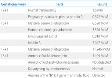
Table 1. Results of the Antenatal Test for the Patient
Gestational week Tests Results
12+1 Nuchal translucency 1.6 mm
Pregnancy-associated plasma protein A 0.392 MoM
16+1 Maternal serum α-fetoprotein 8.520 MoM
Human chorionic gonadotropin 3.526 MoM
Unconjugated estriol 0.818 MoM
Inhibin A 7.047 MoM
17+1 Maternal serum α-fetoprotein 11.246 MoM
18+1 Amniotic fluid α-fetoprotein 41.09 MoM
Amniotic fluid acetylcholine esterase Not detected Karyotyping by amniocentesis Normal Analysis of the NPHS1 gene in amniotic fluid Detected Abbreviations: MoM, multiples of the median; NPHS1, mutations in the gene encoding nephrin.
2020 June;31(2):99-102
www.e-kjp.org
https://doi.org/10.14734/PN.2020.31.2.99101
Perinatology
defect and chromosomal abnormalities were excluded from the diagnosis. Followup tests at 18+1 weeks of gestation showed persistent, abnormally elevated MSAFP and amniotic fluid α- fetoprotein levels. Further evaluation for detection of mutations in the NPHS1 gene in the amniotic fluid demonstrated causative mutations of CNS; frameshifting deletion mutation in exon 6 (c.619del, p.Arg207Glyfs*28) and missense mutation in exon 9 (c.1105C>T, pArg369Trp). The compound heterozygosity was confirmed, and each parent contributed one mutation. In com
pound heterozygous forms, some autosomal recessive diseases like hemochromatosis may have lower penetrance because the mutations involved are often less deleterious in com bination than for a homozygous individual with the classic symptoms of the disease.19 In this case, the neonate did not show any defi
nite symptoms or signs of NS, such as edema, proteinuria, or decreased kidney function. However, close monitoring of infant growth and regular blood and urine tests were performed with nutritional support but without medicine.
Although not observed for this case, CNS in the neonate can cause significant morbidity, resulting in kidney transplantation and mortality in many cases. Fortunately, some mutations in the gene expressing CNS have been revealed. Different mutations in the same gene express the disease at various ages with different penetrance. Therefore, prenatal diagnosis of CNS using genetic tests can help to evaluate and manage affected neo nates after birth.
Conflict of interest
No potential conflict of interest relevant to this article was reported.
References
1) Kestilä M, Järvelä I. Prenatal diagnosis of congenital nephrotic syn
drome (CNF, NPHS1). Prenat Diagn 2003;23:3234.
2) Rheault MN. Nephrotic and nephritic syndrome in the newborn. Clin Perinatol 2014;41:60518.
3) Machuca E, Benoit G, Nevo F, Tête MJ, Gribouval O, Pawtowski A, et al.
Genotypephenotype correlations in nonFinnish congenital nephrotic syndrome. J Am Soc Nephrol 2010;21:120917.
than 25% of the infant’s birth weight.2,11 About 80% of affected infants demonstrate edema and other signs of NS within the first week of life, with others presenting within 3 months.12 Severe hypoalbuminemia is present with a serum albumin concentration generally <1.5 mg/dL.2 Typically, renal function is initially nor
mal, and kidney function disruption presents over several years.2 A kidney biopsy is rarely necessary for diagnosis of CNF.2 De
tection of mutations in NPHS1 can confirm diagnosis of CNF.
More than 80 mutations in NPHS1 have been identified in af
fected patients.13 Mutations have been found throughout all 29 exons.13,14
Elevated MSAFP level obtained in a routine second-trimester screening is a suspicious finding for CNS.2 It is common and is found in approximately 1% of all pregnancies using a cutoff of
>2.5 MoM.2 Increased MSAFP can be seen in pregnancies with neural tube defects, gastroschisis, or chromosomal abnormalit
ies as well as CNS.15,16 CNS is a rare cause of elevated MSAFP, with a retrospective study identifying only five infants with CNS among 658 women with elevated MSAFP (<1%).16 Most pregnancies complicated with CNS show persistently elevated MSAFP on follow-up tests and elevated α-fetoprotein in am
niotic fluid obtained by amniocentesis.16 Median MSAFP con
centration was 8.3 MoM in pregnancies affected by CNS due to NPHS1 mutation, while median α-fetoprotein concentration in amniotic fluid was much higher at 33.4 MoM.17 Brady et al.18 suggested that MSAFP values peak between 19 and 21 weeks and then decline in NPHS1 carrier pregnancies, whereas values remain elevated in affected pregnancies. However, limited in
formation is available on the gestational age range of this peak and decline of MSAFP value for carrier pregnancies.
The treatment is generally supportive and includes massive intravenous albumin infusion, close monitoring of neonate growth, hypercaloric diet with added protein, and infection pre
vention.1,2,11 Bilateral nephrectomies are performed when child
ren are nearing endstage kidney disease or if the CNS cannot be managed with medical treatment, usually at 1-3 years old.2,11
In the present case, the pregnant woman presented with ab
normally elevated MSAFP that was determined by integrated test performed at the second trimester. The patient is from the Philippines, and her husband is Korean. Both had no family history of nephropathy including nephrotic syndrome. Through amniocentesis and ultrasonography examination, neural tube
Seo JM, et al. The diagnosis of congenital nephrotic syndrome
102
https://doi.org/10.14734/PN.2020.31.2.99www.e-kjp.org
Perinatology
4) Santín S, GarcíaMaset R, Ruíz P, Giménez I, Zamora I, Peña A, et al.
Nephrin mutations cause childhood and adultonset focal segmental glomerulosclerosis. Kidney Int 2009;76:126876.
5) Hinkes B, Vlangos C, Heeringa S, Mucha B, Gbadegesin R, Liu J, et al.
Specific podocin mutations correlate with age of onset in steroid
resistant nephrotic syndrome. J Am Soc Nephrol 2008;19:36571.
6) Huttunen NP. Congenital nephrotic syndrome of Finnish type. Study of 75 patients. Arch Dis Child 1976;51:3448.
7) Kestilä M, Lenkkeri U, Männikkö M, Lamerdin J, McCready P, Putaala H, et al. Positionally cloned gene for a novel glomerular proteinnephrin
is mutated in congenital nephrotic syndrome. Mol Cell 1998;1:57582.
8) Spahiu L, Merovci B, Jashari H, Këpuska AB, Rugova BE. Congenital nephrotic syndrome finish type. Med Arch 2016;70:2324.
9) Lee BH, Ahn YH, Choi HJ, Kang HK, Kim SD, Cho BS, et al. Two Korean infants with genetically confirmed congenital nephrotic syndrome of Finnish type. J Korean Med Sci 2009;24 Suppl 1:2104.
10) Lee JH, Han KH, Lee H, Kang HG, Moon KC, Shin JI, et al. Genetic basis of congenital and infantile nephrotic syndromes. Am J Kidney Dis 2011;58:
10423.
11) Yoshizawa C, Kobayashi Y, Ikeuchi Y, Tashiro M, Kakegawa S, Watanabe T, et al. Congenital nephrotic syndrome with a novel NPHS1 mutation.
Pediatr Int 2016;58:12115.
12) Patrakka J, Kestilä M, Wartiovaara J, Ruotsalainen V, Tissari P, Lenkkeri U, et al. Congenital nephrotic syndrome (NPHS1): features resulting from
different mutations in Finnish patients. Kidney Int 2000;58:97280.
13) Heeringa SF, Vlangos CN, Chernin G, Hinkes B, Gbadegesin R, Liu J, et al.
Thirteen novel NPHS1 mutations in a large cohort of children with congenital nephrotic syndrome. Nephrol Dial Transplant 2008;23:3527
33.
14) Beltcheva O, Martin P, Lenkkeri U, Tryggvason K. Mutation spectrum in the nephrin gene (NPHS1) in congenital nephrotic syndrome. Hum Mutat 2001;17:36873.
15) Muller F, Forestier F, Dingeon B; ABA Study Group. Second trimester trisomy 21 maternal serum marker screening. Results of a countrywide study of 854,902 patients. Prenat Diagn 2002;22:9259.
16) Spaggiari E, Ruas M, Dreux S, Valat AS, Czerkiewicz I, Guimiot F, et al.
Management strategy in pregnancies with elevated secondtrimester maternal serum alphafetoprotein based on a second assay. Am J Obstet Gynecol 2013;208:303.e17.
17) Patrakka J, Martin P, Salonen R, Kestilä M, Ruotsalainen V, Männikkö M, et al. Proteinuria and prenatal diagnosis of congenital nephrosis in fetal carriers of nephrin gene mutations. Lancet 2002;359:15757.
18) Brady TB, Mitra AG, Hooks J. Maternal serum alphafetoprotein levels peak at 1921 weeks' gestation and subsequently decline in an NPHS1 sequence variant heterozygote; implications for prenatal diagnosis of congenital nephrosis of the Finnish type. Prenat Diagn 2014;34:8124.
19) Waalen J, Nordestgaard BG, Beutler E. The penetrance of hereditary hemochromatosis. Best Pract Res Clin Haematol 2005;18:20320.