Peroxisome Proliferator-Activated Receptor-Gamma Expression in the Lung Tissue of Obese Rats
Su Jin Hwang,
1Jung Ho Kim,
1Jae Won Shim,
1Duk Soo Kim,
1Hye Lim Jung,
1Moon Soo Park,
1Won Young Lee,
2Se-Yeon Kim,
3and Jung Yeon Shim
11Department of Pediatrics, 2Endocrinology and Metabolism, and 3Research Institute of Medical Science, Kangbuk Samsung Hospital, Sungkyunkwan University School of Medicine, Seoul, Korea.
Received: March 3, 2010 Revised: August 10, 2010 Accepted: August 11, 2010
Corresponding author: Dr. Jung Yeon Shim, Division of Pediatric Allergy & Pulmonology, Department of Pediatrics,
Kangbuk Samsung Hospital,
Sungkyunkwan University School of Medicine, 78 Saemunan-gil, Jongno-gu,
Seoul 110-746, Korea.
Tel: 82-2-2001-2484, Fax: 82-2-2001-2199 E-mail: [email protected]
∙ The authors have no financial conflicts of interest.
© Copyright:
Yonsei University College of Medicine 2011 This is an Open Access article distributed under the terms of the Creative Commons Attribution Non- Commercial License (http://creativecommons.org/
licenses/by-nc/3.0) which permits unrestricted non- commercial use, distribution, and reproduction in any medium, provided the original work is properly cited.
Purpose: Obesity is a risk factor for asthma and type II diabetes. Peroxisome prolif- erator-activated receptor (PPAR)-γ has been suggested to regulate inflammatory re- sponses in diabetes and asthma. We investigated whether PPAR-α, PPAR-γ, adipo- nectin receptors (AdipoR1, AdipoR2), leptin, and tumor necrosis factor (TNF)-α are expressed in rat lung tissues and whether the expression differs between obese Otsu- ka Long-Evans Tokushima Fatty (OLETF) and lean Long Evans Tokushima Otsuka (LETO) rats. Materials and Methods: Obese and lean rats were given with a high fat diet or a 30% restricted diet for 32 weeks, and their blood glucose levels and weights were monitored. After 32 weeks, mRNA levels of PPAR-α, PPAR-γ, Adi- poR1, AdipoR2, leptin, and TNF-α in lung tissues were measured using real time PCR. Results: PPAR-α, PPAR-γ, AdipoR1, AdipoR2, leptin, and TNF-α were ex- pressed in both obese and lean rat lung tissues. Increased serum glucose levels on in- traperitoneal glucose tolerance testing and a higher weight gain at 32 weeks were ob- served in OLETF control rats compared to OLETF diet restricted rats. PPAR-γ expression was markedly elevated in obese control and diet restricted rats compared to lean rats, although PPAR-γ expression in obese rats was not affected by diet re- striction. Leptin was highly expressed in OLETF rats compared to LETO rats.
TNF-α expression was enhanced in OLETF control rats compared LETO diet re- stricted rats, and decreased by diet restriction. PPAR-α, AdipoR1, and AdipoR2 ex- pression were not significantly different between obese and lean rats. Conclusion:
PPAR-γ was highly expressed in the lung tissues of obese rats and may be a novel treatment target for regulating lung inflammation associated with obesity.
Key Words: Obesity, peroxisome proliferator activated receptor, adiponectin re- ceptor, lung, leptin, TNF-alpha
INTRODUCTION
Obesity is a significant health problem worldwide and its incidence has more than
doubled during the last 20 years.
1Obesity is a leading risk factor for the develop-
ment of diabetes, cardiovascular disease, and malignancies, and also has an impact
lean subjects. The goal of this study was to determine whether PPAR-α, PPAR-γ, adiponectin receptor 1 (Adi- poR1), AdipoR2, leptin, and TNF-α were expressed in rat lung tissues and whether their mRNA levels were different between obese and lean rats.
MATERIALS AND METHODS
Animals
Animal experiments were conducted in accordance with Kangbuk Samsung Hospital Guide for the Care and Use of Laboratory Animals. Four week old male obese, prediabetic rats, Otsuka Long-Evans Tokushima Fatty (OLETF) rats and their lean non-diabetic counterparts, Long Evans Tokushima Otsuka (LETO) rats, were supplied by Otsuka Pharmaceu- tical (Tokushima, Japan). The OLETF rats were initially characterized by late onset hyperglycemia and mild obesity, and have been studied as a model of non insulin dependent diabetes mellitus.
23The rats were divided into six groups with approximately equal mean body weights: the LETO control group (n=6);
the LETO diet restriction group (n=6); the OLETF control group (n=6); and the OLETF diet restriction group (n=6).
The control groups were given with a 45% high fat diet, while the diet restriction groups with a 30% restricted diet for 32 weeks.
The animals were housed individually in cages at a con- stant temperature (20-22°C) and humidity (60%) with a 12-hour-light and 12-hour-dark cycle. They had free access to water and rat chow until the age of 32 weeks. Body weight and food intake were checked twice per week and intraperi- toneal glucose tolerance testing was performed at 32 weeks of diet administration. The animals were sacrificed by decap- itation at 32 weeks of treatment, and their lung tissues were rapidly removed.
Intraperitoneal glucose tolerance testing
The serum glucose concentration was monitored using in- traperitoneal glucose tolerance testing (IPGTT) in the OLETF control and diet restricted rats as well as in the LETO control and diet restricted rats. The rats were injected intraper- itoneally with glucose solution (2 g/kg, 50% sterile glucose solution). Blood samples were drawn from the tail vein at 0, 30, 60, 90, and 120 minutes after glucose administration.
Blood glucose levels were measured using a glucometer (Ac- cu-Chek-Performa, Roche Diagnostics, Penzberg, Germany).
on respiratory diseases such as asthma, chronic obstructive pulmonary disease as well as obesity hypoventilation syn- drome and sleep apnea.
2Furthermore, obesity has been re- ported to increase the prevalence and severity of asthma and also influences lung function.
3A recent animal study showed a relationship between obesity and asthma.
4The influence of obesity on lung tissue involves the in- flammatory changes in the lung microenvironment. It induc- es a chronic, low-grade systemic inflammation, characterized by an increase in circulating leukocytes and proinflammatory cytokines such as interleukin (IL)-6, tumor necrosis factor (TNF)-α, resistin, free fatty acids, and leptin.
1,5-7These pro- teins can originate from the adipose tissue itself, leading to systemic inflammation as well as airway inflammation.
8Adiponectin, a fat tissue-induced hormone, is negatively associated with obesity, while leptin is positively related to obesity. In children with obesity, serum leptin levels are ele- vated, which is decreased by weight reduction.
9On the oth- er hand, serum adiponectin levels decrease in obese chil- dren, and adiponectin mRNA expression from adipose tissue has been shown to be inversely associated with obe- sity.
10Adiponectin has also been reported to have anti-in- flammatory effects and down-regulates vascular smooth muscle cell proliferation in the presence of obesity.
11Peroxisome proliferator-activated receptors (PPARs) be- long to a family of ligand-activated transcription factors and include the nuclear hormone receptor family related to reti- noid, glucocorticoid, and thyroid hormone receptors.
12PPAR-α is highly expressed in the liver, heart, kidney, and skeletal muscle.
13PPAR-γ is expressed in the lung epitheli- um, submucosa, and airway smooth muscle. PPAR-α and PPAR-γ are also expressed in alveolar macrophages and air- way epithelial cells.
14PPAR-γ ligands have been shown to inhibit the release of pro-inflammatory cytokines from acti- vated macrophages and airway epithelial cells.
15,16Further- more, PPAR-γ ligands inhibit vascular smooth muscle cell proliferation, and induce apoptosis in endothelial cells, vas- cular smooth muscle,
17T lymphocytes and macrophages.
18Inflammation plays a critical role in asthma, and inhaled
corticosteroids, which are the most potent anti-inflammato-
ry medication have been used as the first line asthma treat-
ment during the last 20 years.
19,20However, obesity can al-
ter the asthma phenotype into a more difficult-to-control
and steroid-resistant type of asthma.
21,22This may be caused
by different characteristics of the lung inflammation in
obese asthmatic patients. Thus, it is critical to understand
the differences in lung inflammation between obese and
thermal cycling profile consisted of a preincubation step at 95°C for 10 minutes, followed by 45 cycles of a 95°C dena- turation step for 10 seconds, a 55°C annealing step for 1 min- ute, and a 72°C extension step for 4-9 seconds. At the end of the PCR, a melting curve analysis was performed by gradu- ally increasing the temperature from 65°C to 95°C (0.2°C/s) to confirm the amplification specificity of the PCR products.
The mRNA levels were corrected using the transcription lev- el of the β-actin as an internal standard.
Statistical analysis
Statistical analysis was performed using GraphPad Prism software 5.0 (GraphPad Software, San Diego, CA, USA).
Comparisons of PPAR-α, PPAR-γ, AdipoR1, AdipoR2, leptin, and TNF-α mRNA concentrations between two groups were analyzed using Student t-test. p<0.05 was con- sidered significant.
RESULTS
Body weight
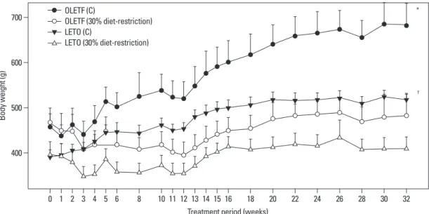
There were no differences in the baseline body weight be- tween OLETF control rats and OLETF diet restricted rats, and between LETO control rats and LETO diet restricted rats at 0-week. The initial mean body weights of the LETO control and diet restricted rats were 394 g and 399 g, re- spectively, and the mean body weights of the OLETF control and diet restricted rats were 460 g and 467 g, respectively.
OLETF control rats had an increase of weight by 48.9%
and the OLETF diet restricted rats had an increase of weight by 5% at 32 weeks of treatment. The LETO control rats had a 31.9% weight gain whereas the LETO diet restricted rats had a 2.2% weight gain at the end of treatment. At 32 weeks of treatment, the mean body weight showed a signif- icant increase in OLETF control rats compared to the OLETF diet restricted rats and in the LETO control rats compared to the LETO diet restricted rats (Fig. 1).
Intraperitoneal glucose tolerance testing
The serum glucose concentrations were significantly higher in the OLETF control rats (p<0.05), then the OLETF diet re- stricted rats and the LETO groups at all study time points (ex- cept for the fasting serum glucose at 0 min), measured after intraperitoneal glucose load. For the OLETF control rats, the mean blood glucose level was 230 mg/dL at 30 minutes after glucose administration and 215 mg/dL at 120 minutes (Fig. 2).
RNA analysis and quantitative real time PCR for PPAR-α, PPAR-γ, AdipoR1, AdipoR2, Leptin, and TNF-α
The lung tissue was ground and homogenized with liquid nitrogen in a mortar. Then, the mRNA expression was ana- lyzed by real time reverse transcriptase (RT) PCR; the ratio of mRNA copy number to β-actin mRNA copy number was determined. The total RNA was extracted, using TRI Reagent (Sigma Chemical Co., St. Louis, MO, USA), ac- cording to the manufacturer’s instructions. To summarize the real time PCR, 1 mL of TRI-reagent was blended in lung tissue and homogenized by ultrasound; 200 µL of chlo- roform was added and the solution was vigorously mixed.
Then, the sample was centrifuged for 15 minutes, and the RNA was subjected to reverse transcription using the cold Strand cDNA Synthesis Kit (ThermoFisher Scientific, Vilni- us, Lithuania) for 60 minutes at 42°C, and for 10 minutes at 70°C. The FastStart Taq DNA polymerase (LightCycler FastStart DNA Master SYBR Green I, Roche Diagnostics, Germany) and 3 mM MgCl
2were added, and 0.4 μM prim- er was amplified in the LightCycler 480 Instrument (Roche Molecular Biochemicals, Penzberg, Germany).
The following primers were used: for the rat AdipoR1, the forward primer was 5’-AGATGGGCTGGTTCTTC CTC-3’, and the reverse primer was 5’-CAGTGCATTT- GCCAGG-3’ (GenBank accession number: NM_207587);
for the rat AdipoR2, the forward primer was 5’-ATGTTT-
GCCACCCCTCAGTA-3’, and the reverse primer was
5’-CAGATGTCACATTTGCCAGG-3’ (GenBank acces-
sion number: NM_001037979); for the rat PPAR-α, the for-
ward primer was 5’-TTCGGAAACTGCAGACCT-3’, and
the reverse primer was 5’-TTAGGAACTCTCGGGT-
GAT-3’ (GenBank accession number: NM_013196); for the
rat PPAR-γ, the forward primer was 5’-TAGGTGTGATC
TTAACTGTCG-3’, and the reverse primer was 5’-GCAT-
GGTGTAGATGATCTCA-3’ (GenBank accession num-
ber: NM_013124); for the rat TNF-α, the forward primer
was 5’-GGGGCCACCACGCTCTTCTGTCTA-3’, and
the reverse primer was 5’-CCTCCGCTTGGTGGTTTGC-
TACG-3’ (GenBank accession number: NM_012675); for
the rat leptin, the forward primer was 5’-TTGTCACCAG-
GATCAATGACATTTG-3’, and the reverse primer was
5’-ACAAACTCAGAATGGGGTGAAG-3’ (GenBank ac-
cession number: NM_013076); for the rat β-actin, the for-
ward primer was 5’-AGGTCATCACTATCGGCAAT-3’,
and the reverse primer was 5’-ACTCATCGTACTCCT-
GCTTG-3’ (GenBank accession number: NM_031144). The
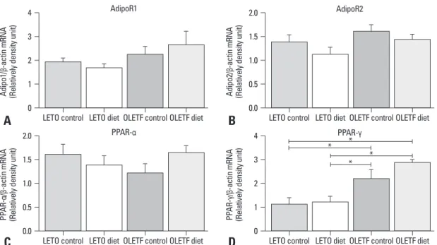
ed by diet restriction. However, the PPAR-γ mRNA levels were significantly elevated in the OLETF control rats com- pared to the LETO control and the LETO diet restricted rats. The OLETF diet restricted rats showed higher PPAR-γ mRNA levels than the LETO diet restriction rats and the LETO control rats. However, there were no significant dif- ferences in the PPAR-γ mRNA levels between the OLETF control and the OLETF diet restricted rats and between the LETO control and LETO diet restricted rats (Fig. 3).
The leptin mRNA levels were highly enhanced in the OLETF control rats compared to the LETO control and the LETO diet restricted rats. There was a decreasing tendency of leptin mRNA levels by diet restriction in OLETF rats, al- though not statistically significant. TNF-α was highly ex- pressed in lung tissue of the OLETF control rats compared to the LETO diet restriction rats, showing significant de- crease by diet restriction in OLETF rats (Fig. 4).
DISCUSSION
The expression of PPAR-α, PPAR-γ, adiponectin receptors (AdipoR1, AdipoR2), leptin, and TNF-α was studied in rat lung tissues to determine whether their expressions were dif- ferent between prediabetic, obese OLETF and lean LETO rat model. And the results demonstrated that PPAR-α, PPAR-γ, adiponectin receptors, leptin, and TNF-α were expressed in rat lung tissue, and that PPAR-γ, leptin, and TNF-α were highly expressed in obese rat lung tissue compared to lean rat AdipoR1, AdipoR2, PPAR-α, PPAR-γ, leptin, and TNF-α
mRNA expression in the lung tissue of the study rats AdipoR1, AdipoR2, PPAR-α, PPAR-γ, leptin, and TNF-α mRNA expression in the lung tissue of control and diet re- stricted OLETF rats and the LETO rats were evaluated. Ad- ipoR1 and AdipoR2 mRNA levels were not significantly different between the OLETF control rats and the LETO control rats, and were not influenced by diet restriction in the OLETF or the LETO rats. In addition, the PPAR-α mRNA levels were not significantly different between the OLETF control rats and the LETO control rats, and were not affect-
Fig. 1. Body weight changes between control rats and rats with 30% diet restriction. The weight gain of the OLETF control group was signif- icantly higher than the other groups (p<0.05). The LETO control group showed a higher weight gain than the LETO diet restricted group. The OLETF diet restricted group had a greater weight gain than the LETO diet restricted group. *p<0.01 compared to the other groups. †p<0.05 compared to LETO (30% diet-restriction). OLETF, Otsuka Long-Evans Tokushima Fatty; LETO, Long-Evans Tokushima Otsuka; C, control.
400 500 600 700
Body weight (g)
0
Treatment period (weeks)
1 2 3 4 5 6 8 10 11 12 13 14 15 16 18 20 22 24 26 28 30 32
*
†
OLETF (C) LETO (C)
OLETF (30% diet-restriction) LETO (30% diet-restriction)
Fig. 2. Intraperitoneal glucose tolerance testing in obese and lean rats at 32 weeks of treatment. The OLETF control rats showed higher serum glucose levels than the other groups at 30, 60, 90 and 120 minutes after an intraperi- toneal glucose challenge. *p<0.05 compared to the other groups. OLETF, Otsuka Long-Evans Tokushima Fatty; LETO, Long-Evans Tokushima Otsuka;
C, control.
50 100 150 200 250 300 350 400
Glucose (mg/dL)
0 30 60 90 120 150
Time (min)
*
* *
*
OLETF (C) LETO (C)
OLETF (30% diet-restriction) LETO (30% diet-restriction)
over, PPAR-γ ligands inhibit PPAR-γ levels in the lung and the release of proinflammatory cytokines from activated macrophages, airway epithelial cells, and eosinophils, and decrease airway hyperreactivity, basement membrane thick- ness, collagen deposition, and eosinophilia in murine models of asthma. Furthermore, PPAR-γ agonists reduced cigarette smoke-induced mucin production in the airway epithelial cells
28and a nebulized PPAR-γ agonist, ciglitazone, signifi- cantly suppressed mucus secretion and collagen deposition in airway epithelial cells of allergen-challenged sensitized mice.
29In this regard, PPAR-γ might be a target for reduc- ing airway inflammation and remodeling.
There are only a few clinical studies for the therapeutic effect of PPAR-γ ligands in asthma.
lung tissue.
The presence of PPARs has been demonstrated in a vari- ety of cells associated with lung inflammation.
24PPAR-γ has been found in lung epithelial cells, smooth muscle cells, fibroblasts, endothelial cells, macrophages, eosinophils, T cells, B cells, and dendritic cells.
14-16,25,26Recent studies have shown that PPAR-γ has some beneficial effects with regard to the regulation of airway inflammation in asthma.
PPAR-γ expression is augmented in the bronchial submu- cosa, airway epithelium and smooth muscle in steroid-un- treated asthma and has been associated with submucosal basement membrane thickening and collagen deposition. In addition, oral or inhaled steroids have been shown to down- regulate PPAR-γ expression and airway remodeling.
27More-
Fig. 3. Adiponectin receptor 1, adiponectin receptor 2, PPAR-α, and PPAR-γ mRNA expressions in lung tissue of OLETF rats and LETO rats:
each mRNA expression was measured by real time PCR. (A) AdipoR1 mRNA levels did not show significant differences among all groups. (B) AdipoR2 mRNA levels were not different in the four groups. (C) PPAR-α mRNA levels in the four groups showed no significant differences. (D) PPAR-γ mRNA levels showed marked increases in the OLETF control rats compared to the LETO control and diet restrict- ed rats. However, PPAR-γ was not affected by diet restriction. *p<0.05. OLETF, Otsuka Long-Evans Tokushima Fatty; LETO, Long-Evans Tokushima Otsuka.
LETO control
LETO control
LETO control
LETO control LETO diet
LETO diet AdipoR1
PPAR-α
AdipoR2
PPAR-γ LETO diet
LETO diet OLETF control
OLETF control
OLETF control
OLETF control OLETF diet
OLETF diet
OLETF diet
OLETF diet 0
0.0
0.0
0 1
0.5
0.5
1 2
1.0
1.0
2 3
1.5
1.5
3 4
2.0
2.0
4
Adipo1/β-actin mRNA (Relatively density unit)PPAR-α/β-actin mRNA (Relatively density unit) Adipo2/β-actin mRNA (Relatively density unit)PPAR-γ/β-actin mRNA (Relatively density unit)
* *
* *
A
C
B
D
Fig. 4. Leptin and TNF-α mRNA expressions in lung tissue of OLETF rats and LETO rats: each mRNA expression was measured by real time PCR. (A) Leptin mRNA levels showed marked increase in the OLETF control rats compared to the LETO control and diet restricted rats.
However, leptin was not affected by diet restriction. (B) TNF-α mRNA levels increased significantly in the OLETF control rats compared to the OLETF and LETO diet restricted rats. *p<0.05. OLETF, Otsuka Long-Evans Tokushima Fatty; LETO, Long-Evans Tokushima Otsuka.
LETO control LETO diet LETO control
Leptin TNF-α
LETO diet
OLETF controlOLETF diet OLETF controlOLETF diet
0 2 4 6 8 10
0.0 0.5 1.0 1.5 2.0 2.5
Leptin/β-actin mRNA (Relatively density unit) TNF-α/β-actin mRNA (Relatively density unit) * *
A B
* *
regulate PPAR-γ expression in mononuclear cells.
40In the present study, leptin expression in the lung of obese rats was highly enhanced compared to lean rats. The origin of leptin and TNF-α in obesity has currently been known to be adi- pose tissue. Our results imply that TNF-α and leptin are also expressed on lung tissue of obese rats. We are not certain whether TNF-α or leptin influences PPAR-γ expression in obesity, since there are no data available. Nevertheless, our results may imply that increased proinflammatory cytokines in lung tissue are related to PPAR-γ expression in obesity.
Adiponectin decreases obesity and may play an anti-in- flammatory role in obesity-related diseases.
11Adiponectin receptors (AdipoR1 and AdipoR2) are reported to be ex- pressed in airway smooth muscle cells.
38However, there have been no reported data which compared the expression of adiponectin receptors in obese and lean rat lung tissues.
The present results showed that adiponectin receptors were expressed in rat lung tissue, although the mRNA levels did not differ in the lean compared to obese states.
The present study was limited by relatively small number of subjects and the absence of an asthma model. Neverthe- less, this study is the first trial to demonstrate that PPAR-γ, leptin, and TNF-α were highly expressed in the lung tissue of obese rats, and that the expression of adiponectin recep- tors and PPAR-α were not affected by obesity. In addition, weight reduction was found to have no effect on PPAR-γ expression, although it reduced TNF-α expression. This sug- gests that leptin, TNF-α, and PPAR-γ are highly expressed in lung tissue of obesity, and that PPAR-γ, but not PPAR-α or adiponectin receptors, might be a novel treatment target for regulating lung inflammation associated with obesity.
ACKNOWLEDGEMENTS
The work was supported by IN-SUNG Foundation of Med- ical Research.
These results were presented at American Thoracic Society International Conference, San Diego, USA, on May 2009.
REFERENCES
1. Shore SA, Fredberg JJ. Obesity, smooth muscle, and airway hy- perresponsiveness. J Allergy Clin Immunol 2005;115:925-7.
2. McClean KM, Kee F, Young IS, Elborn JS. Obesity and the lung:
1. Epidemiology. Thorax 2008;63:649-54.
3. Beuther DA, Weiss ST, Sutherland ER. Obesity and asthma. Am J
Pioglitazone improved wheezing and coughing in pa- tients with asthma and type 2 diabetes and, when the treat- ment with pioglitazine was discontinued because of poor efficacy for the diabetes, asthma symptoms restarted.
30An- other study shows that rosiglitazone improved lung func- tion, compared with inhaled corticosteroid in steroid-resis- tant smokers with asthma.
31More clinical trials are needed to evaluate definite effect of PPAR-γ ligands in asthma. By contrast, PPAR-γ genetic expression has been shown to be reduced in asthmatic patients after an allergen challenge, and inversely related to airway inflammation.
32Therefore, the precise role of PPAR-γ in respiratory disease requires further clarification.
During the last 20 years, the prevalence of asthma has in- creased along with an increase in obesity, or the body mass index (BMI), in children and adults.
33However, there have been only a few studies on PPAR-γ expression in the lung and its association with obesity. The impact of obesity on asthma has not been clearly explained to date. Obesity can cause a reduction in respiratory compliance, lung volumes, and peripheral airflow obstruction,
34and can also lead to an increase in airway hyperresponsiveness, alteration in pul- monary blood flow, and ventilation-perfusion mismatch.
3In addition, obesity leads to a systemic, low-grade proinflam- matory state by enhancing adipose tissue activity.
35Adipose tissue from obese individuals expresses various proinflam- matory mediators such as leptin, TNF-α, IL-6, transforming growth factor-β1 (TGF-β1), and C-reactive protein.
8,36These proinflammatory mediators may induce inflammatory changes in lung tissue and lead to the development of asth- ma or cause preexisting lung inflammation into a more dif- ficult-to-control phenotype in asthma. In this study, we found that TNF-α was highly expressed in lung tissue of obese rats and significantly decreased by diet restriction. In- creased proinflammatory mediators in lung tissue of obesi- ty may play a role in inducing lung inflammation.
Leptin acts as a proinflammatory mediator and its levels
are increased in obesity. It enhances cytokine release in li-
popolysaccharide-stimulated macrophages and monocytes,
and increases CD4+ T cell proliferation.
37Leptin may play a
role in regulating airway inflammation. We previously dem-
onstrated that leptin promoted vascular endothelial growth
factor (VEGF) release by human airway smooth muscle
cells.
38The PPAR-γ ligand decreases leptin levels and in-
creases adiponectin levels in bronchoalveolar lavage (BAL)
fluids from lean mice, whereas it increases BAL leptin levels
in obese mice.
39By contrast, leptin has been shown to down-
Evans Tokushima Fatty) rat: a new NIDDM rat strain. Diabetes Res Clin Pract 1994;24 Suppl:S317-20.
24. Belvisi MG, Hele DJ. Peroxisome proliferator-activated receptors as novel targets in lung disease. Chest 2008;134:152-7.
25. Faveeuw C, Fougeray S, Angeli V, Fontaine J, Chinetti G, Gosset P, et al. Peroxisome proliferator-activated receptor gamma activa- tors inhibit interleukin-12 production in murine dendritic cells.
FEBS Lett 2000;486:261-6.
26. Woerly G, Honda K, Loyens M, Papin JP, Auwerx J, Staels B, et al. Peroxisome proliferator-activated receptors alpha and gamma down-regulate allergic inflammation and eosinophil activation. J Exp Med 2003;198:411-21.
27. Benayoun L, Letuve S, Druilhe A, Boczkowski J, Dombret MC, Mechighel P, et al. Regulation of peroxisome proliferator-activated receptor gamma expression in human asthmatic airways: relation- ship with proliferation, apoptosis, and airway remodeling. Am J Respir Crit Care Med 2001;164:1487-94.
28. Belvisi MG, Hele DJ, Birrell MA. Peroxisome proliferator-acti- vated receptor gamma agonists as therapy for chronic airway in- flammation. Eur J Pharmacol 2006;533:101-9.
29. Honda K, Marquillies P, Capron M, Dombrowicz D. Peroxisome proliferator-activated receptor gamma is expressed in airways and inhibits features of airway remodeling in a mouse asthma model. J Allergy Clin Immunol 2004;113:882-8.
30. Hashimoto Y, Nakahara K. Improvement of asthma after adminis- tration of pioglitazone. Diabetes Care 2002;25:401.
31. Spears M, Donnelly I, Jolly L, Brannigan M, Ito K, McSharry C, et al. Bronchodilatory effect of the PPAR-gamma agonist rosiglitazone in smokers with asthma. Clin Pharmacol Ther 2009;86:49-53.
32. Kobayashi M, Thomassen MJ, Rambasek T, Bonfield TL, Raychaudhuri B, Malur A, et al. An inverse relationship between peroxisome proliferator-activated receptor gamma and allergic airway inflammation in an allergen challenge model. Ann Allergy Asthma Immunol 2005;95:468-73.
33. Xu B, Jarvelin MR, Pekkanen J. Body build and atopy. J Allergy Clin Immunol 2000;105:393-4.
34. Biring MS, Lewis MI, Liu JT, Mohsenifar Z. Pulmonary physio- logic changes of morbid obesity. Am J Med Sci 1999;318:293-7.
35. Fantuzzi G. Adipose tissue, adipokines, and inflammation. J Aller- gy Clin Immunol 2005;115:911-9; quiz 920.
36. Wellen KE, Hotamisligil GS. Obesity-induced inflammatory changes in adipose tissue. J Clin Invest 2003;112:1785-8.
37. Martín-Romero C, Santos-Alvarez J, Goberna R, Sánchez-Mar- galet V. Human leptin enhances activation and proliferation of hu- man circulating T lymphocytes. Cell Immunol 2000;199:15-24.
38. Shin JH, Kim JH, Lee WY, Shim JY. The expression of adiponec- tin receptors and the effects of adiponectin and leptin on airway smooth muscle cells. Yonsei Med J 2008;49:804-10.
39. Holguin F, Rojas M, Hart CM. The peroxisome proliferator acti- vated receptor gamma (PPARgamma) ligand rosiglitazone modu- lates bronchoalveolar lavage levels of leptin, adiponectin, and in- flammatory cytokines in lean and obese mice. Lung 2007;185:
367-72.
40. Cabrero A, Cubero M, Llaverías G, Alegret M, Sánchez R, Lagu- na JC, et al. Leptin down-regulates peroxisome proliferator-acti- vated receptor gamma (PPAR-gamma) mRNA levels in primary human monocyte-derived macrophages. Mol Cell Biochem 2005;275:173-9.
Respir Crit Care Med 2006;174:112-9.
4. Shore SA, Schwartzman IN, Mellema MS, Flynt L, Imrich A, Johnston RA. Effect of leptin on allergic airway responses in mice. J Allergy Clin Immunol 2005;115:103-9.
5. Rajala MW, Scherer PE. Minireview: The adipocyte--at the cross- roads of energy homeostasis, inflammation, and atherosclerosis.
Endocrinology 2003;144:3765-73.
6. Hotamisligil GS. Inflammatory pathways and insulin action. Int J Obes Relat Metab Disord 2003;27 Suppl 3:S53-5.
7. Nawrocki AR, Scherer PE. The delicate balance between fat and muscle: adipokines in metabolic disease and musculoskeletal in- flammation. Curr Opin Pharmacol 2004;4:281-9.
8. Weisberg SP, McCann D, Desai M, Rosenbaum M, Leibel RL, Ferrante AW Jr. Obesity is associated with macrophage accumula- tion in adipose tissue. J Clin Invest 2003;112:1796-808.
9. Elloumi M, Ben Ounis O, Makni E, Van Praagh E, Tabka Z, Lac G. Effect of individualized weight-loss programmes on adiponec- tin, leptin and resistin levels in obese adolescent boys. Acta Paedi- atr 2009;98:1487-93.
10. Kern PA, Di Gregorio GB, Lu T, Rassouli N, Ranganathan G. Ad- iponectin expression from human adipose tissue: relation to obesi- ty, insulin resistance, and tumor necrosis factor-alpha expression.
Diabetes 2003;52:1779-85.
11. Ouchi N, Kihara S, Funahashi T, Matsuzawa Y, Walsh K. Obesity, adiponectin and vascular inflammatory disease. Curr Opin Lipidol 2003;14:561-6.
12. Evans RM. The steroid and thyroid hormone receptor superfamily.
Science 1988;240:889-95.
13. Chinetti G, Fruchart JC, Staels B. Peroxisome proliferator-activated receptors (PPARs): nuclear receptors at the crossroads between lipid metabolism and inflammation. Inflamm Res 2000;49:497-505.
14. Patel HJ, Belvisi MG, Bishop-Bailey D, Yacoub MH, Mitchell JA. Activation of peroxisome proliferator-activated receptors in human airway smooth muscle cells has a superior anti-inflamma- tory profile to corticosteroids: relevance for chronic obstructive pulmonary disease therapy. J Immunol 2003;170:2663-9.
15. Jiang C, Ting AT, Seed B. PPAR-gamma agonists inhibit produc- tion of monocyte inflammatory cytokines. Nature 1998;391:82-6.
16. Wang AC, Dai X, Luu B, Conrad DJ. Peroxisome proliferator-ac- tivated receptor-gamma regulates airway epithelial cell activation.
Am J Respir Cell Mol Biol 2001;24:688-93.
17. Bishop-Bailey D, Warner TD. PPARgamma ligands induce pros- taglandin production in vascular smooth muscle cells: indometha- cin acts as a peroxisome proliferator-activated receptor-gamma antagonist. FASEB J 2003;17:1925-7.
18. Chinetti G, Griglio S, Antonucci M, Torra IP, Delerive P, Majd Z, et al. Activation of proliferator-activated receptors alpha and gam- ma induces apoptosis of human monocyte-derived macrophages.
J Biol Chem 1998;273:25573-80.
19. British Thoraoic Society. British guideline on the management of asthma. Thorax 2003;58 Suppl 1:i1-94.
20. Barnes PJ. Inhaled glucocorticoids for asthma. N Engl J Med 1995;332:868-75.
21. Leung DY, Bloom JW. Update on glucocorticoid action and resis- tance. J Allergy Clin Immunol 2003;111:3-22.
22. Wenzel S. Severe asthma in adults. Am J Respir Crit Care Med 2005;172:149-60.
23. Kawano K, Hirashima T, Mori S, Natori T. OLETF (Otsuka Long-