A R T I C L E O R I G I N A L
Purpose: Peroxisome proliferator-activated recep- tor gamma (PPARγ) has become a potential target for the prevention and treatment of human cancers.
PPARγligands inhibit cell proliferation of estrogen receptorα(ERα)-positive breast cancer cells.
However, it has recently been shown that ERα-nega- tively inhibits PPARγsignaling in breast cancer cells, indicating that PPARγligand may be more useful for treating ERα-negative breast cancer cells compared to ERα-positive breast cancer cells. In this study, we attempted to elucidate the role of PPARg in ERα-neg- ative breast cancer cells.
Methods: The effect of PPARγligand on the growth of MDA-MB-231 cells was measured by MTT assay and flow cytometric analysis. TUNEL staining and Hoechst 33342 fluorescent staining were used to observe the effects of PPARγligand on cell apoptosis.
The regulatory proteins of the cell cycle were mea- sured by Western blot.
Results: The treatment of MDA-MB-231 human
breast cancer cells with the PPARγligand, trgoglita- zone, was shown to induce inhibition of cell growth in a dose-dependent manner. Cell cycle analysis showed a G1arrest in MDA-MB-231 cells exposed to troglitazone. The apoptotic effect by troglitazone demonstrated that apoptotic cells were elevated from 2.5-fold of the control level at 10 mM, to 3.1-fold at 50μM and to 3.5-fold at 75 mM of troglitazone.
Moreover, troglitazone treatment dose-dependently caused a marked decrease in the pRb, cyclin D1, cyclin D2, cyclin D3, cdk2, Cdk4 and Cdk6 expressions and there was a significant increase in the p21 and p27 expressions.
Conclusion: These results indicate that trgoglita- zone induces cell-cycle G1arrest and apoptosis in ER α-negative MDA-MB-231 breast cancer cells.
Collectively, this paper shows that PPARγligand is an important player as a member of the chemothera- peutic candidates for treating ERα-negative breast cancer. (J Breast Cancer 2006;9: 293-300)
Key Words ER-negative MDA-MB-231 breast cancer cells, PPARγ, Apoptosis, Cell cycle arrest
INTRODUCTION
Breast cancer is the most common cancer and then leads to most of cancer deaths among women world-
Correspondence : Sung Hoo Jung
Department of Surgery, Chonbuk National University Hospital, 634-18 Geumam-dong, Deokjin-ku, Jeonju, 561-712, Korea.
Phone: 063-250-2133, Fax: 063-271-6197, E-mail: [email protected] Received: Aug 3, 2006; Accepted : Oct 10, 2006
Peroxisome proliferator-activated receptor gamma activator inhibits cell growth of MDA-MB-231 breast cancer cells through induction of apoptosis
Eun Jeong Jo, Hyun Jo Youn, Sung Hoo Jung
Division of Breast and Endocrine Surgery, Department of Surgery, Chonbuk National University Medical School, Jeonju, Korea
mone therapy has been in best used for prevention and treatment in women with early breast cancer.(1) However, hormone therapy has little effect on estro- gen receptor α(ERα)-negative tumors.(2) In deed, approximately 30% of breast cancer patients are neg- ative for ERαexpression at diagnosis. Furthermore, approximately 50% of patients with advanced dis- ease do not respond to first-line treatment with hor- mone therapy.(3) Therefore, resistance to hormone therapy causes a major problem in treatment and prevention of breast cancer. Thus, in order to over- come resistance to hormone therapy the novel treat- ment strategies should be explored.
Peroxisome proliferator-activated receptors (PPARs) are ligand-activated nuclear receptors that mediate transcriptional regulation of genes involved in the oxidation, transport, and storage of lipids.(4- 7) Among the three PPAR isoforms (PPARα, β, and γ),(8-10) PPARγinfluences such biological processes as inflammation, cell survival, differentiation, cell proliferation, and tumorigenesis.(11) Several ligands for PPARγhave been identified including endoge- nous 15-deoxy-Δ12,14-prostaglandin J2, linoleic acid, lysophophatidic acid, and the thiazolidinediones class of synthetic antidiabetic drugs such as troglita- zone and rosiglitazone.(12-18)
In the other hand, PPARg ligand has recently been incriminated as a potential target for the prevention and treatment of human cancers.(17) In breast can- cer, PPARγligands also inhibit proliferation and induce apoptosis in ERα-positive breast cancer cells.(19,20) These reports indicate that PPARγlig- and may prove to have a role in breast cancer treat- ment /prevention in the future. However, it has recently been known that ERαnegatively interferes PPARγsignaling in breast cancer cells,(21) indicat- ing that PPARγligand have not full activity for ERα- positive MCF-7 breast cancer cells. In deed, PPARγ activation by PPARγligand has little apparent clini- cal value among patients with treatment?refractory breast cancer.(22) Therefore, it is likely to suggest
ERα-positive MCF-7 breast cancer cells. Moreover, effect of PPARγligand on cell proliferation of ERα- negative MDA-MB-231 breast cancer cells is not known.
Therefore, we attempted to elucidate the role of PPARγin ERα-negative breast cancer carcinogenesis and explore the possibility of using PPARγligand as chemopreventive agent for hormone therapy-resis- tant breast cancer patients. In the current study, we determined if PPARγligand induces cell-cycle arrest and apoptosis in ERα- negative MDA-MB-231 breast cancer cell line. The observed apoptotic activi- ty of PPARγligand was accompanied by a cell cycle regulator induction such as p21. This paper shows the first time evidences that PPARγligand plays as a primary member of chemotherapeutic candidates for ERα-negative breast cancer. So, we expect the clinical use of PPARγligand, which is more useful for in ERα-negative breast cancers compared to ERα- positive cancers.
METHODS
Materials
Anti-CDK (2,4,6), cyclin (A, D1, D2, E), p21, p27, p-Rb, and PPARγwere purchased from Santa Cruz Biotechnology (Santa Cruz, CA, USA). Fetal bovine serum (FBS) and charcoal-dextran treated FBS were obtained from Gibco BRL (Life Technologies, Grand Island, USA). HBSS (Hanks balanced salt solution), MTT, propidium iodide, RPMI-1640, and β‚-actin antibody were obtained from Sigma Chemical Co. (St. Louis, USA).
Ciglitazone, rosiglitazone, and troglitazone were purchased from ALEXIS Biochemicals (Lausen, Switzerland).
Cell culture
A human breast cancer cell line, MDA-MD-231 was obtained from the American Type Culture Collection (Rockville, USA). The cells were cultured in RPMI medium containing 10% fetal calf serum,
2 mM glutamine, antibiotics (Penicillin G 60 mg/L, Streptomycin 100 mg/L, Amphotericin B 50 ㎕/L) under a humid atmosphere (37 °C, 5% CO2).
MTT assay
The effect of PPARγligands on cell viability of MDA-MB-231 cells was determined using MTT assay. Viability of cultured cells was determined by reduction of 3-(4,5-dimethylthiazol-2-yl)-2,5- diphenyltetrazolium bromide (MTT) (Sigma, USA) to formazan. Briefly, cells of 1×10⁴cells/well were inoculated into a 96-well plate, treated with ciglita- zone, rosiglitazone or troglitazone at various concen- trations. After incubation for 72 h, cells were washed twice with phosphate-buffered saline (PBS), and MTT (100 ㎍/0.1 ml PBS) was added to each well.
Cells were incubated at 37 °C for 1 h, and 100 ?l dimethyl sulfoxide (DMSO) was added to dissolve the formazan crystals. The plate was read in a microplate reader (model 3550, BIO-RAD, Richmond, USA) at 570 nm.
Cell cycle analysis
For analysis of cell cycle, 5 × 10⁵cells were seeded onto 6-well plates and treated with troglitazone at various concentrations for 48 h. At indicated times, cells were harvested by trypsinization, centrifuged at 1500 rpm for 3 min, washed with PBS, and fixed 1hour in 70% ethanol at 4 °C (fixed with 70%
ethanol for 1h at 4 °C), and then collected by cen- trifugation, resuspended in PBS containing 5㎍/ml RNase and 50㎍/ml propidium iodide (PI), and incubated at 4 °C for 1h, protected from light. DNA content was analyzed using Becton Dickinson FACScan and Cell Quest software. Subsequent data analysis was performed using ModFit software (Becton Dickinson United Kingdom Ltd., Cowley, UK).
Western blot analysis
After washing with PBS and harvesting, cell pellets were lysed with the lysis buffer (50 mM Tris-HCl, pH 7.6, 1% Triton-X 100, 2 mM EDTA, 0.5%
SDS, 150 mM NaCl, 1 mM sodium orthovana- date, 2 mM EGTA, 4 mM p-nitro-phenyl phos- phate, and 100 mM sodium fluoride) supplemented with protease inhibitors (0.5% leupeptin, 0.5%
aprotinin, and 0.02% phenylmethylslfonyl fluo- ride). After incubation for 30 min at 4 °C, cellular debris was removed by centrifugation at 10,000×g for 30 minute and supernatants were analyzed by 12% SDS-PAGE. Electrophoretic transfer from slab gel to nitrocellulose paper and subsequent immunoblotting was performed by incubation with primary antibodies and followed by further incuba- tion with HRP-conjugated secondary antibody.
Reactive proteins were detected using enhanced chemiluminescence (ECL, Amersham Life Sciences, Arlington Heights, USA).
TUNEL staining
Detection of apoptosis in breast cancer cell was car- ried out using a DNA fragmentation assay based on terminal deoxynucleotidyl transferase (TdT)- mediated dUTP digoxigenin nick-end-labeling (TUNEL). Briefly, MDA-MB-231 breast cancer cells plated on glass coverslips in 24-well culture plates were grown at 37 °C for 24 h, troglitazone was added and incubated for an additional 48 h. The cells were then fixed with 4% paraformaldehyde in phos- phate-buffered saline (PBS) for 15 min, washed with PBS, and incubated in 0.3% H2O2to block endogenous peroxidases and incubated with a TUNEL reaction mixture (terminal deoxynucleotidyl transferase, nucleotide mixture, Roche, Mannheim, Germany) at 37 °C for 1 h, and then the sections were washed with distilled water (D/W). They were then reincubated in anti-fluorescein antibody con- jugated with horse-radish peroxidase at room tem- perature for 30 min, re-washed, and then visual- ized using the ABC technique and 0.05% 3,3’- diamino-benzidine (DAB, Sigma, USA) as a chro- mogen. The slides were counterstained with hemo- toxylin and mounted on cover slip. Condensed and fragmented nuclei with the brown label were con- sidered apoptotic when visualized by light
Journal of Breast Cancer 2006 DECEMBER ; Vol.9, NO.4: 293-300
tion, 30 fields/section. The TUNEL-positive cells were counted separately in the gray and white mat- ter at ten axial levels.
Hoechst staining
For morphological examination of apoptotic changes, cells were stained with Hoechst 33342 (Calbiochem, San Diego, USA). Human breast can- cer cells plated on glass coverslips in 6-well culture plates were grown at 37 °C for 24h. Troglitazone was added and incubated for an additional 48 h. The cells were fixed for 15 min at room temperature in 4%
paraformaldehyde and then washed with PBS. Fixed cells were incubated for 30 min at room temperature with Hoechst 33342 (1㎍/ml) and then washed with PBS. Cells were mounted onto glass slides and examined by fluorescence microscopy. Apoptotic cells were identified by the condensation and frag- mentation of their nuclei. The percentage of apop-
were counted for each treatment.
Statistical Analysis
All experimental data are mean ± standard error (SE). Statistical analysis was performed using Student's t- test, and p < 0.005 was considered to be significant.
RESULTS AND DISCUSSION
Troglitazone induces expression of PPARγin MDA-MB-231 cells
Chemopreventive effect of PPARγligands on human cancers is dependent on PPARγexpression in cancer cells. Breast adenocarcinoma cells from patients expressed high levels of PPARγprotein compared to normal breast epithelial cells. PPARγ protein expression was also identified in breast can- cer cell lines including MCF-7 and MDA-MB- 231.(23) First, we determined whether PPARg ligand induces PPARg expression in ERα-negative breast cancer cells. To evaluate the effects of PPARg ligand on PPARg expression, MDA-MB-231 cells were cultured with various concentrations of troglita- zone, a relatively selective PPARg ligand. Exposure to troglitazone for 48 h produced a dose-dependent increase of PPARg protein, as measured by Western blot (Fig 1). This result was similar to other hepatoma cancer cells responding to troglitazone.(24) These results indicate that PPARg-regulated genes play a pivotal role in the carcinogenesis of ERα-neg- ative MDA-MB-231 breast cancer cells.
Troglitazone inhibits cell growth of MDA-MB-231 cells
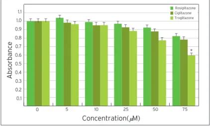
In order to investigate the effect of PPARγlig- ands on cell proliferation of ERα-negative MDA- MB-231 cells, the cells were seeded into 96-well culture plates at a density of 1×10⁴cells/well. MDA- MB-231 breast cancer cells were treated with three types of PPARγligands for 48 h at various concen- trations including rosiglitazone, ciglitazone, and
Fig 1. Troglitazone induces PPARγup-regulation in MDA-MB-231 breast cancer cells.
MDA-MB-231 cells were treated with the indicated concentrations of troglitazone for 48h. Cells were lysed by lysis buffer and the amounts of PPARγwere measured by Western blot. β-actin was used as a loading control. The analysis of electrophoretic band was performed with the LAS-1000 (Fujifilm, Japan). Datas are the means of four separate experiments (bars, SE). p-values are determined using Student’s t-test (* p<0.002 versus zero concentration).
180 160 140 120 100 80 60 40 20
density(% Relative of control) 0
Troglitazone(μM)
0 25 50 75
β-actin PPARγ
T
T T
T*
troglitazone. Cell proliferation was determined by MTT assay. PPARγligands inhibited the cell pro- liferation of MDA-MB-231 breast cancer cells in a dose-dependent manner (Fig 2). Troglitazone strong- ly inhibited the cell proliferation of ERα-negative MDA-MB-231 breast cancer cells. Conversely, rosiglitazone and ciglitazone had weaker effects on the MDA-MB-231 breast cancer cells compared with troglitazone.
Troglitazone induces G1 arrest of MDA-MB-231 cells
To further investigate the inhibitory effect of trogli- tazone on the proliferation of MDA-MB-231, cell cycle analysis was performed. MDA-MB-231 cells were cultured with various concentrations of trogli- tazone for 48 h. DNA content of the nuclei of MDA-MB-231 cells was analyzed by flow cytometry (Fig 3). In troglitazone-treated MDA-MB-231 cells, the percentage of G1phase was maintained in ele- vated levels, indicating that troglitzone induces G1 arrest in ERα-negative MDA-MB-231 cells.
Troglitazone induces G1cell cycle arrest in ERα- positive MCF-7 cells.(19) These results suggest that troglitazone-mediated G1arrest in breast caner cells is independent of estrogen receptor.
Troglitazone-mediated growth inhibition is associ- ated with decreased levels of G1Cdks and D-type cyclins and increased p21 and p27 protein levels in MDA-MB-231 cells
The cell cycle is tightly regulated through a com- plex network of positive and negative regulatory molecules including cyclin dependent kinase (Cdks), cyclin, and Cdk inhibitor (Cdki). To elucidate the role of these molecules in the inhibition of cell cycle induced by troglitazone in MDA-MB-231 breast cancer cells, protein extract was prepared from the cells treated with various concentrations of troglita- zone for 48 h. Western blot was performed using antibodies against pRb, cyclin D1, cyclin D2, cyclin D3, p21, p27, cdk2, Cdk4 and Cdk6. As shown in Fig 4, troglitazone treatment dose-dependently
caused a marked decrease in pRb, cyclin D1, cyclin D2, cyclin D3, cdk2, Cdk4 and Cdk6 expression
Fig 2. MTT assay showing effect of three PPARg agonists, rosiglitazone, ciglitazone, and troglitazone on the growth of MDA-MB-231 breast cancer cells. MDA-MB-231 breast cancer cells were treated with treated with the indicated concentrations of PPARg agonists for 48h. Cell growth of MDA-MB-231 breast cancer cells was deter- mined by MTT assay.
Datas are the means of four separate experiments (bars, SE). p-values are deter- mined using Student’s t-test (* p<0.004 versus zero concentration).
Fig 3. Flow cytometry analysis demonstrating the effect of troglitazone on the cell cycle of MDA-MB-231 breast cancer cells. MDA-MB-231 breast cancer cells treated with troglitazone for 48h at various concentrations. The troglitazone induced the G1
phase proportion and decreased the S phase proportion of MDA-MB-231 breast can- cer cells in a analyzed by flow cytometry.
Journal of Breast Cancer 2006 DECEMBER ; Vol.9, NO.4: 293-300
1.1 1.0 0.9 0.8 0.7 0.6 0.5 0.4 0.3 0.2 0.1
Absorbance
Concentration( M)
0 5 10 25 50 75
Rosiglitazone Ciglitazone Troglitazone
100
80
60
40
20
0
Distribution of cell cycle(%)
Troglitazone(μM)
0 5 10 25 50 75
G1 S G2
T T T T
T T T
T T T
T T T
T
T T
T
T*
cells
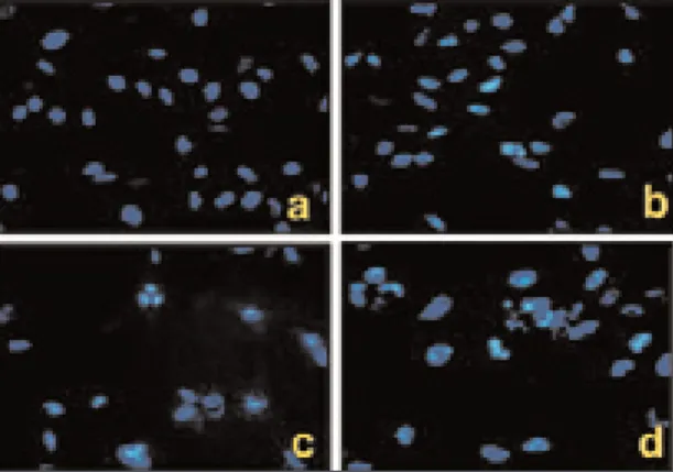
Apoptosis is a distinctive form of cell death that can result in the deletion of specific cell populations during physiologic processes. The growth inhibi- tion by troglitazone treatment in MDA-MB-231 cells appeared to occur dependently of apoptosis. The ability of troglitazone to induce apoptosis in MDA- MB-231 breast cancer cells was initially determined by the DNA fragmentation assay based on TUNEL staining and Hoechst staining. When MDA-MB- 231 breast cancer cells were exposed to various con- centrations of troglitazone for 48 h, they exhibited DNA fragmentation positive cells on TUNEL stain- ing (Fig 5).
Apoptotic cells by TUNEL staining elevated from 2.5-fold of the control level at 10 μM, to 3.1-fold at 50 μM and to 3.5-fold at 75 μM. Furthermore, MDA-MB-231 cells exposed to troglitazone exhib- ited typical morphological changes of apoptosis including cytoplasmic and nuclear shrinkage, chro- matin condensation and fragmentation after staining with Hoechst 33342 (Fig 6).
CONCLUSION
This study addresses for the first time ERα-negative breast cancer cell growth and apoptosis may be mod- ulated through PPARγ. MDA-MB-231 cells exposed to troglitazone showed a G1cell cycle arrest as well as induction of morphological changes characteristic of apoptosis. Moreover, troglitazone caused signifi- cantly increase in p21 and p27 expression.
Our results suggest that troglitazone has potent effect on ERα-negative breast cancer growth and may prove to have a role in hormone therapy-resis- tant breast cancer treatment /prevention in the future. However, the therapeutic implications of the troglitazone growth-inhibitory effect on ERα-neg- ative human breast cancer cells still need to be stud- ied. Further research is necessary to establish possible therapeutic approaches using troglitazone or modi- fied analogs of the thiazolidinedione class of drugs.
Fig 4. Dose-dependent effect of troglitazone on cell cyclins, p21, p27, and pRb in MDA-MB-231 breast cancer cells. Cells were treated with the indicated concentra- tions of troglitazone for 48 h. Cell extracts were separated by SDS/PAGE, followed by Western blot. β-actin was used as a loading control.
The analysis of electrophoretic band was performed with the LAS-1000 (Fujifilm, Japan).
Fig 5. Induction of apoptosis with troglitazone in MDA-MB-231 breast cancer cells.
Detection of apoptosis in breast cancer cell was carried out using a DNA fragmenta- tion assay based on TUNEL staining. MDA-MB-231 breast cancer cells were cultured on 6 well plates and treated with troglitazone for 48h at various concentrations.
Positive nuclei stained brown, and negative nuclei stained blue. Error bars, SE; n=3 in each group (* p<0.005 versus zero concentration).
14 12 10 8 6 4 2
positive TUNEL- cells (%) 0
0 25 50 75
Troglitazone(μM)
pRB CyclinA CyclinD1 Cyclin CyclinE p21 p27 cdk2 cdk4 cdk6 β-actin
0 μM
50 μM 70 μM
25 μM
T
T
T
T**
REFERENCES
1 Houssami N, Cuzick J, Dixon JM. The prevention, detec- tion, and management of breast cancer. Med J Aust 2006;184:230-4.
2 Cuzick J, Warwick J, Pinney E, Warren RM, Duffy SW.
Tamoxifen and breast density in women at increased risk of breast cancer. J Natl Cancer Inst 2004;96:621-8.
3 Muss HB. Endocrine therapy for advanced breast cancer:
a review. Breast Cancer Res Treat 1992;2:15-26.
4 Tontonoz P, Hu E, Graves RA, Budavari AI, Spiegelman BM. mPPAR gamma: tissue specific regulator of an adipocyte enhancer. Genes Dev 1994;8:1224-34.
5 Desvergne B, Michalik L, Wahli W. Transcriptional regula- tion of metabolism. Physiol Rev 2006;86:465-514.
6 Kletzien RF, Clarke SD, Ulrich RG. Enhancement of adipocyte differentiation by an insulin-sensitizing agent.
Mol Pharmacol 1992;41:393-8.
7 Mueller E, Smith M, Sarraf P, Kroll T, Aiyer A, Kaufman DS, et al. Effect of ligand activation of peroxisome
proliferator-activated receptor gamma in human prostate cancer. Proc Natl Acad Sci 2000;97:10990-5.
8 Dreyer C, Krey G, Keller H, Givel F, Helftenbein G, Wahli W.
Control of the peroxisomal beta-oxidation pathway by a novel family of nuclear hormone receptors. Cell 1992;68:879-87.
9 Issemann I, Green S. Activation of a member of the steroid hormone receptor superfamily by peroxisome proliferator.
Nature 1990;347:645-50.
10 Kliewer MW, Forman BM, Blumberg B, Ong ES,
Borgmeyer U, Mangelsdorf DJ, et al. Differential expression and activation of a family of murine peroxisome prolifera- tor-activated receptors. Proc Natl Acad Sci 1994;91:7355-9.
11 Panigraphy D, Huang S, Kieran MW, Kaipainen A.
PPARgamma as a therapeutic target for tumor angiogene- sis and metastasis. Cancer Biol Ther 2005;4:687-93.
12 Forman BM, Tontonoz P, Chen J, Brun RP, Spiegelman BM, Evans RM. 15-Deoxy-delta 12,14-prostaglandin J2 is a lig- and for the adipocyte determination factor PPAR gamma.
Cell 1995;83:803-12.
13 Kliewer SA, Sundseth SS, Jones SA, Brown PJ, Wisely GB, Koble CS, et al. Fatty acids and eicosanoids regulate gene expression through direct interactions with peroxisome proliferator-activated receptors alpha and gamma.
Proc Natl Acad Sci 1997;94:4318-23.
14 Larsen TM, Toubro S, Astrup S. PPARgamma agonists in the treatment of type II diabetes: is increased fatness com- mensurate with long-term efficacy? Int J Obes Relat Metab Disord 2003;27:147-61.
15 McIntyre TM, Pontsler AV, Silva AR, St Hilaire A, Xu Y, Hinshaw JC, et al. Identification of an intracellular receptor for lysophosphatidic acid (LPA): LPA is a transcellular PPARgamma agonist. Proc Natl Acad Sci 2003;100:131-6.
16 Thoennes SR, Tate PL, Price TM, Kilgore MW. Differential transcriptional activation of peroxisome proliferator- activated receptor gamma by omega-3 and omega-6 fatty acids in MCF-7 cells. Mol Cell Endocrinol 2000;160:67-73.
17 Allred CD, Kilgore MW. Selective activation of PPARγin breast, colon, and lung cancer cell lines. Mol Cell Endocrinol 2005;235:21-9.
18 Semple RK, Chatterjee VK, O’Rahilly S. PPARγand human metabolic disease. J Clin Invest 2006;116:581-9.
19 Yin F, Wahino S, Liu Z, Kim S, Hsueh WA, Collins AR, et al.
Triglitazone inhibits growth of MCF-7 breast cancer carcino- ma cells by targeting G1 cell cycle regulators. Biochem Biophys Res Commun 2001;286:916-22.
20 Ferner MH, Elstner E. Peroxisome proliferator-activated receptor gamma ligands for the treatment of breast cancer.
Expert Opin Investig Drugs 2005;14:557-68.
21 Bonofiglio D, Gabriele S, Aquila S, Catalano S, Gentile M, Middea E, et al. Estrogen receptorγbinds to peroxisome proliferator-activated receptor response element and nega-
Journal of Breast Cancer 2006 DECEMBER ; Vol.9, NO.4: 293-300
Fig 6. Fluorescence micrographs of MDA-MB-231 cells stained with Hoechst 33342 (C×400). MDA-MB-231 breast cancer cells were labeled with Hoechst 33342. The cells were exam- ined by fluorescence microscopy. Untreated cells (a) kept a normal chromatin pattern and showed no change in a nuclear morphology. Cells treated with troglitazone, 10 μM (b), 50 μM (c), and 75 μM (d) demonstrated typical findings of apoptosis, marked by nuclear condensation and fragmentation.
Biol 2005;11:6139-47.
22 Burstein HJ, Demetri GD, Mueller E, Sarraf P, Spiegelman BM, Winer EP. Use of the peroxisome proliferator-activated receptor (PPARγ) ligand, troglita- zone as treatment for refractory breast cancer: a phase II study. Breast Cancer Res Treat 2003;79:391-7.
receptor gamma and retinoic acid receptor inhibit growth and induce apoptosis of human breast cancer cells in vitro and in BNX mice. Proc Natl Acad Sci 1998;95:8806-11.
24 Yu J, Qiao L, Zimmermann L, Ebert MP, Zhang H, Lin W, et al. Troglitazone inhibits tumor growth in hepatocellular carcinoma in vitro and in vivo. Hepatology 2006;43:134-43.