의
의
의학
학
학 박
박
박사
사학
사
학
학위
위
위 논
논
논문
문
문
Role
Role
Role
Role of
of
of
of Extracellular
Extracellular
Extracellular S
Extracellular
S
S
Siiiig
gn
g
g
n
n
na
a
a
allll
R
Re
R
R
e
e
eg
g
g
gu
u
u
ulllla
atttte
a
a
e
e
ed
d
d
d Kinase
Kinase
Kinase and
Kinase
and
and
and
Peroxisome
Peroxisome
Peroxisome
Peroxisome P
Pr
P
P
r
r
ro
o
o
olllliiiiffffe
e
e
er
ra
r
r
a
atttto
a
o
or
o
r
r
r
A
A
Ac
A
c
cttttiiiiv
c
v
va
v
a
atttte
a
ed
e
e
d
d Receptor
d
Receptor
Receptor
Receptor gamma
gamma
gamma
gamma
on
on
on
on Transforming
Transforming
Transforming
Transforming Growth
Growth
Growth
Growth F
F
Fa
F
ac
a
a
c
c
ctttto
o
o
or
r
r
r
β
β1
β
β
1
1
1
IIIIn
n
n
nd
d
d
du
uc
u
u
c
c
ce
e
e
ed
d
d
d Human
Human
Human
Human
Endometrial
Endometrial
Endometrial
Endometrial Stromal
Stromal
Stromal
Stromal Cell
Cell
Cell Decidualization
Cell
Decidualization
Decidualization
Decidualization
아
아
아 주
주
주 대
대
대 학
학
학 교
교
교 대
대
대 학
학
학 원
원
원
의
의
의 학
학
학 과
과
과
Role
Role
Role
Role of
of
of Extracellular
of
Extracellular
Extracellular
Extracellular S
S
S
Siiiig
gn
g
g
n
n
na
a
a
allll
R
Re
R
R
e
e
eg
g
g
gu
u
ulllla
u
atttte
a
a
e
e
ed
d
d
d Kinase
Kinase
Kinase and
Kinase
and
and
and
Peroxisome
Peroxisome
Peroxisome
Peroxisome P
P
P
Pr
rr
ro
o
o
olllliiiiffffe
e
e
er
ra
rr
a
atttto
a
o
o
or
rr
r
A
Ac
A
A
c
c
cttttiiiiv
v
v
va
a
a
atttte
ed
e
e
d
d
d Receptor
Receptor
Receptor gamma
Receptor
gamma
gamma
gamma on
on
on
on
Transforming
Transforming
Transforming
Transforming Growth
Growth
Growth F
Growth
F
Fa
F
a
ac
a
ctttto
c
c
o
or
o
rr
r
β
β
β1
β
1
1
1
IIIIn
n
n
nd
d
d
du
uc
u
u
c
c
ce
e
ed
e
d
d Human
d
Human
Human
Human
Endometrial
Endometrial
Endometrial
Endometrial Stromal
Stromal
Stromal
Stromal Cell
Cell
Cell
Cell Decidualization
Decidualization
Decidualization
Decidualization
by
Hye Jin Chang
A Dissertation Submitted to The Graduate School of Ajou University in Partial Fulfillment of the Requirements for the Degree of
DOCTOR OF PHILOSOPHY
Supervised by
Kyung Joo Hwang, M.D., Ph.D.
Department
Department
Department
Department of
of
of
of Medical
Medical
Medical Sciences
Medical
Sciences
Sciences
Sciences
The
The
The
The Graduate
Graduate
Graduate
Graduate School,
School,
School,
School, Ajou
Ajou
Ajou
Ajou University
University
University
University
February,
February,
February, 2007
February,
2007
2007
2007
장
장
장혜
혜
혜진
진
진의
의 의
의
의
의학
학
학 박
박
박사
사
사학
학
학위
위
위 논
논
논문
문
문을
을
을 인
인
인준
준
준함
함
함.
.
.
심
심
심사
사
사위
위
위원
원
원장
장
장
유
유
유 희
희
희 석
석
석
인
인
인
심
심
심 사
사
사 위
위
위 원
원
원
이 영
이
이
영
영 돈
돈
돈
인
인
인
심
심
심 사
사
사 위
위
위 원
원
원
김
김
김 해
해
해 권
권
권
인
인
인
심
심
심 사
사
사 위
위
위 원
원
원
황
황
황 경
경
경 주
주
주
인
인
인
심
심
심 사
사
사 위
위
위 원
원
원
김
김
김 미
미
미 란
란
란
인
인
인
아
아
아 주
주
주 대
대
대 학
학
학 교
교
교 대
대
대 학
학
학 원
원
원
2
2
20
0
00
0
06
6
6년
년
년 1
1
12
2
2월
월
월 2
2
22
2
2일
일
일
- ABSTRACT -
Role of Extracellular Signal-Regulated Kinase (ERK) and
Peroxisome Proliferator-Activated Receptor gamma (PPAR
γγγγ) on
Transforming Growth Factor(TGF)-β
β
β
β1-Induced Human
Endometrial Stromal Cell Decidualization
Objective : To investigate the role of ERK and PPARγ on TGF-β1-induced human endometrial stromal cell decidualization in vitro.
Materials and Methods : Human endometrial tissues obtained by hysterectomy
specimens from patients with conditions other than endometrial diseases such as leiomyoma. Endometrial stromal cells were cultured under the following conditions: DMEM/F-12 (10% FBS, 1 nM E2 and 100 nM P4). 5 ng/ml of TGF-β1, 50 nM of
rosiglitazone (PPARγ agonist), and 20 μM of PD98059 (ERK inhibitor) were added according to experimental purposes.
Trypan-blue and a hemocytometer were utilized to count the viable cell number. Enzyme-linked immunosorbent assay (ELISA) and Western blotting was employed to detect proteins.
Results : TGF-β1 inhibited proliferation of cultured human endometrial stromal
cells and induced expression of prostaglandin E2 (PGE2) and prolactin. This effect
was mediated by Smad and ERK activation. Addition of rosiglitazone, a PPARγ
rosiglitazone inhibited TGF-β1 induced activation of ERK, consequently reducing PGE2 and prolactin production.
Conclusion: TGF-β1-induced decidualization of endometrial stromal cells through
Smad and ERK phosphorylation. PPARγ acts as a negative regulator of human endometrial cell decidualization in vitro.
TABLE OF CONTENTS
ABSTRACT ··· i
TABLE OF CONTENTS ··· iii
LIST OF FIGURES ··· iv
. Ⅰ INTRODUCTION ··· 1
. Ⅱ MATERIALS AND METHODS ··· 4
A. MATERIALS ··· 4
1. Clinical subjects and endometrial biopsies ··· 4
2. Reagents and antibodies ··· 4
B. METHODS ··· 5
1. Endometrial stromal cell isolation and primary cell culture ··· 5
2. Cell count by using trypan-blue dye ··· 6
3. Western blot analysis of prolactin, pSmad2/3, PPARγ, COX-2 and pERK ··· 6
4. Measurement of Prostaglandin E2 concentrations in the conditioned Media ··· 7 5. Statistical analysis ··· 7 . Ⅲ RESULTS ··· 8 . Ⅳ DISCUSSION ··· 14 . Ⅴ CONCLUSION ··· 17 REFERENCES ··· 18 국문요약 ··· 24
LIST OF FIGURES
Fig. 1. Effect of TGF-β1 on the endometrial stromal cell proliferation ··· 8
Fig. 2. Expression of prolactin, PPARγ and pSmad 2/3 in cultured endometrial stromal cells ··· 9
Fig. 3. Effect of TGF-β1 on the cell proliferation was prevented by rosiglitazone (PPARγ agonist) treatment ··· 10
Fig. 4. Rosiglitazone reduced expression of pSmad, pERK, COX-2 and PGE2 in cultured endometrial stromal cells ··· 12
Fig. 5. PD98059 reduced expression of pERK, COX-2 and PGE2 in cultured
I. INTRODUCTION
The human uterine endometrium is a dynamic organ that undergoes remarkable periodic growth, remodeling and breakdown under hormonal control. The decidualization of human endometrial stromal cells, which develops in the late secretory stage of the menstrual cycle, is characterized by morphological and functional differentiation. The most obvious features of decidualization are the stromal cell transformation. It is characterized by the transformation of the elongated fibroblast-like phenotype of endometrial stromal cell to the larger, rounder phenotype of the decidual cell (Verma, 1983; Tang et al, 1994; Giudice & Ferenczy, 1996). These differentiation processes are mediated by changes of numerous gene expressions of the endometrial stromal cells in vivo. The processes can be induced in
vitro by progesterone in estradiol-treated cultures (Huang et al., 1987; Classen-Linke
et al., 1998), by ligands which are coupled to the cAMP pathway such as PGE2
(Frank et al., 1994), gonadotropins (Frank & Gurpide, 1993), and by cAMP alone (Tank et al., 1993). It was also recently shown that heparin-binding epidermal growth factors and their receptors (Chobotova et al., 2005) participate in the decidualization process.
Transforming Growth factor-β (TGF-β) was first discovered in the fibroblast of white mice's sarcoma (DeLarco & Todaro, 1978). The TGF-β superfamily, which includes various TGF-βs, activin, bone morphogenetic proteins (BMPs), inhibin and growth/differentiation factors, is known as a multifunctional polypeptide cytokine. They are secreted in a latent form and activated by plasmin (Odekone et al., 1994). In
endometrium, TGF-β is a multifunctional cytokine that regulates many biological functions ranging from growth/differentiation to apoptosis of various cell types (Sporn & Roberts, 1990). Because of their powerful effects on the cellular and molecular processes, they are associated with cellular proliferation (Moses et al., 1987) and differentiation (Moses & Serra, 1996), angiogenesis (Renner et al., 2002), extracellular matrix modification and immunomodulation (Akhurt & Derynck, 2001).
Peroxisome proliferator-activated receptors (PPARs) are ligand-dependent transcription factors of the nuclear hormone receptor superfamily. Three subtypes of PPARs, which are activated by polyunsaturated fatty acid and eicosanoid metabolites
have been characterized (PPARα, PPARβ and PPARγ). After binding a ligand (e. g. thizolidinediones, arachidonate, or others), it forms a heterodimer with the cis-retinoid acid receptor; retinoic X receptor (RXR), binds to a peroxisome proliferator responsive elements (PPRE), and activates transcription of selected genes. However, their specific endogenous functions are yet to be identified. Of the three PPAR isoforms, PPARγ has been implicated in the control of a broad range of cellular responses, such as differentiation, proliferation, cell death and inflammation (Murphy & Holder, 2000; MacDougald & Mandrup, 2002). It has been reported that PPARs can suppress the growth of different types of human cancer cells isolated from colon, breast and prostatic cancer through distinctive ways (Grommes et al., 2004). Overexpression of PPARγ significantly inhibits TGF-β1-mediated Smad activation in human liver cancer cells (Han et al., 2004), and also PPARγligandscan repress vascular endothelial cell growth factor (VEGF) gene expression via a PPARγ
-responsive element (PPRE)in the VEGF gene promoter in human endometrial cells (Peeters et al., 2006).
It was previously reported that progesterone indirectly induced stromal cell decidualization via enhancing the expression and secretion of TGF-β1 from epithelial cells in vitro. The secreted epithelial-derived TGF-β1 acted on adjacent stromal cells, at least in part, to turn on Smad signaling that may lead to stromal decidualization. This was reversed by anti-TGF-β1 antibody (Kim et al., 2005).
Extracellular signal-regulated kinase (ERK) is one of the mitogen-activated protein kinase (MAPK) and its major roles are cell proliferation and differentiation (Torii et al., 2004). Also it is known to induce differntiation/G0 arrest in the differentiation of HL-60 cells (Yen et al., 2006).
In this study, by using cultured human endometrial stromal cells, I investigated the role of ERK and PPARγ on the TGF-β1-induced human endometrial stromal cell decidualization in vitro.
II. MATERIALS AND METHODS
A. Materials
1.
Clinical subjects and endometrial biopsies
Human endometrial tissues were obtained by curettage of hysterectomy specimens from patients undergoing hysterectomy for conditions other than endometrial diseases (leiomyoma or adenomyosis), according to protocols approved by the Institutional Review Board, Ajou University Hospital. Tissue specimens from patients of ages 30 to 45, who were estimated to be in the mid- or late proliferative phase of the menstrual cycle, were taken for the experiment.
2.
Reagents and antibodies
Human recombinant TGF-β1 and reagents for ELISA analysis were purchased from Sigma (St. Louis, MO, USA). The PPARγagonist, rosiglitazone, was purchased from Alexin (San Diego, CA, USA). The ERK inhibitor PD98059 was purchased from Calbiochem (San Diego, CA, USA). The antibodies against human prolactin (goat), p-Smad2 (goat), p-ERK (mouse), PPARγ(rabbit), and COX-2 (goat) were purchased from Santa Cruz Biotechnology (Santa Cruz, CA, USA). The antibody against β-actin was purchased from Sigma. The antibodies against goat horseradish peroxidase (HRP), mouse HRP, rabbit HRP were purchased from Chemicon (Temecula, CA, USA).
B. Methods
1. Endometrial stromal cell isolation and primary cell culture
The protocol for cell isolation was identical to that of a previously reported method (Park et al., 2001).Briefly, the endometrial tissues obtained were placed in a conical tube (Falcon, Becton Dickinson, NJ, USA) containing DMEM/F-12 supplemented with 10 % fetal bovine serum (FBS, Gibco Life Technologies) and transported to the aseptic laboratory, where the tissue was again washed several times with phosphate-buffered saline (PBS) to remove any residual blood clots and FBS. Then the tissues were minced in 2-3 ml of Dulbecco's modified Eagle's medium (DMEM, USA). The minced tissues were centrifuged at 85×g in a conical tube and 10 ml of trypsin-EDTA (Gibco Life Technologies, Grand Island, NY, USA) was added to the pellet and incubated in a shaker at 37 ℃. After 30 min of incubation, 1 ml of 10% heat-inactivated FBS was added to stop the enzymatic reaction. The tissues were centrifuged and the upper portion was removed. The cell pellet was suspended with 5ml of PBS and centrifuged again. After trypsinizing and washing with PBS, the cells were resuspended in 10ml DMEM/F-12 to a final concentration of 5 x 10⁶ cells/ml, and 8 ml of the cell suspension was seeded on a 100mm culture dish (Corning, NY, USA). After incubation for 24 hr, cells and red blood cell (RBC) unattached but suspended in the media were washed with PBS, attached cells were cultured for 72 hr in DMEM/F-12 medium (10 % FBS; 1 nM E2; 100 nM P4) under
rosiglitazone , and 20 μM of phosphorylated-ERK inhibitor, PD98059. Unless stated elsewhere, all reagents were purchased from Sigma.
2. Cell count by using trypan-blue dye
Cell growth was determined by using trypan-blue (Gibco, USA) stain. Viable cells are able to repel the dye and thus can not be stained. After trypsinizing and washing with PBS buffer, the stromal cells were resuspended in 10ml DMEM/F-12 and the same volume of trypan-blue was added. Viable stromal cells that were not stained with trypan-blue dye, were counted by a hemocytometer.
3. Western blot analysis of prolactin, pSmad2/3, PPAR
γ, COX-2 and
pERK
At the end of each treatment, cells were scraped off the plates and centrifuged, resuspended with PBS buffer containing 40 nM Tris-HCl (pH 8.0), 120 mM NaCl, 0.1 % Nonidet P-40 and protease inhibitors. After sonication, the whole cell lysate was collected by centrifugation at 14,000 rpm at 4 ℃ for 20 min. Total proteins were separated by sodium dodecyl sulphate-polyacrylamide gel electrophoresis (SDS-PAGE; 8, 10, 15 %) and transferred onto nitrocellulose blotting membrane (Satorius AG, Germany). The membranes were blocked with 5 % non-fat dry milk in Tris-buffered saline and incubated with primary antibodies for 1 hour at 4 ℃. Blots were developed by peroxidase-conjugated secondary antibody, and proteins were visualized by enhanced chemiluminescence (ECL) procedure (Santa Cruz
4. Measurement of Prostaglandin E
2concentrations in the conditioned
media
To measure the secretion of prostaglandin E2 (PGE2) by cells during culture in
various culture media for 72 hr, cells were removed by centrifugation, the cell-free conditioned media was collected and was stored at -75 ℃ until use. After thawing at room temperature, the medium was analyzed for immunoreactive PGE2 by
enzyme-linked immunosorbent assay (ELISA) using anti-PGE2 antibodies (ELISA
kit) according to the manufacturer's specifications with absorbance at 450 nm in a plate reader (SpectraMax 190, Molecular Device, Sunnyvale, CA, USA). Experiments were repeated at least three times with different media under the same condition to minimize intra- and interassay variations.
5. Statistical analysis
Student's t-test was used for the statistical analysis of concentrations of PGE2 in
the culture media. P values of < 0.05 were considered to be statistically significant. Mann-Whitney test was used for the statistical analysis of cell count by trypan blue staining. Unless otherwise indicated, P < 0.05 was considered to be statistically significant.
III. RESULTS
A. The effect of TGF-
β
1 on the human endometrial stromal cell
proliferation
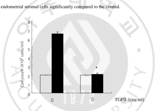
Endometrial stromal cells were treated with or without 5 ng/ml of TGF-β1 for 72 hr. After that cells were stained with trypan-blue dye and counted using a hemocytometer. As shown in Fig. 1, TGF-β1 inhibited in vitro cell proliferation of endometrial stromal cells significantly compared to the control.
Fig. 1. Effect of TGF-βββ1 on the endometrial stromal cell proliferation. Endometrial stromal cells β
were treated with or without 5 ng/ml of TGF-β1 for 72 hr. Cells were stained with trypan-blue and
counted using hemocytometer. TGF-β1 inhibited proliferation of cultured endometrial stromal cells.
Data are the means ± SE of three independent experiments. Open bar; 0 hr, Black bar; 72 hr. * p
value < 0.05 0 1 2 3 4 5 6 7 8 C e ll c o u nt ( X 1 0 6 c e lls /m l) TGFβ-1(ng/ml) 0 5 *
B. The effect of TGF-β1 on the expression of prolactin, PPAR
γ, and
pSmad2/3 by endometrial stromal cells during decidualization
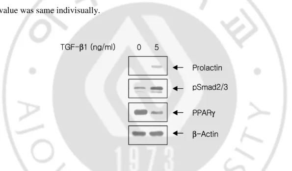
To confirm the effects of TGF-β1 on the stromal cell decidualization, stromal cells were maintained under serum-free condition for 24 hr, and then treated with TGF-β1 at concentration of 5 ng/ml for 72 hr. As shown in Fig. 2, 5 ng/ml of TGF-β1 induced expression of prolactin, and Smad 2/3 phosphorylation after 72 hr treatment. On the other hand, at the same time, expression of PPARγ was significantly decreased by treatment of TGF-β1. We didn’t show total Smad 2/3 here, because the value was same indivisually.Prolactin β-Actin TGF-β1 (ng/ml) PPARγ pSmad2/3 0 5 Prolactin β-Actin TGF-β1 (ng/ml) PPARγ pSmad2/3 0 5
Fig. 2. Expression of prolactin, PPARγγγγ and pSmad 2/3 in cultured endometrial stromal cells.
Endometrial stromal cells were treated with or without 5 ng/ml of TGF-β1 for 72hr. Cell lysates were
subjected to western blot analysis using anti-prolactin, phosphorylated Smad2/3 and PPARγ antibodies.
TGF-β1 induced expression of prolactin and pSMAD2/3 after 72 hr treatment. Expression of PPARγ
C. Rosiglitazone restores the mitosis-inhibitory effect of TGF-β1 on
endometrial stromal cells
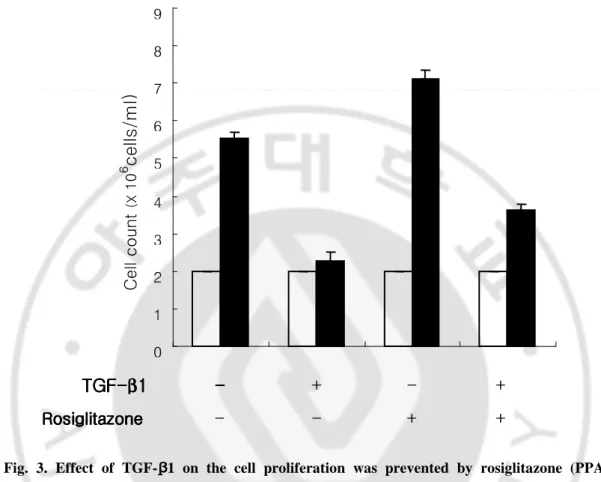
Fig. 3. Effect of TGF-ββββ1 on the cell proliferation was prevented by rosiglitazone (PPARγγγγ
agonist) treatment. Endometrial stromal cells were treated with 5 ng/ml of TGF-β1 and/or 50 nM
of rosiglitazone for 72 hr. Cells were stained with trypan-blue and counted using hemocytometer. Cell
proliferation was increased by co-treatment of TGF-β1 and rosiglitazone compared with TGF-β1
treatment alone. Data are the means ± SE of three independent experiments. Open bar; 0 hr, Black
bar; 72 hr. 0 1 2 3 4 5 6 7 8 9 - - + + TGF TGF TGF TGF----ββββ11 11 Rosiglitazone RosiglitazoneRosiglitazone Rosiglitazone -- -- + - + C e ll c o u nt (X 1 0 6 c e lls /m l)
It was also examined whether the signaling pathway of TGF-β1 was controlled by PPARγ. To do this, endometrial stromal cells were cultured with 5 ng/ml of TGF-β1 and/or 50 nM of rosiglitazone, the specific ligand of PPARγ for 72 hr. As shown in Fig. 3, the mitosis-inhibitory effect of TGF-β1 was reversed by combined treatment with rosiglitazone.
D. The changes of prolactin, pSmad2/3, PPAR
γ, pERK, COX-2 and
PGE
2expression by rosiglitazone
Rosiglitazone treatment alone resulted in the considerable decrease of prolactin expression and Smad2/3 phosphorylation compared with the TGF-β1 treatment alone. Also, COX-2 and pERK expression was significantly inhibited by combined treatment with rosiglitazone (Fig. 4A). COX-2 is known as a key enzyme of prostaglandin production. And thus concentration of PGE2 in stromal cell cultured
medium was analyzed using an ELISA assay. Concentration of PGE2 in the cell
culture conditioned medium was decreased by combined treatment with TGF-β1 and rosiglitazone, whereas rosiglitazone did not affect on PGE2 concentration in the same
medium (Fig. 4B). Total ERK was not shown here, because the vaule was same indivisually.
- - + + TGF-β1 Rosiglitazone - + - + 0 50 100 150 200 250 P G E 2 (p g /m l) B pSmad2/3 Prolactin PPARγ COX-2 A - - + + TGFβ-1 Rosiglitazone - + - + pERK β-Actin - - + + TGF-β1 Rosiglitazone - + - + 0 50 100 150 200 250 P G E 2 (p g /m l) B - - + + TGF-β1 Rosiglitazone - + - + 0 50 100 150 200 250 P G E 2 (p g /m l) 0 50 100 150 200 250 P G E 2 (p g /m l) B pSmad2/3 Prolactin PPARγ COX-2 A - - + + TGFβ-1 Rosiglitazone - + - + pERK β-Actin pSmad2/3 Prolactin PPARγ COX-2 A - - + + TGFβ-1 Rosiglitazone - + - + pERK β-Actin
Fig. 4. Rosiglitazone reduced expression of pSmad, pERK, COX-2 and PGE2 in cultured endometrial stromal cells. Endometrial stromal cells were treated with 5 ng/ml of TGF-β1 and/or 50
nM of rosiglitazone for 72 hr. Cell lysates were subjected to Western blot analysis using anti-prolactin,
pSmad2/3, PPARγ, pERK, COX-2 antibodies. TGF-β1 induced expression of prolactin, pSmad2/3,
pERK, and COX-2 were prevented by combined treatment of rosiglitazone (A). Concentration of
PGE2 in culture medium was measured by ELISA. TGF-β1 induced PGE2 releasing from cultured
endometrial stromal cells was inhibited by combined treatment of rosiglitazone. Data are the mean ±
SE of three independent experiments (B).
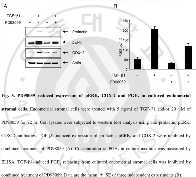
E. The change of prolactin, pERK, COX-2 and PGE
2expression by
PD98059
To verify the effect of ERK on the COX-2 expression and the prostaglandin production in cultured endometrial stromal cells, endometrial stromal cells were
hr. As shown in Fig. 5A, PD98059 inhibited the TGF-β1-induced prolactin, ERK phosphorylation and COX-2 expression simultaneously. Concentration of PGE2 in
culture medium was decreased by combined treatment of TGF-β1 and PD98059 (Fig. 5B). 0 50 100 150 200 250 P G E 2 (p g /m l) - - + + TGF-β1 PD98059 - + - + B COX-2 pERK Actin Prolactin - - + + TGF-β1 PD98059 - + - + A 0 50 100 150 200 250 P G E 2 (p g /m l) - - + + TGF-β1 PD98059 - + - + B 0 50 100 150 200 250 P G E 2 (p g /m l) 0 50 100 150 200 250 P G E 2 (p g /m l) - - + + TGF-β1 PD98059 - + - + B COX-2 pERK Actin Prolactin - - + + TGF-β1 PD98059 - + - + A COX-2 pERK Actin Prolactin - - + + TGF-β1 PD98059 - + - + A
Fig. 5. PD98059 reduced expression of pERK, COX-2 and PGE2 in cultured endometrial
stromal cells. Endometrial stromal cells were treated with 5 ng/ml of TGF-β1 and/or 20 μM of
PD98059 for 72 hr. Cell lysates were subjected to western blot analysis using anti-prolactin, pERK,
COX-2 antibodies. TGF-β1-induced expression of prolactin, pERK, and COX-2 were inhibited by combined treatment of PD98059 (A). Concentration of PGE2 in culture medium was measured by
ELISA. TGF-β1-induced PGE2 releasing from cultured endometrial stromal cells was inhibited by
IV. DISCUSSION
Generally, cell differentiation is known to occur when cell proliferation stops. Previous reports have shown that progesterone induces endometrial stromal cell decidualization via triggering expression and secretion of TGF-β1 by epithelial cells (Kim et al., 2005). In the present study, it was investigated whether TGF-β1 could affect on the endometrial stromal cell proliferation, and that TGF-β1 might inhibit cell proliferation. The results suggest that a possible linkage between the intracellular signaling pathway via TGF-β1-mediated Smad phosphorylation and the molecules associated with cell proliferation and differentiation of endometrial stromal cells might exist.
PPARγ is widely expressed in various tissues including adipose tissue, placenta, amnion and choriodecidual tissue, although the exact role of PPARγin these tissues remains unclear (Marvin et al., 2000). The present study shows that PPARγ expression is decreased in TGF-β1-induced decidualization of endometrial stromal cells. Consequently it was attempted further to elucidate the role of PPARγ in
β1-mediated stromal cell decidualization process. To ascertain the change in TGF-β1-mediated cellular response by PPARγ activation, cell proliferation of cultured stromal cells was investigated after treatment with TGF-β1 and/or PPARγ ligand, rosiglitazone. The result showed that TGF-β1-mediated mitoinhibition could be restored by adding rosiglitazone. PPARγ activation by rosiglitazone inhibited not only mitoinhibition, but also Smad phosphorylation as well. Based on these
observation, it is suggested as follows ; TGF-β1-induced decidualization takes place via Smad phosphorylation. Subsequently, cell proliferation is suppressed, and endometrial stromal cells differentiate into decidual cells simultaneously. However these mechanisms are regulated by activation of PPARγ. Similar findings was made in the previous reports in which the PPARγ inhibited the TGF-β-induced differentiation in pulmonary myofibroblasts (Burgess et al., 2005). To further confirm the above conclusions, whether intracellular signaling transmission of
TGF-β1 might influence cell growth factor activity was examined.
ERK is a type of MAPK, which is known to have an important role in cell proliferation and differentiation. Enhanced ERK activation affects differentiation and G0 arrest in specific cell differentiation (Yen et al., 2006). Present study shows that TGF-β1 could induce not only decidualization of endometrial stromal cells, but also inhibition of cell proliferation, and enhancement of ERK phosphorylation. Overall, these results suggests that TGF-β1 could induce ERK phosphorylation, thereby resulting in the differentiation/G0 arrest of endometrial stromal cells, which again could be prevented by PPARγ activation.
Prostaglandins (PGs) are ubiquitous compounds involved in various homeostatic and inflammatory processes throughout the body. They are formed by the combined action of phospholipase A2, which liberates arachidonic acid from the sn-2 position
of cellular membrane phospholipids, and cyclooxygenase (COX), which converts arachidonic acid into the endoperoxide intermediate, PGH2. PGH2 is subsequently
converted to various PGs by the action of cell-specific synthases (Vane et al., 1998). It is well known that PGs have an important role in ovulation, implantation, and
parturition (Poyser, 1995). The suppression of PG biosynthesis is known to inhibit endometrial stromal cell decidualization, which can be induced by artificial manipulation of the mouse uterus (Rankin et al., 1979). Thus, it was investigated whether TGF-β1 might affect PG synthesis, and the process could be prevented by activation of PPARγ in human endometrial stromal cells in vitro. The results shows that TGF-β1 could increase the expression of COX-2, in order to possibly induce PG biosynthesis and PGE2 release from cultured endometrial stromal cells. In addition to
this, activation of PPARγ by rosiglitazone prevented this mechanism. From the results, it was found that TGF-β1-induced decidualization occurred through Smad and ERK phosphorylation. In murine fibroblasts, activation of the Ras/Raf-1/ERK signal pathway is required for COX-2 induction (Xie et al., 1994). To further confirm whether the ERK phosphorylation affect PGE2 biosynthesis, it was examined the
COX-2 expression and PGE2 release from the cultured stromal cells after treatment
with ERK phosphorylation inhibitor, PD98059. It was observed that COX-2 expression and PGE2 concentration in medium significantly decreased by PD98059
treatment, and prolactin expression of stromal cells also decreased. These findings demonstrate the role of ERK-mediated COX-2 activation in the TGF-β1-induced PGE2 synthesis, and that PPARγ is the counteracting part of TGF-β1-mediated
V. CONCLUSION
In conclusion, TGF-β1 potently induces decidualization of cultured human endometrial stromal cells in vitro. The study also demonstrates the expression of pSmad 2/3 and pERK, which are intracellular mediators of TGF-β1 signaling in stromal cells. This proteins would subsequently increase PGE2 production and finally
induce stromal cell mitoinhibition and prolactin expression during decidualization. PPARγ could reverse these processes. It is evident in the present study that PPARγ is linked to crosstalk between ERK1/2 and Smad signaling cascades activated by
REFERENCES
1. Akhurt RJ, Derynck R: TGF-beta signaling in cancer-a double-edged sword.
Trends Cell Biol 11: 44-51, 2001
2. Burgess HA, Daugherty LE, Thatcher TH, Lakatos HF, Ray DM, Redonnet M, Phipps RP, Sime PJ: PPAR gamma agonists inhibit TGF-beta induced pulmonary myofibroblast differentiation and collagen production: implications for therapy of lung fibrosis. Am J Physiol Lung Cell Mol Physiol 288(6): L1146-1153, 2005
3. Chobotova K, Karpovich N, Carver J, Manek S, Gullick WJ, Barlow DH, Mardon HJ: Heparin-binding epidermal growth factor and its receptors mediate decidualization and potentiate survival of human endometrial stromal cells. J
Clinical Endocrinol Metabol 90(2): 913–919, 2005
4. Classen-Linke I, Alfer J, Hey S, Krusche CA, Kusche M, Beier H: Marker molecules of human endometrial differentiation can be hormonally regulated under in-vitro conditions as in-vivo. Human Reprod Update 5: 539-549, 1998
5. DeLarco JE, Todaro GJ: Growth factors form murine sarcoma virus-transfected cells. Proc Nath Acad USA 75: 4001-4005, 1978
6. Frank GR, Brar AK, Cedars MI, Handwerger S: Prostaglandin E2 enhances human
endometrial cell differentiation. Endocrinology 34: 258-263, 1994
7. Frank GR, Gurpide E: Direct effect of gonadotropins on decidualization of human endometrial stromal cells. J Steroid Biochem Mol Biol 47: 115-121, 1993
8. Giudice LC, Ferenczy A: The endometrial cycle. In Reproductive Endocrinology : Surgery and Technology (ed. Adashi EY, Rock JA and Rosenwaks Z) Lippincott-Raven Publishers, Philadelphia, PA, pp.272-301, 1996
9. Grommes C, Landreth GE, Heneka MT: Antineoplastic effects of peroxisome proliferator-activated receptor gamma agonists. Lancet Oncol 5: 419-429, 2004
10. Han C, Demetris AJ, Liu Y, Shelhamer JH, Wu T: Transforming Growth Factor (TGF) Activates Cytosolic Phospholipase A2α (cPLA2α)-mediated Prostaglandin
E2 (PGE)2/EP1 and Peroxisome Proliferator-activated Receptor (PPAR)/Smad
Signaling Pathways in Human Liver Cancer Cells. J Biol Chem 279(43): 44344-44354, 2004
11. Huang JR, Tseng L, Bischof P, Janne OA: Regulation of prolactin production by progestin, estrogen, and relaxin in human endometrial stromal cells.Endocrinology 121: 2011-2017, 1987
12. Kim MR, Park DW, Lee JH, Choi DS, Hwang KJ, Ryu HS, Min CK. Progesterone-dependent release of transforming growth factor-beta1 from epithelial cells enhances the endometrial decidualization by turning on the Smad signalling in stromal cells. Mol Hum Reprod 11(11): 801-808, 2005
13. MacDougald OA, Mandrup S: Adipogenesis: forces that tip the scales. Trends
Endocrinol Metab 13: 5-11, 2002
14. Marvin KW, Eykholt RL, Keelan JA, Sato TA, Mitchell MD: The 15-deoxy-delta (12,14)-prostaglandin J(2)receptor, peroxisome proliferator activated receptor-gamma (PPARreceptor-gamma) is expressed in human gestational tissues and is functionally active in JEG3 choriocarcinoma cells. Placenta 21(4): 436-440, 2000
15. Moses HL, Coffey RJ Jr, Leof EB, Lyons RM, Keski-Oja J: Transforming growth factor beta regulation of cell proliferation. J Cell Physiol 5: 1-7, 1987
16. Moses HL, Serra R: Regulation of differentiation by TGF-beta. Curr Opin Genet
Dev 6: 581-586, 1996
17. Murphy GJ, Holder JC: PPAR-gamma agonists: therapeutic role in diabetes, inflammation and cancer. Trends Pharmacol Sci 21(12): 469-474, 2000
18. Odekone S, Matt D, Stom S, Rifkin DB: Requirement for receptor-bound urokinase in plasmin-dependent cellular conversion of latent TGF-beta to TGF beta. J Cell Physiol 158: 398-407, 1994
19. Park DW, Ryu HS, Choi DS, Park YH, Chang KH, Min CK: Localization of matrix metalloproteinases on endometrial cancer cell invasion in vitro. Gynecol
Oncol 82: 442-449, 2001
20. Peeters LL, Vigne JL, Tee MK, Zhao D, Waite LL, Taylor RN: PPAR represses VEGF expression in human endometrial cells: Implications for uterine angiogenesis. Angiogenesis 8(4):373-379, 2006
21. Poyser NL: The control of prostaglandin production by the endometrium in relation to luteolysis and menstruation. Prostaglandins Leukot Essent Fatty Acids. 53(3): 147-195, 1995
22. Rankin JC, Ledford BE, Jonsson HT Jr, Baggett B: Prostaglandins, indomethacin and the decidual cell reaction in the mouse uterus. Biol Reprod 20(2): 399-404, 1979
23. Renner U, Lohrer P, Schaaf L, Feirer M, Schmitt K, Onofri C, Arzt E, Stalla GK: Transforming growth factor-beta stimulate vascular endothelial growth factor
production by folliculostellate pituitary cells. Endocrinology 143: 3759-3765, 2002
24. Sporn MB, Roberts AB: TGF-beta: problems and prospects. Cell Regul 1(12): 875-882, 1990
25. Tang B, Guller S, Gurpide E: Mechanism of human endometrial stromal cells decidualization. Ann NY Acad Sci 734: 19-25, 1994
26. Tank B, Guller S and Gurpide E: Cyclic adenosine 3′, 5′-monophosphate induces prolactin expression in stromal cells isolated from human proliferative endometrium. Endocrinology 133: 2197–2203, 1993
27. Torii S, Nakayama K, Yamamoto T, Nishida E: Regulatory mechanisms and function of ERK MAP kinases. J Biochem (Tokyo) 136(5): 557-561, 2004
28. Vane JR, Bakhle YS, Botting RM: Cyclooxygenases 1 and 2. Annu Rev
Pharmacol Toxicol 38: 97–120, 1998
29. Verma V: Ultrastructural changes in human endometrium at different phases of the menstrual cycle and their functional significance. Gynecol Obstet Invest 15: 193-212,1983
30. Xie W, Fletcher BS, Andersen RD, Herschman HR: v-src induction of the TIS10/PGS2 prostaglandin synthase gene is mediated by an ATF/CRE transcription response element. Mol Cell Biol 14(10): 6531–6539, 1994
31. Yen A, Varvayanis S, Smith JL, Lamkin TJ: Retinoic acid induces expression of SLP-76: expression with c-FMS enhances ERK activation and retinoic acid-induced differentiation/G0 arrest of HL-60 cells. Eur J Cell Biol 85(2): 117-132, 2006
- 국문요약 -
Transforming Growth Factor-
ββββ
1 에
에
에
에 의해
의해
의해
의해 유도된
유도된 인간
유도된
유도된
인간
인간 자궁내막
인간
자궁내막
자궁내막
자궁내막
기질세포의
기질세포의
기질세포의
기질세포의 탈락막화에서
탈락막화에서
탈락막화에서
탈락막화에서 Extracellular Signal-Regulated
Kinase 와
와
와
와 Peroxisome Proliferator-Activated Receptor
γγγγ의
의
의 역할
의
역할
역할
역할
아주대학교 대학원의학과 장 혜 진 (지도교수 : 황 경 주) 자궁내막 탈락막화는 수정란의 착상 및 임신의 유지에 중요한 자궁 내막의 분화과정으로 TGF-β1 가 관여한다고 알려져 있다. 본 연구를 통해 TGF-β1 에 의해 유도된 인간자궁내막의 탈락막화 과정에서 ERK 와 PPARγ의 역할을 규명하고자 하였다. 자궁내막 기질세포는 DMEM/F12 (10% FBS, 1nM E2 and 100nM P4) 조건에서 배양하였다. 연구 목적에 따라 TGF-β1 (5 ng/ml), Rosiglitazone (50 nM)과 PD98059 (20 µM)를 배양액에 첨가하였다. Trypan-Blue 와 hemocytometer 를 이용하여 현미경하에서 세포의 개수를 측정하였다. Enzyme-linked immunosorbent assay (ELISA)와 Western blotting 방법을 사용하여 Prostaglandin E2 (PGE2) 및 prolactin 단백질의 발현 정도를 관찰하였다. 배양액에 TGF-β1 을 첨가하여 세포의 증식정도를 측정한 결과 TGF-β1 이 세포의 증식을 억제하는 것을 알 수 있었다. 또한
이러한 TGF-β1 의 작용은 Smad 및 ERK 의 활성화를 통하여 일어남을 알 수 있었다. PPARγ의 기질인 rosiglitazone 을 배양액에 첨가한 결과 TGF-β1 에 의한 세포 증식의 억제가 역전되는 것을 알 수 있었다. 뿐만 아니라, 세포 내 ERK 의 활성 역시 억제 시켰으며 이 결과 PGE2 와 prolactin 의 발현이 감소 되는 것을 관찰할 수 있었다. 따라서 본 연구를 통해 TGF-β1 에 의한 자궁내막 기질세포의 탈락막화는 Smad 와 ERK 의 활성화를 통하여 이루어지며 이러한 과정은 PPAR γ 에 의해 억제됨을 확인하였다.