Relaxin Receptor RXFP1 and RXFP2 Expression in Ligament, Tendon, and Shoulder Joint Capsule of Rats
Numerous musculoskeletal disorders are caused by thickened ligament, tendon stiffness, or fibrosis of joint capsule. Relaxin, a peptide hormone, can exert collagenolytic effect on ligamentous and fibrotic tissues. We hypothesized that local injection of relaxin could be used to treat entrapment neuropathy and adhesive capsulitis. Because hormonal effect depends on the receptor of the hormone on the target cell, it is important to confirm the presence of such hormonal receptor at the target tissue before the hormone therapy is initiated. The aim of this study was to determine whether there were relaxin receptors in the ligament, tendon, and joint capsular tissues of rats and to identify the distribution of relaxin receptors in these tissues. Transverse carpal ligaments (TCLs), inguinal ligaments, anterior cruciate ligaments (ACLs), Archilles tendons, and shoulder joint capsules were obtained from male Wistar rats. Western blot analysis was used to identify relaxin receptor isoforms RXFP1 and RXFP2. The distribution of relaxin receptors was determined by immunohistochemical staining. The RXFP1 isoform was found in all tissues examined. The RXFP2 isoform was present in all tissues but the TCLs. Its expression in ACLs tissues was relatively weak compared to that in other tissues. Our results revealed that RXFP1 and RXFP2 were distributed in distinctly different patterns according to the type of tissue (vascular endothelial cells, fibroblast-like cells) they were identified.
Keywords: Relaxin Receptor; Ligament; Joint Capsule; Rat; Tendon; Fibrosis Jae Hyung Kim,1,2 Sang Kwang Lee,3
Seong Kyu Lee,4 Joo Heon Kim,5 and Michael Fredericson1
1Division of Physical Medicine and Rehabilitation, Department of Orthopaedic Surgery, Stanford University, Stanford, CA, USA; 2Department of Physical Medicine & Rehabilitation, Eulji University Hospital and Eulji University School of Medicine, Daejeon, Korea; 3Eulji Medi-Bio Research Institute, Daejeon, Korea; 4Department of Biochemistry, Eulji University School of Medicine, Daejeon, Korea;
5Department of Pathology, Eulji University School of Medicine, Daejeon, Korea
Received: 4 December 2015 Accepted: 19 March 2016 Address for Correspondence:
Michael Fredericson, MD
Division of Physical Medicine and Rehabilitation, Department of Orthopaedic Surgery, Stanford University, 450 Broadway Street, Pavilion A, 2nd Floor MC 6120, Redwood City, CA 94063, USA E-mail: [email protected]
Funding: This research was supported by EMBRI (Eulji Medi-Bio Research Institute) grant 2013-EMBRI DJ0003 from the Eulji University School of Medicine in Daejeon, Republic of Korea.
http://dx.doi.org/10.3346/jkms.2016.31.6.983 • J Korean Med Sci 2016; 31: 983-988
INTRODUCTION
Numerous musculoskeletal disorders such as carpal tunnel syndrome, Achilles’ tendinopathy, and adhesive capsulitis are caused by thickened ligament, tendon stiffness, or fibrosis of the joint capsule. Entrapment neuropathy has been suggested to be caused by increasing compartment pressure due to stiff and thickened ligament structures surrounding the nerve. In- creased pressure on a nerve can compress the neural microvas- culature and alter blood flow dynamics. High pressures can lead to epineurial arterial ischemia and impaired venous out- flow, resulting in venous stasis. This can cause capillary leakage, intraneural edema, or extraneural edema. Consequently, chron- ic compression can result in inflammation, fibrosis, demyelin- ation, and ultimately axonal loss (1). Pathophysiologic charac- teristics of adhesive capsulitis include fibrotic tissue changes due to decreased collagen length and fibrofatty infiltration into capsular recess (2). The usual treatments for those conditions include local steroid injections, physical therapy, and adminis- tration of nonsteroidal anti-inflammatory drugs. However, these treatments do not decrease compartment pressure or ligament stiffness. They can only provide symptomatic relief. If conserva-
tive treatments are ineffective, surgical treatment may be nec- essary (2,3).
Relaxin, a peptide hormone, can exert collagenolytic effect on ligamentous and fibrotic tissues (4). In 2002, Hsu and col- leagues (5) reported that orphan G-protein receptors LGR7 and LGR8 were relaxin receptors. LGR7 and LGR8 are now known as relaxin family peptide receptors 1 (RXFP1) and 2 (RXFP2), respectively (6). It has been demonstrated that relaxin can bind and activate both RXFP1 and RXFP2 in in vitro cell models (7).
Because relaxin can decrease compartment pressure and re- lax ligament, tendon, and fibrotic tissue, we hypothesized that relaxin could be used to treat local entrapment neuropathy, tendon stiffness, and adhesive capsulitis. Since hormonal effect depends on the receptor of the hormone at target cells, it is im- portant to confirm the presence of hormonal receptor at target tissues. The effect of relaxin on ligament tissue of knee has been described in ovariectomized adult female rats (8). However, there is limited research on the presence or the distribution of relaxin receptors in various ligaments, tendons, or fibrous tis- sues of young male Wistar rats. Therefore, the objective of this study was to determine whether relaxin receptors were present in the ligaments, Archilles tendons, or shoulder capsules of young
male Wistar rats and to identify the distribution of relaxin re- ceptors in these issues.
MATERIALS AND METHODS Animals and biological samples
Six 120-day-old male Wistar rats with weight of 180-220 g were obtained from Oriental-Bio Co. (Seoul, Korea). They were euth- anized with anesthetic overdose to obtain transverse carpal lig- aments, inguinal ligaments, patellar ligaments, anterior cruci- ate ligaments (ACLs), Archilles tendons, and shoulder joint capsules. Western blot analysis was used to identify relaxin re- ceptor isoforms RXFP1 and RXFP2. The distribution of RXFP1 or RXFP2 in those tissues was determined by immunohisto- chemical staining.
Protein expression using western blot analysis
After removing the bony attachment to the left forearm and left leg of each rat, tissues described above were snap-frozen in liq- uid nitrogen and stored at -80°C until analysis. Protein was ex- tracted from 50 mg of tissue (wet weight) using PRO-PREP (In- tron Biotech., Seongnam, Korea). An equal amount of protein from each tissue lysate was mixed with a loading dye, boiled for 5 minutes, and subjected to sodium dodecylsulfate-polyacryl- amide gel electrophoresis, 10%. Separated proteins were then transferred onto polyvinylidene fluoride membrane (Bio-Rad, Hercules, CA, USA) and blocked with 5% nonfat milk overnight at 4°C. Rabbit anti-Insig1 (RXFP1) polyclonal antibody (Bioss, GA, USA) and rabbit anti-GPR106 (RXFP2) polyclonal antibody (Bioss) were used as primary antibodies with common reactivi- ty to rat and mouse. The membrane was then incubated with rabbit anti-Insig1 (RXFP1) polyclonal antibody (1:200, Bioss), rabbit anti-GPR106 (RXFP2) polyclonal antibody (1:200, Bioss), and rabbit monoclonal anti-glyceraldehyde-3-phosphate de- hydrogenase (GAPDH) antibody (1: 1,000, Cell Signaling Tech- nology Inc., Danvers, MA, USA) diluted in phosphate-buffered saline (PBS) (Sigma-Aldrich, Ontario, Canada) containing 1%
bovine serum albumin and Tween-20 (Sigma-Aldrich) and in- cubated at 4°C for 2 hours. Blots were then washed 3 times for 5 minutes each time and incubated with horseradish peroxidase conjugated anti-rabbit IgG secondary antibodies (1:2,000, San- ta Cruz, TX, USA) at room temperature for 1 hour. The mem- brane was then washed and examined with Pico EPD Western blot detection kit (ELPIS biotech Inc., Daejeon, Korea) to en- able visualization of protein bands. Photographs of blots were captured with a ChemiDocTM MP system (Bio-Rad). The density of each band was determined with Image LabTM Software (Bio- Rad). The ratio of each target-adjusted intensity of band was cal- culated and considered as the relative expression level of target proteins.
Immunohistochemistry
After removing the bony attachment of the forearm and right leg of each rat, each tissue sample was removed and frozen in liquid nitrogen. Sections (8 μm each) of these frozen tissues were stored in a cryostat at -20°C and transferred to silanized slides, kept at room temperature for 30 minutes, fixed in 4% parafor- maldehyde buffered with PBS for 20 minutes, incubated in 0.1 M glycine for 5 minutes, and treated with 0.3% hydrogen perox- ide for 10 minutes to remove endogenous peroxidase activity.
These sections were then incubated in blocking buffer (0.01%
saponin and 3% albumin in PBS) and placed in a humidified chamber at room temperature for 1 hour. Endogenous biotin was blocked with Avidin/Biotin Blocking Kit (Vector Laborato- ries, Burlingame, CA, USA).
The sections were incubated with primary antibodies diluted at 1:100 in blocking buffer at 4°C overnight. Rabbit anti-Insig1 polyclonal antibody (Bioss) was used as the primary antibody against RXFP1. Rabbit anti-GPR106 polyclonal antibody (Bioss) was used as the primary antibody against RXFP2. Both of these antibodies could recognize rat RXFP proteins.
These sections were rinsed 3 times for 5 minutes each time with blocking buffer and incubated for 1 hour with goat anti- rabbit IgG secondary antibody (1:300, Thermo Fisher Scientific Inc., Rockford, IL, USA). These sections were then rinsed again 3 times for 30 minutes each time with blocking buffer and incu- bated at room temperature with avidin-biotin-peroxidase com- plex (Vector Laboratory, Burlingame, CA, USA) for 1.5 hours.
Peroxidase activity was revealed by incubation the sections with 3, 3´-diaminobenzidine (0.5 mg/mL) in 0.01% H2O2 for 3 min- utes. These sections were then washed and counterstained with hematoxylin using standard protocols. Photographs were taken with Q-imaging Retig 200R camera connected to a Nikon Eclipse 50i microscope (Nikon, Melville, NY, USA).
Ethics statement
All procedures involving experimental animal subjects were approved by the Eulji University institutional animal care and use committee (EUIACUC) (The approval number, EUIACUC 13-07).
RESULTS
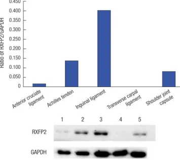
RXFP1 and RXFP2 proteins expression in each tissue The intensity of RXFP1/GAPDH ratio in each tissue samples is summarized in Table 1. RXFP1 protein was detected in all tis- sues examined. The protein level of RXFP1 was the highest in ACL sample, followed by transverse carpal ligament, Archilles tendon, shoulder joint capsule, and inguinal ligament samples (Fig. 1). The intensity of RXFP1/GAPDH ratio in each tissue sam- ple is shown in Table 2. RXFP2 protein was not detected in trans- verse carpal ligament samples. However, it was identified in oth-
er samples collected. Its level was the highest in inguinal liga- ment samples, followed by Archilles tendon, the shoulder joint capsule, and the ACL samples (Fig. 2).
Immunolocalization of RXFP1 and RXFP2 proteins in each tissue
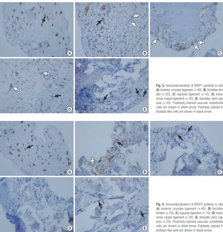
Immunohistochemical analyses were performed to reveal the distribution of RXFP1 or RXFP2 in each tissue sample. RXFP1 was identified in vascular endothelial cells in the inguinal liga- ment samples. It was also found in fibroblast-like cells of ACL and shoulder joint capsule samples as well as in mixed cells of
the Archilles tendon and transverse carpal ligament samples (Fig. 3). RXFP2 was detected in vascular endothelial cells and fibroblast-like cells of the Archilles tendon samples. It was also found in fibroblast-like cells of the inguinal ligament, shoulder joint capsule, and ACL samples (Fig. 4). In shoulder joint cap- sule and ACL samples, immunostaining revealed that relaxin receptors were mainly distributed in fibroblast-like cells.
DISCUSSION
The effect of relaxin on ligament laxity has been previously re- Table 1. The ratio of RXFP1 to GAPDH
Tissue RXFP1 intensity GAPDH intensity RXFP1/GAPDH ratio
ACLs 1,605,813.57 (18,321.25) 929,146.09 (17,944.75) 1.728
Achilles tendons 2,453,456.09 (24,041.38) 3,501,927.17 (14,181.33) 0.701
Inguinal ligament 1,563,577.87 (19,657.47) 5,398,032.00 (17,678.98) 0.290
TCLs 3,320,293.35 (22,481.62) 3,221,980.39 (18,962.31) 1.031
Shoulder joint capsule 2,141,760.83 (23,373.74) 4,080,341.28 (18,915.13) 0.525
Values are mean (SD).
RXFP1, relaxin family peptide receptors 1; GAPDH, glyceraldehyde-3-phosphate dehydrogenase; ACLs, anterior cruciate ligaments; TCLs, transverse carpal ligaments.
Table 2. The ratio of RXFP2 to GAPDH
Tissue RXFP2 intensity GAPDH intensity RXFP2/GAPDH ratio
ACLs 103,244.00 (1,701.35) 6,333,155.33 (24,814.75) 0.016
Achilles tendons 977,737.33 (5,714.08) 7,101,984.00 (24,006.32) 0.138
Inguinal ligaments 1,933,960.00 (1,040.14) 4,803,963.00 (22,315.90) 0.403
TCLs 393.33 (3,030.59) 2,570,707.00 (18,138.24) 0.000
Shoulder joint capsules 544,578.67 (3,107.11) 6,669,186.33 (24,012.00) 0.082
Values are mean (SD).
RXFP2, relaxin family peptide receptors 2; GAPDH, glyceraldehyde-3-phosphate dehydrogenase; ACLs, anterior cruciate ligaments; TCLs, transverse carpal ligaments.
Fig. 1. Ratio of RXFP1 to GAPDH protein in tissue samples studied.
GAPDH, glyceraldehyde-3-phosphate dehydrogenase.
1, anterior cruciate ligament; 2, achilles tendon; 3, inguinal ligament; 4, transverse carpal ligament; 5, shoulder joint capsule.
Ratio of RXFP1/GAPDH
Anterior crucia te
ligament Achilles tendon Inguinal ligament Transverse carpal ligament Shoulder joint capsule 2.000
1.800 1.600 1.400 1.200 1.000 0.800 0.600 0.400 0.200 0
RXFP1 GAPDH
1 2 3 4 5
Fig. 2. Ratio of RXFP2 to GAPDH protein in tissue samples studied.
GAPDH, glyceraldehyde-3-phosphate dehydrogenase.
1, anterior cruciate ligament; 2, achilles tendon; 3, inguinal ligament; 4, transverse carpal ligament; 5, shoulder joint capsule.
Ratio of RXFP2/GAPDH
Anterior crucia te
ligament Achilles tendon Inguinal ligament Transverse carpal ligament Shoulder joint capsule 0.450
0.400 0.350 0.300 0.250 0.200 0.150 0.100 0.050 0
RXFP2 GAPDH
1 2 3 4 5
ported. In 1926, Hisaw found an aqueous extract of the corpora lutea of pregnant sows caused by interpubic ligament forma- tion in estrogen-primed guinea pigs (9). The hormone contained in that extract that relaxed symphysis pubis was called “relaxin”.
The relaxin subfamily consists of the following seven peptides:
relaxin-1, relaxin-2, relaxin-3, and 4 other insulin-like peptides (INSL3, INSL4, INSL5, and INSL6). They are polypeptide hor- mones that possess structural similarity to insulin (10). The re- laxin family peptides possess their physiologic effect by activat- ing a group of 4 G-protein–coupled receptors (GPCRs), i.e., the
relaxin family peptide receptors 1-4 (RXFP1-4). Human relaxin H2 and INSL3 are leucine-rich repeat-containing GPCRs. They are cognate ligands for RXFP1 and RXFP2, respectively (11). Hu- man relaxins H1 and H2 can activate both RXFP1 and RXFP2 while rat relaxin-1 can only weakly bind to RXFP2. RXFP1 and RXFP2 are the two main relaxin receptor isoforms (12). Relax- in-3/RXFP3 system is thought to function primarily in mouse brain (13) while INSL5/RXFP4 system stimulates appetite and activates colon motility to control food intake and glucose ho- moeostasis in mouse intestine (14).
Fig. 3. Immunolocalization of RXFP1 proteins in rats.
(A) Anterior cruciate ligament (× 40), (B) Archilles ten- don (× 20), (C) inguinal ligament (× 10), (D) trans- verse carpal ligament (× 20), (E) shoulder joint cap- sule (× 20). Positively stained vascular endothelial cells are shown in white arrow. Positively stained fi- broblast-like cells are shown in black arrow.
A
D
B
E
C
Fig. 4. Immunolocalization of RXFP2 proteins in rats.
(A) Anterior cruciate ligament (× 40), (B) Archilles tendon (× 20), (C) inguinal ligament (× 10), (D) trans- verse carpal ligament (× 20), (E) shoulder joint cap- sule (× 20). Positively stained vascular endothelial cells are shown in white arrow. Positively stained fi- broblast-like cells are shown in black arrow.
A
D
B
E
C
The primary focus of our study is on RXFP1 and RXFP2 re- ceptors associated with ligaments, tendons, and fibrotic joint capsules. A few reports have revealed the association between ligaments and relaxin receptors. In humans, relaxin receptors have been identified in ACL of men and women, with higher expression in women. This gender-specific difference suggests that estrogen priming might increase the release of endogenous relaxin, the response of target organs to relaxin, and the num- ber of relaxin-binding sites on human smooth muscle or rat myometrium (15). Lubahn and colleagues have reported that relaxin can bind to the volar oblique ligament with specificity (16), suggesting the presence of either cellular or extracellular matrix receptors and a receptor-mediated process. Relaxin re- ceptors are also found in the ligaments, cartilage, and synovium of temporomandibular joint, indicating that these sites might be potential targets for relaxin therapy (17). Recently, the ex- pression of relaxin receptor RXFP1 and RXFP2 has been shown to be regulated by sex steroids in the ligaments and tendons of ovariectomized adult female rat knee (8). However, the studies that yielded those results were restricted to the association of ligaments and joint instability. To further expand those find- ings, we determined whether relaxin receptors were present in shoulder joint capsules or the tendinous and ligamentous tis- sues affected by musculoskeletal disorders. Because the expres- sion of relaxin receptors could be controlled by female sex hor- mones (8), only male rats were chosen in order to minimize the effect of female sex hormone on relaxin of the target tissue. We could not completely control regulating factors associated with relaxin receptor expression, including nutritional conditions, systemic hormonal feedback mechanisms, or the interactions with other hormones such as insulin and glucagon. This study also has some limitations. We found that RXFP1 was distribut- ed only in the transverse carpal ligaments of rats, suggesting that RXFP1 might be a main target for local relaxin administra- tion to treat carpal tunnel syndrome in animal models. We also found that both RXFP1 and RXFP2 were present in the shoul- der joint capsules of rats, suggesting that relaxin might be able to loosen fibrotic joint capsules in human patients with adhe- sive capsulitis. In addition, our immunohistochemical findings failed to reveal whether RXFP1 and RXFP 2 on vascular endo- thelial cells of inguinal ligament, Achilles tendon, and transverse carpal ligament were involved in regulating the secretion of re- laxin. The presence of RXFP1 and RXFP 2 around arterioles sug- gests that relaxin and the RXFP receptor system might have a role in the regulation of local arterial function as indicated in a previous study (18).
Systemic administration of relaxin has an effect on tissue ex- tracellular matrix components, including collagen and hyaluro- nan. It can increase the concentration of hyaluronan by upreg- ulating the expression of hyaluronic acid synthetase. Hyaluro- nan can interfere with the assembly of fibrillar extracellular ma-
trix components, which can results in loosening of the dense collagen fiber network (19). Relaxin has also been shown to have antifibrotic action by downregulating fibroblast activity, increas- ing collagenase synthesis, and inhibiting transforming-growth- factor–stimulated collagen-1 lattice concentration in rat kidney (20). In addition, relaxin can up-regulate the expression and activities of matrix metalloproteinases (MMPs), a family of ex- tracellular proteases that interfere with matrix remodeling by degrading extracellular matrix components (21). Furthermore, relaxin can downregulate tissue inhibitors of metalloproteinas- es (TIMPs) (21). Therefore, MMPs can be used to treat fibrotic disease by reducing collagen production and increasing the clearance of TIMPs. Those mechanisms have been explained by systemic administration of relaxin for the treatment of fibrot- ic diseases. However, local administration of relaxin for muscu- loskeletal disorders has not been reported yet. Our study is a precedent study on local injection of relaxin for the treatment of stiff ligaments, tendons, and fibrotic joint capsules. Further research is needed to identify the exact mechanisms and inter- actions between local relaxin hormones and relaxin receptors.
Mechanisms of gender-specific responses to relaxin in many target tissues and the value of local administration of relaxin in musculoskeletal tissue remain unclear.
In conclusion, we demonstrated that relaxin receptors RXFP1 and RXFP2 were present in rat musculoskeletal tissues such as ligaments, tendons, and shoulder joint capsules. Studies of re- laxin receptors in animal musculoskeletal tissues may provide valuable information for studies on human subjects. In order to understand whether local administration of relaxin is effective and safe treatment for entrapment neuropathies, tendon stiff- ness, and adhesive capsulitis in humans, animal studies should be preceded. After that, the correct dosage and potential adverse effects of local relaxin injections should be determined before it can be administered to patients in a clinical setting.
DISCLOSURE
The authors have no potential conflicts of interest to disclose.
AUTHOR CONTRIBUTION
Study concepts and design: Kim JH, Lee SK, Lee SK, Kim JH, Fredericson M. Data collection: Kim JH, Lee SK, Lee SK. Analy- sis and interpretation of result: Kim JH, Lee SK, Lee SK, Kim JH, Fredericson M. Writing draft: Kim JH. Revision: Fredericsion M.
Approval of final manuscript and agreement of submission: all authors.
ORCID
Jae Hyung Kim http://orcid.org/0000-0002-7005-0113
Sang Kwang Lee http://orcid.org/0000-0001-8315-0458 Seong Kyu Lee http://orcid.org/0000-0002-5446-3233 Joo Heon Kim http://orcid.org/0000-0002-0999-4131 Michael Fredericson http://orcid.org/0000-0002-4827-9026
REFERENCES
1. Rempel DM, Diao E. Entrapment neuropathies: pathophysiology and pathogenesis. J Electromyogr Kinesiol 2004; 14: 71-5.
2. Neviaser AS, Neviaser RJ. Adhesive capsulitis of the shoulder. J Am Acad Orthop Surg 2011; 19: 536-42.
3. Bouche P. Compression and entrapment neuropathies. Handb Clin Neu- rol 2013; 115: 311-66.
4. Bennett RG. Relaxin and its role in the development and treatment of fi- brosis. Transl Res 2009; 154: 1-6.
5. Hsu SY, Nakabayashi K, Nishi S, Kumagai J, Kudo M, Sherwood OD, Hsueh AJ. Activation of orphan receptors by the hormone relaxin. Science 2002;
295: 671-4.
6. Bathgate RA, Ivell R, Sanborn BM, Sherwood OD, Summers RJ. Interna- tional Union of Pharmacology LVII: recommendations for the nomencla- ture of receptors for relaxin family peptides. Pharmacol Rev 2006; 58: 7-31.
7. Zimmermann S, Steding G, Emmen JM, Brinkmann AO, Nayernia K, Hol- stein AF, Engel W, Adham IM. Targeted disruption of the Insl3 gene causes bilateral cryptorchidism. Mol Endocrinol 1999; 13: 681-91.
8. Dehghan F, Muniandy S, Yusof A, Salleh N. Sex-steroid regulation of re- laxin receptor isoforms (RXFP1 & RXFP2) expression in the patellar ten- don and lateral collateral ligament of female WKY rats. Int J Med Sci 2014;
11: 180-91.
9. Weiss G. Relaxin. Annu Rev Physiol 1984; 46: 43-52.
10. Bathgate RA, Halls ML, van der Westhuizen ET, Callander GE, Kocan M, Summers RJ. Relaxin family peptides and their receptors. Physiol Rev 2013;
93: 405-80.
11. Scott DJ, Rosengren KJ, Bathgate RA. The different ligand-binding modes of relaxin family peptide receptors RXFP1 and RXFP2. Mol Endocrinol 2012; 26: 1896-906.
12. Scott DJ, Layfield S, Riesewijk A, Morita H, Tregear GW, Bathgate RA. Char- acterization of the mouse and rat relaxin receptors. Ann N Y Acad Sci 2005; 1041: 8-12.
13. McGowan BM, Stanley SA, Donovan J, Thompson EL, Patterson M, Sem- jonous NM, Gardiner JV, Murphy KG, Ghatei MA, Bloom SR. Relaxin-3 stimulates the hypothalamic-pituitary-gonadal axis. Am J Physiol Endo- crinol Metab 2008; 295: E278-86.
14. Belgi A, Hossain MA, Shabanpoor F, Chan L, Zhang S, Bathgate RA, Tre- gear GW, Wade JD. Structure and function relationship of murine insu- lin-like peptide 5 (INSL5): free C-terminus is essential for RXFP4 receptor binding and activation. Biochemistry 2011; 50: 8352-61.
15. Dragoo JL, Lee RS, Benhaim P, Finerman GA, Hame SL. Relaxin receptors in the human female anterior cruciate ligament. Am J Sports Med 2003;
31: 577-84.
16. Lubahn J, Ivance D, Konieczko E, Cooney T. Immunohistochemical de- tection of relaxin binding to the volar oblique ligament. J Hand Surg Am 2006; 31: 80-4.
17. Clifton KB, Rodner C, Wolf JM. Detection of relaxin receptor in the dorso- radial ligament, synovium, and articular cartilage of the trapeziometacar- pal joint. J Orthop Res 2014; 32: 1061-7.
18. Novak J, Parry LJ, Matthews JE, Kerchner LJ, Indovina K, Hanley-Yanez K, Doty KD, Debrah DO, Shroff SG, Conrad KP. Evidence for local relaxin li- gand-receptor expression and function in arteries. FASEB J 2006; 20: 2352- 62.
19. Soh YM, Tiwari A, Mahendroo M, Conrad KP, Parry LJ. Relaxin regulates hyaluronan synthesis and aquaporins in the cervix of late pregnant mice.
Endocrinology 2012; 153: 6054-64.
20. Masterson R, Hewitson TD, Kelynack K, Martic M, Parry L, Bathgate R, Darby I, Becker G. Relaxin down-regulates renal fibroblast function and promotes matrix remodelling in vitro. Nephrol Dial Transplant 2004; 19:
544-52.
21. Chow BS, Chew EG, Zhao C, Bathgate RA, Hewitson TD, Samuel CS. Re- laxin signals through a RXFP1-pERK-nNOS-NO-cGMP-dependent path- way to up-regulate matrix metalloproteinases: the additional involvement of iNOS. PLoS One 2012; 7: e42714.