413 http://dx.doi.org/10.4196/kjpp.2012.16.6.413
ABBREVIATIONS: BSA, bovine serum albumin; DPH, 1,6-Diphenyl- 1,3,5-hexatriene; PBS, phosphate-buffered saline; Py-3-Py, 1,3-Di (1-pyrenyl) propane; RET, radiationless energy transfer; SPMV, synaptosomal plasma membrane vesicles isolated from bovine cerebral cortex; SPMVTL, liposome of total lipids extracted from SPMV; SPMVPL, liposome of phospholipids extracted from SPMV;
TNBS, 2,4,6-Trinitrobenzenesulfonic acid.
Received August 24, 2012, Revised October 2, 2012, Accepted October 10, 2012
*Correspondence to: Moon-Kyoung Bae, Hye-Ock Jang and Il Yun, Departments of Dental Pharmacology and Biophysics and Oral Physiology and Molecular Biology, School of Dentistry and Research Institute for Oral Biotechnology, Yangsan Campus of Pusan National University, Beomeo-ri, Mulgeum-eup, Yangsan 626-870, Korea. (Tel) 82-51-510-8236, (Fax) 82-51-510-8233, (E-mail) [email protected] (I Yun), [email protected] (HO Jang) and [email protected] (MK Bae).
‡
Co-first authors.
This is an Open Access article distributed under the terms of the Creative Commons Attribution Non-Commercial License (http://
creativecommons.org/licenses/by-nc/3.0) which permits unrestricted non-commercial use, distribution, and reproduction in any medium, provided the original work is properly cited.
The Effect of Lidocaine
ㆍHCl on the Fluidity of Native and Model Membrane Lipid Bilayers
Jun-Seop Park
1,‡, Tae-Sang Jung
1,‡, Yang-Ho Noh
1, Woo-Sung Kim
1, Won-Ick Park
1, Young-Soo Kim
1, In-Kyo Chung
2, Uy Dong Sohn
3, Soo-Kyung Bae
1, Moon-Kyoung Bae
4,*, Hye-Ock Jang
1,*, and Il Yun
1,*
1
Department of Dental Pharmacology and Biophysics,
2Department of Oral and Maxillofacial Surgery and Clinical Pharmacology,
3
Department of Pharmacology, College of Pharmacy, Chung-Ang University, Seoul 156-756,
4Department of Oral Physiology and Molecular Biology, School of Dentistry and Research Institute for Oral Biotechnology, Yangsan Campus of Pusan National University, Yangsan 626-870, Korea
The purpose of this study is to investigated the mechanism of pharmacological action of local anesthetic and provide the basic information about the development of new effective local anesthetics.
Fluorescent probe techniques were used to evaluate the effect of lidocaineㆍ HCl on the physical properties (transbilayer asymmetric lateral and rotational mobility, annular lipid fluidity and protein distribution) of synaptosomal plasma membrane vesicles (SPMV) isolated from bovine cerebral cortex, and liposomes of total lipids (SPMVTL) and phospholipids (SPMVPL) extracted from the SPMV. An experimental procedure was used based on selective quenching of 1,3-di(1-pyrenyl)propane (Py-3-Py) and 1,6-diphenyl-1,3,5-hexatriene (DPH) by trinitrophenyl groups, and radiationless energy transfer from the tryptophans of membrane proteins to Py-3-Py. Lidocaineㆍ HCl increased the bulk lateral and rotational mobility of neuronal and model membrane lipid bilayes, and had a greater fluidizing effect on the inner monolayer than the outer monolayer. Lidocaineㆍ HCl increased annular lipid fluidity in SPMV lipid bilayers. It also caused membrane proteins to cluster. The most important finding of this study is that there is far greater increase in annular lipid fluidity than that in lateral and rotational mobilities by lidocaineㆍ HCl. Lidocaineㆍ HCl alters the stereo or dynamics of the proteins in the lipid bilayers by combining with lipids, especially with the annular lipids. In conclusion, the present data suggest that lidocaine, in addition to its direct interaction with proteins, concurrently interacts with membrane lipids, fluidizing the membrane, and thus inducing conformational changes of proteins known to be intimately associated with membrane lipid.
Key Words: Annular lipid fluidity, LidocaineㆍHCl, Membrane protein clustering, Neuronal and model membranes, Transbilayer lateral and rotational mobility
INTRODUCTION
Two general theories have been proposed to explain the action of local anesthetics on sodium channel: one considers a direct binding of local anesthetic molecules to specific re- ceptors on sodium channels [1-4] and the other proposes
the general perturbation of the bulk membrane structure by anesthetics and its consequences on channel function [3-9]. There is a large amount of evidence in support of the specific receptor hypothesis [10]. General membrane per- turbation may also contribute to an explanation of anes- thetic actions [10]. However, the precise location of molec- ular mechanism of action of local anesthetics has continued to be a subject of controversy to the present day.
Effects of local anesthetics on motion, order and phase
transitions of bulk bilayer systems of native or model mem-
branes have received considerable attention in past deca-
des. This is due to the interest in biological membranes as
well as the unique information on intermolecular inter-
actions that can be derived from the investigation of volume
changes. It is known that the potency of an anesthetic in-
creases roughly in proportion with its lipid/water partition coefficient, strongly suggesting an amphiphilic site for anes- thetic molecules [3,4,8,11-13]. Yun et al. [8] reported that local anesthetics decreased microviscosity of synaptosomal plasma membrane vesicles isolated from the bovine cere- bral cortex (SPMV). In addition, differential scanning ther- mograms of dimyristoylphosphatidylcholine multilamellar liposomes showed that local anesthetics significantly low- ered the phase transition temperature, broadened the ther- mogram peaks, and reduced the size of the cooperative unit.
If local anesthetics cause expansion of neuronal and mod- el membranes, this expansion is probably due to the in- creased fluidity in neuronal and model membrane lipid bi- layers induced by local anesthetics. Our questions were what the role of local anesthetics (which is believed to have more interaction with protein than other lipids) was and to what degree the neuronal and model membrane lipid bi- layers was expanded by local anesthetics.
More specifically, our questions were; first, how much of an increase do local anesthetics bring to rotational and lat- eral mobilities of the neuronal and model membrane lipid bilayers; second, whether such increasing effects were shown evenly on both lipid bilayers or differently between inner and outer monolayers; third, it the degree of increase is different between the inner and outer monolayers, then which monolayer has been mostly affected; fourth, whether annular lipid fluidity of the neuronal membrane lipid bi- layers is increased or decreased by local anesthetics, and whether the degree of such increase or decrease is approx- imately the same as or much greater than the degree of changes in rotational and lateral mobilities; and fifth, to what degree the neuronal and model membrane is ex- panded by local anesthetics.
We are here to present the results of our study on how we solved the aforementioned questions by employing fluo- rescence technique, including the fluorescence quenching technique which Prof. Yun first developed specifically for the study to measure the rate and range of asymmetrical lateral mobilities between inner and outer monolayers of the lipid bilayers of neuronal and model membranes.
METHODS Materials
The fluorescent probes, 1,3-di(1-pyrenyl)propane (Py-3- Py), 1-annilinonaphthalene-8-sulfonic acid (ANS) and 1,6- diphenyl-1,3,5-hexatriene (DPH) were purchased from Molecular Probes, Inc. (Junction City, OR, USA). Lidocaine ㆍHCl, N-2-hydroxyethyl-piperazine-N'-2-ethanesulfonic acid (Hepes) and bovine serum albumin (BSA) were purchased from Sigma Chemical (St. Louis, MO, USA). 2,4,6-Trinitro- benzenesulfonic acid (TNBS) was obtained from Fluka (Switzerland). All other reagents were purchased commer- cially and were of the highest quality available. Water was deionized.
Preparation of synaptosomes
Synaptosomes were prepared as described previously [12,14].
Membrane isolation
SPMV were isolated according to the procedure reported from earlier studies [3,4,12,14-18]. Their protein concen- tration was determined by the method of Lowry et al. [19]
with BSA as a standard.
Liposomes preparation and TNBS labeling
Total lipids were extracted from the SPMV as previously described [12]. Cholesterol content of the extracted total lip- ids was determined according to the Liebermann-Buchard reaction [20]. Phospholipids were quantitated by measuring the amounts of inorganic phosphate [21] after hydrolysis of the phospholipids at 180
oC in 70% HClO
4[22]. The SPMV had a high lipid to protein ratio (0.942 mg total lipids/1 mg protein) and a low cholesterol to phospholipid molar ra- tio (0.593±0.011: cholesterol 0.208±0.010, phospholipids 0.702±0.025). An average molecular weight of 775 for phos- pholipids is assumed and the molecular weight of cholester- ol is 387 for the calculation. Phospholipids were composed (mol%) of phosphatidylcholine (41.55±0.91), phosphatidyle- thanolamine (36.83±0.48), phosphatidylserine (13.60±0.26), sphingomyelin (4.15±0.16), phosphatidylinositol (2.90±0.09) and lysophosphatidylcholine (0.97±0.03).
Stock solutions of total lipids and phospholipids were made in chloroform. The concentration of the phospholipid stock solutions was 0.2 mg/ml. Giant unilamellar vesicles (GUVs: SPMVTL and SPMVPL) with a mean diameter of 45 μm were prepared by the method developed by Angelova and Dimitrov [23,24] and Angelova et al. [25]. To grow the GUVs, a special temperature-controlled chamber, which was previously described [26,27], was used. The experi- ments were carried out in the same chamber after the vesi- cle formation, using an inverted microscope (Axiovert35:
Zeiss, Thornwood, NY). The following steps were used to prepare the GUVs: 1) ∼3 μl of the lipid and phospholipid stock solution was spread on each Pt wire under a stream on N
2. To remove the residues of organic solvent we put the chamber in a liophilizer for ∼2 h. 2) To add the aqueous solvent inside the chamber (Millipore water 17.5 MΩ/cm), the bottom part of the chamber was sealed with a coverslip.
The Millipore water was previously heated to the desired temperature (80
oC), and then sufficient water was added to cover the Pt wires. Just after this step, the Pt wires were connected to a function generator (Hewlett-Packard, Santa Clara, CA), and a low-frequency AC field (sinusoidal wave function with a frequency of 10 Hz and an amplitude of 3 V) was applied for 90 min. After the vesicle formation, the AC field was turned off.
To determine the fluorescence parameters of probe mole- cules in each of the membrane monolayers, TNBS labeling reactions were performed as described [12,13,15-17,28,29]
with a few modifications. The synaptosomal pellet was gen-
tly resuspended in 50 ml of 4 mM TNBS in buffer A for
80 min (in the case of asymmetric lateral mobility) or 50
ml of 2 mM TNBS in buffer A for 40 min (in the case of
asymmetric rotational mobility) or in buffer A alone. Model
membranes were gently resuspended in 50 ml of 0.5 mM
TNBS in buffer A for 20 min buffer A alone. CO
2was bub-
bled through the solution. Buffer A composed of 30 mM
NaCl, 120 mM NaHCO
3, 11 mM glucose and 1% bovine se-
rum albumin (BSA), and its pH was adjusted to 8.5 with
NaOH. To assure complete exposure of all synaptosomal
and model membrane outer monolayers to TNBS, the pellet was passed slowly through an Eberbach tissue grinder (3 up and down strokes). Unless otherwise specified, treatment was carried out at 4
oC. The TNBS labeling reaction was terminated by adding an equal volume of 1% BSA in phos- phate buffered saline (PBS; 8 g/l NaCl, 0.2 g/l KCl, 0.2 g/l KH
2PO
4, 1.15 g/l Na
2HPO
4ㆍ7H
2O, 0.48 g/l Hepes, pH 7.4).
Fluorescence measurements
The fluorescence measurements were taken using a modified method of earlier studies [13,15-17,29].
All fluorescence measurements were obtained with a Multi Frequency Cross-Correlation Phase and Modulation Fluorometer (Model; ISS K2-003). Cuvette temperature was maintained at 37.0±0.1
oC in a circulating water bath (pH 7.4). Bandpass slits were 10 nm on excitation and 5 nm on emission. Blanks, prepared under identical con- ditions without fluorescent probes, served as controls.
Py-3-Py was incorporated by adding aliquots of a 5×10
-5M stock solution absolute ethanol to the SPMV, SPMVTL and SPMVPL, such that the final probe concentration was less than 5×10
-7M [13,15-17,29]. Mixtures were initially vigorously vortexed for 10 s at room temperature and then incubated at 4
oC for 18 h with gentle stirring [13,15-17,29].
DPH was dissolved in tetrahydrofuran, and 0.5 μl tetra- hydrofuran per ml of PBS was added directly to the mem- brane suspension to a concentration of 0.01 μg/50 μg membrane protein or membrane lipid (fluorescent probe DPH 2: membrane protein or lipid 10,000) as described pre- viously [13,15-17,28,29]. After probe incorporation the membrane suspension was placed in cuvettes, and control fluorescence was determined. Concentrated solutions of li- docaineㆍHCl were prepared in 10 mM Tris-HCl (pH 7.4) and added to the labeled membrane suspension (or un- treated SPMV, SPMVTL and SPMVPL suspension) to give the desired concentration of lidocaineㆍHCl (in this case, for 30 min incubation).
Excitation wavelengths were 286 nm for tryptophan and 330 nm for Py-3-Py. Emission wavelengths were 335 nm for tryptophan, 379 nm for Py-3-Py monomer and 480 nm for Py-3-Py excimer. For Py-3-Py excimer emission, a GG- 455 cut-off filter was used. The excimer to monomer fluo- rescence intensity ratio, I'/I, was calculated from the 480 nm to 379 nm signal ratio. The excitation wavelength for DPH was 362 nm and emission wavelength was 424 nm.
Effect of lidocaine
ㆍHCl on the structure of the indivi- dual monolayers of SPMV, SPMVTL and SPMVPL:
selective quenching of Py-3-Py
The method used is based on the assumption that the system is composed of fluorescing compartments that are differentially accessed by TNBS. The excimer to monomer fluorescence intensity ratios, I'/I, of Py-3-Py in bulk (inner plus outer), and in the inner and outer monolayers were calculated from the following equations:
(I'/I)
t=I'
t/ I
t ………equation 1 (I'/I)
i=I'
i/ I
i ………equation 2 (I'/I)
o=(I'
t-I'
i) / (I
t-I
i)
………equation 3 where (I'/I)
t, (I'/I)
iand (I'/I)
oare the excimer to mono- mer fluorescence intensity ratios of Py-3-Py (I'/I) in bulk, and in the inner and outer monolayers, respectively. The
values of I'
t(excimer fluorescence intensity for inner plus outer monolayers) and I'
i(excimer fluorescence intensity for the inner monolayer) were determined for Py-3-Py from SPMV, SPMVTL and SPMVPL incubated with buffer A and buffer A plus TNBS, respectively, at 4
oC (pH 8.5) (non-pene- trating conditions).
Determination of annular lipid fluidity in SPMV The experimental determination of the annular lipid flu- idity in SPMV is based on a method previously established for synaptic plasma membrane [30,31] and SPMV [15-17].
Incorporated Py-3-Py in the SPMV was excited by radia- tionless energy transfer (RET) from tryptophan (excitation at 286 nm) and the excimer to monomer fluorescence in- tensity ratio (I'/I) of Py-3-Py was calculated from the ratio 480 nm to 379 nm signal. Taking into account that the Förster radius (the RET-limiting distance) for the trypto- phan-Py-3-Py donor-acceptor pair is 3 nm [15-17,32], only Py-3-Py located in annular lipids (close to proteins) was ex- cited, and the fluidity of annular lipids was considered pro- portional to I'/I [13,15-17,26,27].
Determination of protein distribution in the SPMV llipid bilayers
This was based on a method previously established for membranes [15-17,30,31]. The fluorescence intensity of en- dogenous tryptophan in SPMV was determined. Thereafter the Py-3-Py probe was incorporated at a concentration of 5×10
-7M (in absolute ethanol), and after 10 min, trypto- phan emission fluorescence intensity was measured again.
The efficiency of RET from tryptophan to Py-3-Py was cal- culated from the equation:
RET=(I
d-I
da) / I
d…
…………… equation 4 where I
dand I
darepresent the fluorescence intensities of donor (in this case, endogenous tryptophan) in the absence and presence, respectively, of acceptor (in this case, Py-3-Py). The wavelengths of excitation and emission of tryptophan were 286 and 335 nm, respectively.
Effect of lidocaine
ㆍHCl on the rotational mobility of bulk SPMV, SPMVTL and SPMVPL
The intensities of the components of fluorescence parallel (I
∥) and perpendicular (I
┴) to the direction of the vertically polarized excitation light were determined by measuring the light emitted through polarizers oriented vertically and horizontally. The polarization (P) was obtained from the in- tensity measurements using P=(I
∥-GI
┴) / (I
∥+GI
┴) where G is a grating correction factor for the transmission effi- ciency of the monochromator for vertically and horizontally polarized light. It is given by the ratio of the fluorescence intensities of the vertical to horizontal components when the exciting light is polarized in the horizontal direction.
The polarization was expressed as the anisotropy [r=2P / (3-P)] (equation 5).
Effect of lidocaine
ㆍHCl on the separate monolayers of SPMV, SPMVTL and SPMVPL: selective quen- ching of DPH
The experimental determination of the separate mono-
layer structure in SPMV, SPMVTL and SPMVPL is based on a method by the method developed for tumor cell plasma membranes by Schroeder [33]. And a method previously es- tablished for LM fibroblast plasma membrane [34], for syn- aptic plasma membrane [35-38] and for synaptosomal plas- ma membrane vesicles [3,4,12,15-18] for the plasma mem- brane vesicles of Chinese hamster ovary cells, for Mar 18.5 hybridoma cells [29] and for the myeloma cell line Sp2/0- Ag14 [13]. It does not simply provide a theoretically calcu- lated or average value: instead it is based on the assump- tion that the system is composed of fluorescing compart- ments of different accessibility to TNBS. If fluorescence in- tensity, F, and anisotropy, r, are measured simultaneously, then
r=∑F
jr
j……… equation 6 where F
jis the fraction of fluorescence intensity in com- partment j. For a binary system composed of the outer and inner monolayers of the SPMV, SPMVTL and SPMVPL, this leads to
r=
……… equation 7 where F and F
iare the DPH fluorescence obtained for SPMV incubated with buffer A and buffer A plus 2 mM TNBS for 40 min at 4ºC (pH 8.5) (non-penetrating con- ditions), (in the case of model membranes, incubated with buffer A and buffer A plus 0.5 mM TNBS for 20 min) respectively. The values of fluorophore concentration-in- dependent parameter anisotropies, r (anisotropy for both monolayers) and r
i(inner monolayer anisotropy), were also determined for DPH in SPMV, SPMVTL and SPMVPL in- cubated with buffer A and buffer A plus TNBS at 4ºC, respectively. The equation was then solved for r
o(outer monolayer anisotropy).
RESULTS
In order to determine the effect of the lidocaineㆍHCl on the bulk and asymmetric rotational and lateral mobilities of the SPMV, SPMVTL and SPMVPL monolayers, it is, first, necessary to demonstrate that this local anesthetic does not interact directly with Py-3-Py and DPH and there- by quench its fluorescence. No quenching of absorbance- corrected fluorescence intensity of both Py-3-Py and DPH by the lidocaineㆍHCl was observed at all tested concen- trations.
In our judgment, it would be rational to use liposome that is made up of total lipids extracted from neuronal mem- branes and liposome that is made up of phospholipids ex- tracted from neuronal and model membranes as samples in the study of the structure-activity relationship of local anesthetics.
The purity of SPMV
We assessed the purity of SPMV by enzymatic and mor- phological criteria. The specific activities of Na
+, K
+- ATPase, acetylcholinesterase and 5'-nucleotidase were en- riched about 4-, 2.5- and 3-fold, respectively, in the plasma membrane fraction with respect to crude homogenates. The transmission electron microscopic examination of the SPMV
indicated very high purity. The vesicles, which were sepa- rated according to size demonstrated homogeneous dis- tribution and no longer showed the presence of intracellular organelles or leakage. The protein concentration was de- termined by the method of Lowry et al. [19] using BSA as a standard.
Effect of lidocaine
ㆍHCl on the rate and range of lateral mobility in SPMV, SPMVTL and SPMVPL bulk bilayer
The I'/I value in intact SPMV, SPMVTL and SPMVPL (lidocaineㆍHCl-untreated) was 0.412±0.007, 0.606±0.007, 0.790±0.010, respectively (at 37
oC, pH 7.4). Incubation with lidocaineㆍHCl increased the range and rate of lateral mo- bility of bulk (inner+outer monolayer) SPMV, SPMVTL and SPMVPL lipid bilayers at concentrations as low as 0.02 mM (n=5, p<0.05), respectively, and above as demonstrated in Fig. 1.
The I'/I value of Py-3-Py in bulk SPMV, SPMVTL and SPMVPL incubated with 1 mM lidocaineㆍHCl was 0.441±
0.004, 0.639±0.009 and 0.828±0.007 (n=5, p<0.01), re- spectively, and the change in I'/I value before and after adding the 1 mM lidocaineㆍHCl was 0.028, 0.032 and 0.037. The I'/I values of Py-3-Py in the SPMV, SPMVTL and SPMVPL bilayer were 0.356±0.006, 0.524±0.005 and 0.715±0.007 (n=5) at 25
oC (pH 7.4), respectively. Hence the effect of 1 mM lidocaineㆍHCl was equivalent to that pro- duced by a temperature increase of approximate 6.0, 4.7and 5.9
oC, respectively.
Effect of lidocaine
ㆍHCl on the rate and range of transbilayer asymmetric lateral mobility of SPMV, SPMVTL and SPMVPL monolayers
The effect of increasing concentrations of lidocaineㆍHCl on the I'/I values in the individual SPMV, SPMVTL and SPMVPL monolayers is shown in Fig. 1. LidocaineㆍHCl increased the rate and range of lateral mobility of the SPMV, SPMVTL, SPMVPL inner monolayer to a significant extent (0.390±0.013, 0.582±0.006 and 0.748±0.011, p<0.01, p<0.05 and p<0.05, n=5) at 0.02, 0.05 and 0.02 mM lido- caineㆍHCl, respectively (Fig. 1). It had a greater increas- ing effect on the lateral mobility of the inner monolayer (Fig. 1, filled triangles) than that of the outer monolayer (Fig. 1, filled circles). Since the changes in I'/I values is due primarily from the effect on the inner monolayer, we studied the selective effect of lidocaineㆍHCl on the rate and range of mobility of the probe. To the best of our knowl- edge, the results for asymmetric lateral mobility presented here are the first to demonstrate that the Sheetz-Singer hypothesis [39] is valid in neuronal membranes.
Effect of lidocaine
ㆍHCl on annular lipid fluidity in the SPMV lipid bilayers
I'/I measurements showed that the annular lipid fluidity of SPMV (intact membrane) was 0.245±0.006 (37ºC, pH 7.4), and in this increased in response to concentration of 0.02 mM lidocaineㆍHCl and above (Fig. 2).
The I'/I values of Py-3-Py in the bilayer are 0.245±0.006 (n=5) and 0.199±0.002 (n=5) at 37ºC and 25ºC, respectively.
Thus the effect by 1 mM lidocineㆍHCl was the same as
that produced by a temperature increase of approximate
9.6ºC. The important finding is that there was much great-
A B C
Fig. 1. The effect of lidocaineㆍHCl on excimer to monomer fluorescence intensity ratio (I'/I) of Py-3-Py in SPMV (A), SPMVTL (B) and SPMVPL (C). The excitation wavelength of Py-3-Py was 330 nm and the I'/I values were calculated from the 480 nm to 379 nm signal ratio. SPMV was treated±4mM TNBS, pH 8.5, at 4
oC for 80 min. SPMVTL and SPMVPL were treated±0.5 mM TNBS, pH 8.5, at 4
oC for 20 min. Py-3-Py was incorporated into SPMV, SPMVTL and SPMVPL and fluorescence measurements were performed at 37
oC (pH 7.4). Untreated (inner and outer monolayers, ■); TNBS treated (inner monolayer, ▲); calculated for outer monolayer (●) by eq. 3 as described in Materials and Methods. Each point represents the mean±SEM of 5 determinations. An asterisk and double asterisks signify p<0.05 and p<0.01, respectively, compared to control by Student's t-test.
Fig. 2. The effect of lidocaineㆍHCl on annular lipid fluidity in SPMV. Py-3-Py was excited through RET from tryptophan (exci- tation wavelength, 286 nm) and the excimer to monomer fluore- scence intensity ratio (I'/I) was calculated from the 480 nm to 379 nm signal ratio. Fluorescence measurements were performed at 37
oC (pH 7.4). Each point represents the mean±SEM of 5 determinations. An asterisk and double asterisks signify p<0.05 and p<0.01, respectively, compared to control by Student's t-test.
Fig. 3. The effect of lidocaineㆍHCl on protein distribution in SPMV.
Efficiency of RET from tryptophan to Py-3-Py was taken as a measure of protein clustering and calculated by eq. 4. Fluorescence measurements were performed at 37
oC (pH 7.4). Each point repre- sents the mean±SEM of 5 determinations. An asterisk and double asterisks signify p<0.05 and p<0.01, respectively, compared to control by Student's t-test.
er increase in annular lipid fluidity in the lateral and rota- tional mobilities.
Effect of lidocaine
ㆍHCl on protein distribution in SPMV
We evaluated protein distribution by RET from trypto- phan to Py-3-Py. The RET value of untreated SPMV was 0.295±0.003 (37
oC, pH 7.4) and it was lowered by concen-
trations of lidocaineㆍHCl of 0.02 mM or more (Fig. 3).
Protein clustering is probably caused by interaction among phospholipids, especially annular lipids, whose movement is increased by lidocaineㆍHCl and proteins around them.
Effect of lidocaine
ㆍHCl on the range of rotational
mobility of SPMV, SPMVTL and SPMVPL bulk bilayer
The anisotropy (r) of DPH in the intact SPMV, SPMVTL
and SPMVPL was 0.202±0.004, 0.183±0.002 and 0.149±
A B C
Fig. 4. The effect of lidocaineㆍHCl on the anisotropy (r) of DPH in SPMV (A), SPMVTL (B) and SPMVPL (C). The excitation wavelength for DPH was 362 nm and fluorescence emission was read at 424 nm. SPMV was treated±2 mM TNBS, pH 8.5, at 4
oC for 40 min. SPMVTL and SPMVPL were treated±0.5 mM TNBS, pH 8.5, at 4
oC for 20 min. DPH was incorporated into SPMV, SPMVTL and SPMVPL and fluorescence measurements were performed at 37
oC (pH 7.4). Untreated (inner and outer monolayers, ■); TNBS treated (inner monolayer,
▲); calculated for outer monolayer (●) by eq. 7 as described in Materials and Methods. Each point represents the mean±SEM of 5 determinations. An asterisk and double asterisks signify p<0.05 and p<0.01, respectively, compared to control by Student's t-test.
0.001 (at 37
oC, pH 7.4), respectively (Fig. 4). Lidocaineㆍ HCl inceased rotational mobility, with a significant de- crease in anisotropy (r) values in SPMV, SPMVTL and SPMVTL even at 0.1, 0.2 and 0.1 mM, respectively (Fig.
4). The difference in anisotropy of the bulk SPMV, SPMVTL and SPMVPL lipid bilayers before and after adding 1 mM lidocaineㆍHCl was 0.031, 0.023 and 0.027, respectively.
This can be evaluated by comparison with the effect of tem- perature on this parameter. The anisotropy of DPH in the SPMV, SPMVTL and SPMVPL bilayer was 0.257±0.002, 0.242±0.003 and 0.201±0.001 (n=5) at 25
oC (pH 7.4), respectively. The effect of 1 mM lidocaineㆍHCl was thus the same as that of a temperature increase of approximate 6.8, 4.7 and 6.2
oC, respectively.
Effect of lidocaine
ㆍHCl on the range of transbilayer asymmetric rotational mobility of SPMV monolayers The structures of the intact SPMV, SPMVTL and SPMVPL (inner plus outer monolayers), and the outer (extracellular) and inner (intracellular) monolayers sepa- rately, were evaluated with DPH as a fluorescent reporter and trinitrophenyl groups as quenching agents. Trinitro- phenylation of the intact synaptosome at 4
oC (non-penetrat- ing conditions) results in covalent attachment of trinitro- phenyl quenching agents to the outer monolayers. Approxi- mately half of the DPH fluorescence was quenched in the treated SPMV outer monolayer. When TNBS labeling was conducted in penetrating conditions (37
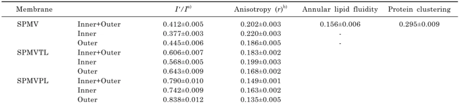
oC), greater than 80% of the fluorescence of the DPH was quenched. Values of fluorescence parameters in intact SPMV, SPMVTL and SPMVPL (both monolayers) and in TNBS- treated SPMV, SPMVTL and SPMVPL (inner monolayer) are listed in Table 1. The anisotropy of DPH in SPMV, SPMVTL and SPMVPL of the inner monolayer was 0.034, 0.031, 0.028 significantly greater than that calculated for the outer mon- olayer, as demonstrated in Table 1.
Fig. 4 shows that the anisotropy (r) of DPH in the TNBS untreated membrane (inner plus outer monolayers) de-
creased gradually (fluidization) with increasing lidocaineㆍ HCl concentrations (Fig. 4, filled squares). There was a sim- ilar, but more gradual, decrease in the calculated aniso- tropy of the inner monolayer at any lidocaineㆍHCl concen- tration (Fig. 4, filled triangles). However, there was no stat- istically significant decrease in the anisotropy (the rota- tional mobility range) of the outer monolayer of SPMV, SPMVPL at 0.01∼0.5 mM (in case of SPMVTL 0.01∼1 mM) of the lidocaineㆍHCl concentrations used. These re- sults suggest that the fluidizing effect (range of rotational mobility) of lidocaineㆍHCl is selective.
DISCUSSION
We have reported through our earlier study developed by prof. Yun in our laboratory the facile method to measure transbilayer asymmetric lateral mobility of native mem- brane lipid bilayers [40]. In this study, we report here de- veloped by prof. Yun in the lavoratory the facile method to measure transbilayer asymmetric lateral and rotational mobilities of model membrane lipid bilayers.
We used Py-3-Py, a pyrene derivative that has been used to quantify lateral mobility within native and model mem- branes [13,28,29,41,42], to determine the rate and range of lateral mobility in the SPMV, SPMVTL and SPMVPL.
With this probe one monitors emission of both the monomer
(I) and the excimer (I') components in such a way that a
ratio can be derived and used as a measure of lateral mobi-
lity [13,28,29,41,42]. As the probe mobility increases emis-
sion from the excimer predominates, since formation of the
intramolecular excimer is dependent upon lateral move-
ment of its two components. Therefore, an increase in the
I'/I ratio indicates increased lateral mobility of the probe
within the membranes. The excimer fluorescence technique
using Py-3-Py has the advantage over its counterpart based
on intermolecular excimerization that very low probe con-
centrations can be used (<10
-7M) and perturbation of the
SPMV, SPMVTL and SPMVPL by the probe molecule is
Membrane I'/I
a)Anisotropy (r)
b)Annular lipid fluidity Protein clustering
SPMV Inner+Outer
Inner Outer
0.412±0.005 0.377±0.003 0.445±0.006
0.202±0.003 0.220±0.003 0.186±0.005
0.156±0.006 - -
0.295±0.009
SPMVTL Inner+Outer Inner Outer
0.606±0.007 0.568±0.005 0.643±0.009
0.183±0.002 0.199±0.003 0.168±0.002
SPMVPL Inner+Outer
Inner Outer
0.790±0.010 0.742±0.009 0.838±0.012
0.149±0.001 0.163±0.002 0.135±0.005
a)