http://e-nrp.org
Estrogen deprivation and excess energy supply accelerate
7,12-dimethylbenz(a)anthracene-induced mammary tumor growth in C3H/HeN mice
Jin Kim
1, Yoon Hee Lee
1, Jung Han Yoon Park
2and Mi-Kyung Sung
1§1Department of Food and Nutrition, Sookmyung Women’s University, 100 Chungpa-ro 47-gil, Yongsan-gu, Seoul, 140-742, Korea
2Department of Food Science and Nutrition, College of Natural Sciences, Hallym University, 39 Hallymdaehak-gil, Chuncheon, 200-702, Korea
BACKGROUND/OBJECTIVES: Obesity is a risk factor of breast cancer in postmenopausal women. Estrogen deprivation has been suggested to cause alteration of lipid metabolism thereby creating a cellular microenvironment favoring tumor growth. The aim of this study is to investigate the effects of estrogen depletion in combination with excess energy supply on breast tumor development.
MATERIALS/METHODS: Ovariectomized (OVX) or sham-operated C3H/HeN mice at 4 wks were provided with either a normal diet or a high-fat diet (HD) for 16 weeks. Breast tumors were induced by administration of 7,12-dimethylbenz(a)anthracene once a week for six consecutive weeks.
RESULTS: Study results showed higher serum concentrations of free fatty acids and insulin in the OVX+HD group compared to other groups. The average tumor volume was significantly larger in OVX+HD animals than in other groups. Expressions of mammary tumor insulin receptor and mammalian target of rapamycin proteins as well as the ratio of pAKT/AKT were significantly increased, while pAMPK/AMPK was decreased in OVX+HD animals compared to the sham-operated groups. Higher relative expression of liver fatty acid synthase mRNA was observed in OVX+HD mice compared with other groups.
CONCLUSIONS: These results suggest that excess energy supply affects the accelerated mammary tumor growth in estrogen deprived mice.
Nutrition Research and Practice 2015;9(6):628-636; doi:10.4162/nrp.2015.9.6.628; pISSN 1976-1457 eISSN 2005-6168
Keywords: Breast cancer, obesity, estrogen, postmenopause
INTRODUCTION
9)Despite many undeniable therapeutic successes, breast cancer remains a major health issue in both developed and developing countries. Obesity, as reflected by increased body mass index (BMI), is associated with an increased risk of more aggressive breast cancer, as well as reduced survival [1]. The association between BMI and breast cancer risk becomes particularly strong for postmenopausal women [2] and weight gain after meno- pause is the most important risk factor for breast cancer in postmenopausal women [3,4]. However, the 2007 WCRF/AICR Second Expert Report [5] followed by the Updated Breast Cancer 2010 Report [6] found limited evidence on the associa- tion between breast cancer risk and dietary components which may contribute to body fat mass. The association between dietary fat has been a topic of debate for more than 20 years, however a recent meta-analysis reported no significant associa-
tion [7,8].
Estrogen is a known modulator of lipid and glucose metabolism.
Systemic loss of estrogen in postmenopausal women is typically associated with increased abdominal fat tissue. Studies have indicated that estradiol (E2) replacement can prevent meno- pause induced gains in adipose tissue mass [9,10]. In addition, ovariectomized (OVX) rodents rapidly become obese; however estrogen administration prevents the increase in body fat [11].
Estrogen activates AMP-activated protein kinase (AMPK), promo- ting fat oxidation and decreased expression of sterol regulatory element-binding protein 1c (SREBP1c) in adipose tissue, muscle, and the liver [12,13]. SREBP1c stimulates expression of lipogenic genes, such as fatty acid synthase (FAS) [12,14]. In addition to lipid storage in adipose tissue, estrogen deprivation disturbs glucose homeostasis. For example, premenopausal women are more insulin sensitive, have insulin-associated improved glucose tolerance, and are less likely to develop insulin resistance than
This research was supported by the Basic Science Research Program (20110011669) of the National Research Foundation of Korea (NRF) grant funded by the Korean government (MSPI), the SRC program (Center for Food & Nutritional Genomics: grant number 2015R1A5A6001906) of the NRF grant funded by the Korean government (MSIP), and the High Value-added Food Technology Program, Ministry of Food, Agriculture and Forestry, Republic of Korea (312006-3).
§Corresponding Author: Mi-Kyung Sung, Tel. 82-2-710-9395, Fax. 82-2-710-9395, Email. [email protected] Received: January 22, 2015, Revised: March 18, 2015, Accepted: June 17, 2015
This is an Open Access article distributed under the terms of the Creative Commons Attribution Non-Commercial License (http://creativecommons.org/licenses/by-nc/3.0/) which permits unrestricted non-commercial use, distribution, and reproduction in any medium, provided the original work is properly cited.
AIN93G ND1) HD2) Macronutrient composition
Carbohydrate, % of energy 63.95 65.71 35.69
Protein, % of energy 19.33 19.30 19.30
Fat, % of energy 16.72 14.99 45.00
Ingredient, g/kg
Cornstarch 397.4 404.0 266.5
Dextrin 132 134.2 88.5
Sucrose 100 101.6 67.1
Fiber 50 50 50
Casein 200 198.0 240.4
L-cystine 3.0 3.0 3.7
Corn oil - 12.44 45.36
Lard - 49.76 181.44
Mineral Mix (AIN-93G-MX) 35.0 34.6 42.1
Vitamin (AIN-93G-VX) 10.0 9.9 12.0
Choline bitartrate 2.50 2.50 3.10
Tert-butylhydroquinone 0.01 0.01 0.02
1)ND: Normal diet (15% energy as fat)
2)HD: High-fat diet (45% energy as fat) Table 1. Composition of experimental diets
men [15,16]. OVX rodents were reported to show dyslipidemia, impaired glucose tolerance, and impaired insulin-mediated glucose uptake in skeletal muscle [17]. In addition, estradiol replacement therapy improved glucose tolerance and insulin sensitivity and reduced lipid accumulation in the liver of leptin- deficient ob/ob mice [18]. One study suggested postmeno- pausal women with increased risk of hyperinsulinemia, insulin- resistant type 2 diabetes, and metabolic syndrome [19].
Association of postmenopausal weight gain with hormone- dependent breast cancer development has also been demons- trated [20]. Previous studies have reported overexpression of the insulin receptor (IR) in breast cancer cells [21]. Increased insulin binding to the IR leads to stimulation of the pho- sphoinositide 3-kinase (PI3K)/AKT signaling pathway, which plays an important role in breast cancer progression [22]. AKT is transported to both cytosolic and nuclear compartments, where it phosphorylates various proteins involved in the regulation of cell growth and apoptosis. Abnormal AKT activation has been observed in various types of cancer [23,24].
Therefore, it can be hypothesized that defects in insulin sensitivity in postmenopausal women and an increase in fat mass may play a role in breast cancer development. However, few mechanistic studies have provided evidence to explain the association between excess energy intake in breast cancer development under conditions with different estrogen availability.
In this study, we evaluate the effects of estrogen deprivation and/or excess energy intake on mammary tumor development and growth in a rodent model. Possible molecular mechanisms of action are also suggested.
MATERIAL AND METHODS
Animals and experimental designThree-week old C3H/HeN female mice were obtained from Central Laboratory (Seoul, Korea). Animals were housed in polycarbonate cages and maintained at a room temperature of 26 ± 1˚C, with a relative humidity of 60 ± 5%, and 12 h light/12 h darkness exposure. Fresh food was provided every 2-3 days and food intake was monitored throughout the experiment. Body weight was monitored once a week.
After acclimation for one week, animals were either ovarecto- mized or sham-operated and then randomly assigned to different experimental groups. Group 1 consisted of sham- operated animals fed a normal diet (15% energy as fat) (SHAM+
ND, n = 14); Group 2 consisted of sham-operated animals fed a high-fat diet (45% energy as fat) (SHAM+HD, n = 8); Group 3 consisted of ovariectomized animals fed a normal diet (OVX+ND, n = 6); and Group 4 consisted of ovariectomized animals fed a high-fat diet (OVX+HD, n = 7). The composition of the experimental diet was based on AIN-93G diet (25) as shown in Table 1, and soybean oil was substituted with corn oil because isoflavones present in soybean oil may act as anticarcinogens. A mixture of lard and corn oil was used as a source of dietary fat to mimic the fatty acid composition in the human diet as used in our previous study (26). Mineral mix, vitamins, choline, and tert-butylhydroquinone were adjusted to provide an equal amount for each experimental group based on difference in daily food consumption of animals in ND and
HD. At 7 weeks of age, all animals were provided with a dose of 1mg 7,12-dimethylbenz(a)anthracene (DMBA) (Sigma, MO, USA) dissolved in sesame oil. This dosage was repeated once a week for six consecutive weeks. After the last DMBA exposure, mice were subjected to regular examination for mammary tumors by palpation. Tumor size was measured using a digital caliper, and the length and width of each tumor were used in the formula [volume = length
2× width/2] to approximate volume (cm
3). Mice were sacrificed at the age of 19 weeks. All mammary tumors were weighed and the size was measured.
All procedures were approved by the Institutional Animal Care and Use committee of Sookmyung Women’s University (SMU-IACUC-2010-0625-009).
Preparation of blood and tissue samples
At the end of the experiment, animals were sacrificed and the mammary tumor, mammary fat pad, liver, spleen, and abdominal fat pad were removed, rinsed in normal saline, and weighed. Blood was collected from the inferior vena cava into EDTA-free tubes and centrifuged at 1,550 × g for 20 minutes.
All samples were stored at -80˚C until assayed.
Serum measurements
Serum insulin concentration was determined using a commercially-available enzyme-linked immunosorbent assay (ELISA) kit (Millipore, MA, USA). Serum free fatty acid (FFA) concentration was also determined using an ELISA kit (Wako, Osaka, Japan) according to the manufacturer’s instructions.
Western blots
Protein expression of IR, mammalian target of rapamycin (mTOR), p-mTOR, AKT, pAKT, AMPK, and phosphorylated AMPK (pAMPK) was measured in mammary tumor tissue samples.
Because only two animals in the OVX+ND group developed
tumors, statistical analyses were performed in SHAM+ND, SHAM
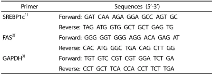
Primer Sequences (5’-3’) SREBP1c1) Forward: GAT CAA AGA GGA GCC AGT GC
Reverse: TAG ATG GTG GCT GCT GAG TG FAS2) Forward: GGG GGT GGG AGG ACA GAG AT
Reverse: CAC ATG GGC TGA CAG CTT GG GAPDH3) Forward: TGT GTC CGT CGT GGA TCT GA Reverse: CCT GCT TCA CCA CCT TCT TGA
1)SREBP1c: sterol regulatory element-binding transcription factor 1
2)FAS: fatty acid synthase
3)GAPDH: glyceraldehyde-3-phosphate dehydrogenase Table 2. Gene Primers
Body (g)
Liver (% of B.W.)
Spleen (% of B.W.)
Abdominal fat (% of B.W.)
Mammary fat pad (% of B.W.)
SHAM+ND 22.04 ± 2.73 4.43 ± 0.77 0.61 ± 0.76 2.27 ± 1.39 0.64 ± 0.53
SHAM+HD 21.50 ± 2.47 4.92 ± 0.79 0.87 ± 0.78 2.74 ± 1.50 0.77 ± 0.66
OVX+ND 22.26 ± 1.65 4.35 ± 0.62 0.34 ± 0.17 2.21 ± 1.46 0.97 ± 0.52
OVX+HD 20.50 ± 1.23 4.19 ± 1.30 0.33 ± 0.09 2.73 ± 2.02 0.91 ± 1.25
Diet NS NS NS NS NS
OVX NS NS NS NS NS
Values are presented as a mean ± SD. Data were analyzed by one way-analysis of variance (ANOVA) followed by Duncan’s multiple range test. Two-way ANOVA was used to determine the interactions between ovariectomy and diet. Liver, spleen, abdominal fat, and mammary fat pad weight were calculated as percentage of body weight. SHAM:
sham-operated fed normal diet; SHAM+HD: sham-operated fed high-fat diet; OVX+ND: ovariectomized fed normal diet; OVX+HD: ovariectomized fed high-fat diet.
Table 3. Effects of estrogen deprivation and excess energy supply on body and organ weights of experimental animals
+HD, and OVX+HD animals. Tumor tissue was homogenized with a PRO-PREP
TMprotein extraction solution (Intron Biotechn- ology Inc., Gyeonggi, Korea), left on ice for 20 minutes, and centrifuged (16,600 × g, 10 min, 4°C). Protein content was determined against a standardized control, using a Bio-Rad Protein Assay kit (Bio-Rad Laboratories, Inc., CA, USA); 50 μg of protein from each sample was separated by 4-12% and 6%
sodium dodecyl sulfate-polyacrylamide gel electrophoresis and then transferred to PVDF membranes (Koma Biotechnology, Seoul, Korea). The membranes were blocked with 2% skim milk (Amersham Corp., IL, USA) and incubated with specific antibo- dies for IR (Santa Cruz Biotech, CA, USA), pAKT (Ser473) (Cell Signaling, MA, USA), AKT (Cell Signaling, MA, USA), mTOR (Cell Signaling, MA, USA), pAMPK (Cell Signaling, MA, USA), AMPK (Cell Signaling, MA, USA), cyclin D1 (Cell Signaling, MA, USA), CDK4 (Cell Signaling, MA, USA), and β-actin (Sigma, MO, USA).
The membranes were washed with PBS/Tween 20 (PBST) containing 0.1% Tween 20 (Sigma, MO, USA). Reactive bands were visualized using an enhanced chemiluminescence (ECL) system (Amersham Corp., IL, USA). Stripping was checked by re-exposure to enhanced chemiluminescence (ECL), and was detected using a LAS 3000 (Fujifilm, Tokyo, Japan). The mem- branes were subsequently blocked and reprobed. The intensity of the bands was quantified using a Bio-Rad GS-800 densitometer equipped with the Quantity One program (Bio-Rad Laboratories, Inc., CA, USA).
Real-time quantitative PCR
Total RNA was extracted from liver tissue using TRIzol reagent, according to the manufacturer’s instructions (Invitrogen, CA, USA). Total RNA (1 μg) from liver tissue was reverse-transcribed using a cDNA Synthesis kit (PhileKorea Technology, Seoul, Korea) according to the manufacturer’s instructions. First strand cDNA was generated from 1 μg RNA using both oligo (DT)
18primer mix and random hexamer primer mix. Real-time quantitative PCR was performed on a 7500 Fast Real time PCR system (Applied Biosystems, CA, USA) using a QuantiMix SYBR Kit (PhileKorea Technology, Seoul, Korea). Primers for SREBP1c, FAS, and Glyceraldehyde-3-phosphate dehydrogenase (GAPDH) were synthesized by Bioneer (Bioneer, Daejeon, Korea) (Table 2). Relative fold-changes in expression were determined using the 2
-△△CT(relative quantification) analysis protocol. Expression of the GAPDH housekeeping gene was used to normalize PCR reactions. Each experiment was repeated three times.
Statistical analysis
Statistical analysis was performed using the SAS package (release 9.1, SAS Institute Inc., NC, USA). Data are expressed as the mean ± SD. One-way analysis of variance (ANOVA) and Duncan’s multiple test were used to determine statistical differences between the treatment groups. Interactions between two variables were examined using two-way ANOVA. P-values less than 0.05 were considered significant. Only two mice developed mammary tumors in the OVX+ND group; therefore, statistical analyses on tumor tissue were performed for animals in the SHAM+ND, SHAM+HD, and OVX+HD groups using one-way ANOVA.
RESULTS
Body and organ weights
There was no significant difference in body weights between experimental groups (Table 3). No significant difference in the weights of liver, spleen, abdominal adipose tissue, or mammary fat pad was observed. Neither diet nor OVX affected body weight and organ weights was observed between groups.
Mammary tumor development
At the end of the study period, the percentage of tumor-
bearing mice in each group was 57.1% in the SHAM+ND group,
75% in the SHAM+HD group, 33.3% in the OVX+ND group, and
42.9% in the OVX+HD group (Table 4). Number of tumors of
tumor bearing mice in each group was 1.63, 1.50, 1.50, and
1.67, respectively. The average mammary tumor volume was
significantly higher in the OVX+HD group than in the other
groups. Both diet and OVX showed significant association with
tumor size, and there was no significant interaction between
ovariectomy and diet.
Group (n) Tumor number Number of TBM
(% animals) Tumor number
/TBM Average tumor volume (cm3)
SHAM+ND (14) 13 8 (57.14) 1.63 0.09 ± 0.07b
SHAM+HD (8) 9 6 (75) 1.50 0.22 ± 0.21b
OVX+ND (6) 3 2 (33.33) 1.50 0.17 ± 0.22b
OVX+HD (7) 5 3 (42.86) 1.67 0.84 ± 0.60a
Values are presented as a mean ± SD. abMeans with different superscripts are significantly different at (P< 0.05). SHAM: sham-operated fed normal diet; SHAM+HD:
sham-operated fed high-fat diet; OVX+ND: ovariectomized fed normal diet; OVX+HD: ovariectomized fed high-fat diet; TBM: tumor-bering mouse.
Table 4. Effects of estrogen deprivation and excess energy supply on mammary tumor number, multiplicity, and volume
Group Insulin (ng/mL) FFA (mEq/L)
SHAM+ND 0.65 ± 0.29b 0.60 ± 0.16b
SHAM+HD 0.98 ± 0.54b 0.85 ± 0.42ab
OVX+ND 0.76 ± 0.42b 0.75 ± 0.15ab
OVX+HD 2.73 ± 1.71a 1.00 ± 0.24a
Significance P < 0.05 P < 0.05
OVX P < 0.01 N.S.
Diet P < 0.01 P < 0.01
OVX × Diet P < 0.05 N.S.
Values are presented as a mean ± SD Data were analyzed by one way-analysis of variance (ANOVA) followed by Duncan’s multiple range test. abMeans with different superscripts are significantly different at P< 0.05. Two-way ANOVA was used to determine the interactions between ovariectomy and diet. SHAM:
sham-operated fed normal diet; SHAM+HD: sham-operated fed high-fat diet;
OVX+ND: ovariectomized fed normal diet; OVX+HD: ovariectomized fed high-fat diet; FFA: free fatty acids.
Table 5. Effects of estrogen deprivation and excess energy supply on serum insulin and FFA concentrations
Group SREBP1c FAS
SHAM+ND 1.00 1.00b
SHAM+HD 1.49 ± 0.94 1.19 ± 0.64b
OVX+ND 1.48 ± 0.79 1.01 ± 0.59b
OVX+HD 2.32 ± 3.41 2.55 ± 2.41a
Significance N.S. P < 0.05
OVX N.S. N.S.
Diet N.S. P < 0.05
OVX × Diet N.S. N.S.
Relative expression of the indicated lipogenic genes was detected by real-time PCR in liver tissue. abMeans with different superscripts are significantly different at P<
0.05. Two-way ANOVA was used to determine the interactions between ovariectomy and diet. SHAM: sham-operated fed normal diet; SHAM+HD: sham-operated fed high-fat diet; OVX+ND: ovariectomized fed normal diet; OVX+HD, ovariectomized fed high-fat diet; SREBP1c: sterol regulatory element-binding protein 1; FAS: fatty Table 6. Effects of excess energy supply and estrogen deprivation on lipogenic gene expression
(A) (B) (C) (D)
Fig. 1. Effects of excess fat and estrogen deprivation on expression of IR(A), AKT & pAKT (B), mTOR & pmTOR (C), and AMPK & pAMPK (D) protein in mammary tumor tissue samples. Because only two animals developed tumors in the OVX+ND group, statistical analyses were performed in SHAM+ND, SHAM+HD, and OVX+HD animals. Tumors tissue protein was extracted, separated, and incubated with respective antibodies. Reactive bands were visualized using enhanced chemiluminescence (ECL). The intensity of the bands was quantified using a Bio-Rad GS-800 densitometer. Values with different letters are significantly different based on “one way-analysis of variance (ANOVA) followed” by Duncan’s multiple range test (P< 0.05). SHAM+ND: sham-operated fed normal diet; SHAM+HD: sham-operated fed high-fat diet; OVX+HD, ovariectomized fed high-fat diet.
Serum concentration of insulin and free fatty acids
Significantly higher circulating concentrations of insulin were observed in the OVX+HD group compared with the other groups (Table 5). A significant difference in FFA concentration was observed between the OVX+HD group and SHAM+ND group. Two-way ANOVA indicated significant association of FFA concentrations with diet, while insulin concentrations showed significant association with both ovariectomy and diet. A significant interaction was observed between ovariectomy and diet.
Liver SREBP1c and FAS mRNA expression
Liver SREBP1c and FAS are involved in regulation of lipoge-
nesis. The relative expression of SREBP1c was increased in the
OVX+HD group, although without statistical significance (Table
6). In addition, significantly higher FAS expression was observed
in the OVX+HD group. These results indicate that the coexis-
tence of OVX and HD affected the expression of lipogenic
genes. However, no statistically significant interaction was
found between OVX and diet.
(A) (B)
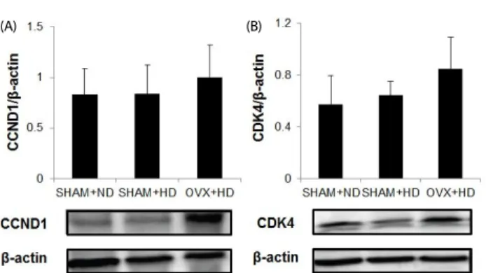
Fig. 2. Effects of excess fat and estrogen deprivation on expression of cell cycle regulatory CCND1 and CDK4 protein in mammary tumor tissue samples. Because only two animals developed tumors in the OVX+ND group, statistical analyses were performed in SHAM+ND, SHAM+HD, and OVX+HD animals. Tumor tissue protein was extracted, separated, and incubated with respective antibodies. Reactive bands were visualized using enhanced chemiluminescence (ECL). The intensity of the bands was quantified using a Bio-Rad GS-800 densitometer. SHAM+ND: shamoperated fed normal diet; SHAM+HD: sham-operated fed high-fat diet; OVX+HD: ovariectomized fed high-fat diet.
Tumor tissue IR, AKT, mTOR, and AMPK expression
To determine the molecular mechanisms responsible for tumor growth, expression of IR-mediated signaling molecules including AKT and pAKT was measured in mammary tumor tissues. Significantly higher tissue protein expression levels of IR were observed in the OVX+HD group compared to the SHAM+ND and SHAM+HD groups (Fig. 1A), and significantly higher pAKT/AKT levels were observed in the OVX+HD group compared to the SHAM+ND group (Fig. 1B). Significantly higher protein expression of mTOR was observed in the OVX+HD group, while significantly lower expression of pAMPK/AMPK was observed in the OVX+HD group compared to expression in other groups (Fig. 1C and 1D).
Cell cycle regulation
Activation of AMPK and mTOR was involved in regulation of the cell cycle, including cyclin D1 (CCND1) and CDK4 protein.
Elevated expression levels of CCND1 and CDK4 were observed in the OVX+HD group compared to other groups, but without statistical significance (Fig. 2A, B).
DISCUSSION
Despite an inverse relationship between BMI and breast cancer risk in premenopausal women, obesity is a recognized risk factor in postmenopausal breast cancer development [27].
Although it is presumed that the interaction between obesity and breast cancer risk is altered by the availability of estrogen, evidence supporting the association between dietary compo- nents including dietary fat and the risk of breast cancer is limited. Because estrogen is known to regulate fat metabolism, including lipogenesis and lipolysis, it is presumable that interactions between dietary fat intake, energy metabolism, and estrogen availability play a critical role in mammary tumor development.
Estrogen deprivation often leads to diminished insulin sensi- tivity, possibly due to accelerated fat accumulation. Previous studies have shown that estradiol repletion can overcome
high-fat diet-induced or FFA-induced insulin resistance [28,29].
Accordingly, high circulating concentrations of insulin in postmenopausal women may be a possible connection between postmenopausal obesity and breast cancer risk [30].
However, a limited number of studies have examined the interactive effects of estrogen deprivation and excess dietary energy supply on mammary tumor development. Genetically obese ovariectomized Zucker rats showed higher susceptibility to DMBA-induced mammary tumor development compared to sham-operated rats [31]. However, in another study high-fat diet stimulated mammary tumor development was not ovarian- dependent [32]. In this study, we evaluated the effects of estrogen deprivation on breast tumor growth in mice fed either a 45% fat diet or 15% fat diet. The animal model of DMBA- induced mammary gland tumors was used because this carcinogen has been implicated in mammary tumorigenesis that is histologically similar to hormone-dependent human breast adenocarcinomas [33]. Results showed that there was no difference in body weight among experimental groups. Previous studies have shown that body weight of C3H/HeN mice fed HD was higher than that of ND supplemented C3H/HeN mice [34,35]. In many other studies, OVX rodents became obese rapidly with body fat accumulation [12,36,37]. However, mice treated with DMBA had significantly lower final body weights, body fat weights, and carcass energy contents compared to mice that received the corn oil placebo [38]. Another study also reported that DMBA treatment resulted in suppression of body weight gain of the animals [39] possibly due to rapid growth of tumor tissues, which accelerates tissue wasting, indicating that the absence of body weight gain in animals with either high-fat feeding or OVX might be due to tumor-induced tissue wasting. Estrogen depletion is known to be associated with dysregulation of lipid metabolism, which may contribute to the accumulation of intra-abdominal fat in postmenopausal women [40]. Estradiol reduces fatty acid and triglyceride synthesis through the down-regulation of lipoprotein lipase and fatty acid synthase [12]. We have determined the liver tissue expression of SREBP1c and FAS, both of which are involved in lipogenesis.
Results showed higher liver tissue expression of SREBP1c and FAS in the OVX+HD group compared to the other groups, although no statistical significance was found in SREBP1c expression. The fact that cancer cachexia and tissue wasting are often associated with decreased insulin sensitivity and hyperglycemia has also been pointed out [41], indicating that tumor growth and hypercatabolism can lead to hyperinsu- linemia.
Estradiol depletion is associated with insulin resistance in humans and rodents [29,30,42]. The increased circulating concentration of FFA in OVX rodents contributes to develop- ment of insulin resistance [43,44]. Postmenopausal insulin resistance is also associated with an elevated blood concen- tration of inflammatory markers, TG, LDL-cholesterol, and FFA [45,46]. Our study results clearly showed significantly higher concentrations of circulating insulin in the OVX+HD group than other groups.
In addition, the levels of IR increased significantly in tumor
tissues of the OVX+HD group. Because insulin has been
suggested as a key molecule to explain obesity-related cancers
including colon and breast, we hypothesized that the larger tumor size in OVX+HD may be mediated through the higher availability of insulin as well as the up-regulation of IR in tumor tissues. Another recent study reported that tumor weight was significantly higher in OVX mice fed a high-fat diet, compared to OVX mice fed a low fat diet in a mouse breast cancer model [47]. In addition, obese mice inoculated with mouse mammary tumor virus (MMTV) Wnt-1 mammary tumor cells did not exhibit tumor growth in the presence of estrogen; however, in the absence of the ovaries, obese mice exhibited higher Wnt-1 tumor growth [48]. These results suggest a strong association of excess energy intake with tumor growth in the absence of ovarian estrogen. Therefore, it is presumable that excess energy intake of postmenopausal women may pose greater risk of developing tumors compared to that of premenopausal women.
The tumor incidence was 75% in the SHAM+HD group, 57.1%
in the SHAM+ND group, 33.3% in the OVX+ND group, and 42.9% of the OVX+HD group, indicating no clear association between the number of tumors and OVX or dietary energy.
Although further investigations are necessary, the higher concentrations of circulating insulin due to OVX and the excess energy supply may not be directly associated with DMBA- induced cancer initiation. Because only two animals developed tumors in the OVX+ND group, no statistical comparisons for tumor tissue biomarkers of cell growth were made with animals in this group. It was reported that breast tumor incidence in Zucker rats, where 30% of the lean sham-operated group, 59%
of the obese sham-operated group, 0% of the lean ovariec- tomized group, and 36% of the obese ovariectomized group developed mammary tumors [31], implying that DMBA-induced mammary tumor formation is dependent on estrogen levels as other studies have also indicated [49-51]. However, it has been suggested that estrogen synthesized from adipose tissue is sufficient to promote mammary tumor development [52]. In this study, since OVX+ND mice were shown to have little adipose tissue, the level of systemic and local estrogen production may not be sufficient for development of tumors. In another study examining incidence of mammary cancer induced by DMBA combined with different concentrations of estradiol in 8-week- old Sprague-Dawley rats, it was found that mammary tumors appear for the first time between the 12th -17th week [28].
Fifty percent of mice had tumors by the 36th, 19
th, and 18th week, with estradiol doses of 1, 2, and 3 mg, respectively. At the 36th week, the incidence rate of breast tumors was 50%, 73%, and 100%, for estradiol doses of 1, 2, and 3 mg, respec- tively. These results imply that estrogen plays an important role in the initiation of mammary tumor formation induced by DMBA. In our study, OVX was performed at 4 weeks, which may have caused depletion of circulating estrogen required for initiation of carcinogenesis.
Hyperinsulinemia can indirectly affect tumorigenesis through activation of insulin/AKT signaling. A previous study reported that larger tumor size in obese women is the result of growth stimulation, and IR-mediated AKT signaling is a major growth pathway [53]. Another study indicated that IR was overex- pressed in human breast cancer cells [54]. To investigate involvement of IR-mediated AKT pathway signaling in mammary
tumor growth, we measured protein expression levels of IR and AKT in tumor tissues. IR protein level was significantly higher in the OVX+HD group compared to the other groups, and AKT phosphorylation and mTOR expression were also significantly higher in the OVX+HD group. mTOR, which regulates essential cell growth signals through cell cycle progression, is an important downstream effector of AKT [55]. A previous study reported that in breast cancer, activation of the AKT/mTOR pathway is responsible for cell survival [56]. Therefore, it is presumable that IR-mediated AKT activation is responsible for the larger tumor size in the OVX+HD group. Epidemiological evidences have suggested that excess body fat is a risk factor for development of breast cancer in postmenopausal women, but not in premenopausal women [2,3]. Results from this study showing an interactive effect of high-fat diet and OVX on circulating concentration of insulin support higher breast cancer risk among postmenopausal women with excess body weight.
We found that OVX+HD suppressed the activation of AMPK, which is potentially associated with tumor cell growth. Interes- tingly, it is reported that the levels of AMPK phosphorylation are reduced by OVX and excess fat mass [57,58]. In addition, AMPK is known as a major tumor suppressor kinase that acts through p53-dependent cell cycle regulation [59,60]. AMPK is also known to suppress cell proliferation through down regula- tion of mTOR [61]. Metformin, a well-known AMPK activator, has been consistently shown to inhibit breast cancer cell growth [56]. In this study, we found that the OVX+HD group showed higher AMPK expression, which may have led to down- regulated expression of mTOR, thereby suppressing tumor growth. Estrogen has been implicated in maintenance of insulin sensitivity [62], and stimulated AMPK phosphorylation by17β -estradiol through estrogen receptor α in 3T3-L1 adipocytes has also been reported [63]. In addition, injections of estradiol resulted in activation of AMPK in ovariectomized mice [64].
Therefore, it is possible that the significantly larger tumor size observed in the OVX+HD group resulted from the combined effects of OVX and excess energy supply. A previous study reported that mTOR facilitates CCND1, which is required for the G1 to S phase transition of the cell cycle [65]. We measured protein expression of CCND1 and CDK4 as downstream targets of mTOR in mammary tumor tissue. Although expression levels of CCND1 and CDK4 were not significantly different, the OVX+HD group tended to show higher expression levels of cell cycle-controlling molecules.
In conclusion we demonstrate that a high-fat diet in OVX
animals leads to development of insulin resistance, which may
accelerate mammary tumor growth through the IR-mediated
AKT pathway and inactivation of AMPK in vivo. The current
study clearly showed that dietary fat induces systemic insulin
resistance and mammary tumor growth in estrogen-deprived
animals in the absence of body weight gain. High circulating
insulin in combination with increased IR in tumor tissues may
result in stimulation of AKT/mTOR signaling and inactivation of
AMPK leading to the acceleration of solid tumor growth. The
current results suggest that a high-fat diet can stimulate breast
cancer progression in postmenopausal women even those
maintaining normal BMI.
REFERENCES
1. Grossmann ME, Ray A, Nkhata KJ, Malakhov DA, Rogozina OP, Dogan S, Cleary MP. Obesity and breast cancer: status of leptin and adiponectin in pathological processes. Cancer Metastasis Rev 2010;29:641-53.
2. Rohan TE, Heo M, Choi L, Datta M, Freudenheim JL, Kamensky V, Ochs-Balcom HM, Qi L, Thomson CA, Vitolins MZ, Wassertheil- Smoller S, Kabat GC. Body fat and breast cancer risk in postmeno- pausal women: a longitudinal study. J Cancer Epidemiol 2013;2013:
754815.
3. Calle EE, Rodriguez C, Walker-Thurmond K, Thun MJ. Overweight, obesity, and mortality from cancer in a prospectively studied cohort of U.S. adults. N Engl J Med 2003;348:1625-38.
4. Eliassen AH, Colditz GA, Rosner B, Willett WC, Hankinson SE. Adult weight change and risk of postmenopausal breast cancer. JAMA 2006;296:193-201.
5. World Cancer Research Fund (GB); American Institute for Cancer Research. Food, Nutrition, Physical Activity, and the Prevention of Cancer: a Global Perspective. Washington D.C.: American Institute for Cancer Research; 2007.
6. World Cancer Research Fund International, Continuous Update Project (GB). Diet, Nutrition, Physical Activity, and the Breast Cancer Survivors. London: World Cancer Research Fund International; 2014.
7. Brennan SF, Woodside JV, Lunny PM, Cardwell CR, Cantwell MM.
Dietary fat and breast cancer mortality: a systematic review and meta-analysis. Crit Rev Food Sci Nutr Forthcoming 2015..
8. Mourouti N, Kontogianni MD, Papavagelis C, Panagiotakos DB. Diet and breast cancer: a systematic review. Int J Food Sci Nutr 2015;66:1-42.
9. Demir B, Ozturkoglu E, Solaroglu A, Baskan B, Kandemir O, Karabulut E, Haberal A. The effects of estrogen therapy and estrogen combined with different androgenic progestins on carbohydrate and lipid metabolism in overweight-obese younger postmeno- pausal women. Gynecol Endocrinol 2008;24:347-53.
10. Jensen LB, Vestergaard P, Hermann AP, Gram J, Eiken P, Abraha- msen B, Brot C, Kolthoff N, Sørensen OH, Beck-Nielsen H, Nielsen SP, Charles P, Mosekilde L. Hormone replacement therapy dissociates fat mass and bone mass, and tends to reduce weight gain in early postmenopausal women: a randomized controlled 5-year clinical trial of the Danish Osteoporosis Prevention Study.
J Bone Miner Res 2003;18:333-42.
11. Cooke PS, Naaz A. Role of estrogens in adipocyte development and function. Exp Biol Med (Maywood) 2004;229:1127-35.
12. D'Eon TM, Souza SC, Aronovitz M, Obin MS, Fried SK, Greenberg AS. Estrogen regulation of adiposity and fuel partitioning. Evidence of genomic and non-genomic regulation of lipogenic and oxidative pathways. J Biol Chem 2005;280:35983-91.
13. Field FJ, Born E, Murthy S, Mathur SN. Polyunsaturated fatty acids decrease the expression of sterol regulatory element-binding protein-1 in CaCo-2 cells: effect on fatty acid synthesis and triacyl- glycerol transport. Biochem J 2002;368:855-64.
14. Horton JD. Sterol regulatory element-binding proteins: transcrip- tional activators of lipid synthesis. Biochem Soc Trans 2002;30:
1091-5.
15. Kuhl J, Hilding A, Ostenson CG, Grill V, Efendic S, Båvenholm P.
Characterisation of subjects with early abnormalities of glucose tolerance in the Stockholm Diabetes Prevention Programme: the
impact of sex and type 2 diabetes heredity. Diabetologia 2005;48:
35-40.
16. Macotela Y, Boucher J, Tran TT, Kahn CR. Sex and depot differences in adipocyte insulin sensitivity and glucose metabolism. Diabetes 2009;58:803-12.
17. Saengsirisuwan V, Pongseeda S, Prasannarong M, Vichaiwong K, Toskulkao C. Modulation of insulin resistance in ovariectomized rats by endurance exercise training and estrogen replacement.
Metabolism 2009;58:38-47.
18. Gao H, Bryzgalova G, Hedman E, Khan A, Efendic S, Gustafsson JA, Dahlman-Wright K. Long-term administration of estradiol decreases expression of hepatic lipogenic genes and improves insulin sensitivity in ob/ob mice: a possible mechanism is through direct regulation of signal transducer and activator of transcription 3. Mol Endocrinol 2006;20:1287-99.
19. Vona-Davis L, Howard-McNatt M, Rose DP. Adiposity, type 2 diabetes and the metabolic syndrome in breast cancer. Obes Rev 2007;8:395-408.
20. Jouyandeh Z, Nayebzadeh F, Qorbani M, Asadi M. Metabolic syndrome and menopause. J Diabetes Metab Disord 2013;12:1.
21. Frasca F, Pandini G, Sciacca L, Pezzino V, Squatrito S, Belfiore A, Vigneri R. The role of insulin receptors and IGF-I receptors in cancer and other diseases. Arch Physiol Biochem 2008;114:23-37.
22. Kim HJ, Lee HO, Min DB. Effects and prooxidant mechanisms of oxidized alpha-tocopherol on the oxidative stability of soybean oil.
J Food Sci 2007;72:C223-30.
23. Hong J, Holcomb VB, Kushiro K, Núñez NP. Estrogen inhibits the effects of obesity and alcohol on mammary tumors and fatty liver.
Int J Oncol 2011;39:1443-53.
24. Blume-Jensen P, Hunter T. Oncogenic kinase signalling. Nature 2001;411:355-65.
25. Reeves PG, Nielsen FH, Fahey GC Jr. AIN-93 purified diets for laboratory rodents: final report of the American Institute of Nutrition ad hoc writing committee on the reformulation of the AIN-76A rodent diet. J Nutr 1993;123:1939-51.
26. Park SY, Kim JS, Seo YR, Sung MK. Effects of diet-induced obesity on colitis-associated colon tumor formation in A/J mice. Int J Obes (Lond) 2012;36:273-80.
27. Stephenson GD, Rose DP. Breast cancer and obesity: an update.
Nutr Cancer 2003;45:1-16.
28. Healy LA, Ryan AM, Carroll P, Ennis D, Crowley V, Boyle T, Kennedy MJ, Connolly E, Reynolds JV. Metabolic syndrome, central obesity and insulin resistance are associated with adverse pathological features in postmenopausal breast cancer. Clin Oncol (R Coll Radiol) 2010;22:281-8.
29. Nadal A, Alonso-Magdalena P, Soriano S, Ropero AB, Quesada I.
The role of oestrogens in the adaptation of islets to insulin resistance. J Physiol 2009;587:5031-7.
30. Hong J, Stubbins RE, Smith RR, Harvey AE, Núñez NP. Differential susceptibility to obesity between male, female and ovariectomized female mice. Nutr J 2009;8:11.
31. Hakkak R, MacLeod S, Shaaf S, Holley AW, Simpson P, Fuchs G, Jo CH, Kieber-Emmons T, Korourian S. Obesity increases the incidence of 7,12-dimethylbenz(a)anthracene-induced mammary tumors in an ovariectomized Zucker rat model. Int J Oncol 2007;30:
557-63.
32. Sylvester PW, Ip C, Ip MM. Effects of high dietary fat on the growth and development of ovarian-independent carcinogen-induced
mammary tumors in rats. Cancer Res 1986;46:763-9.
33. Costa I, Solanas M, Escrich E. Histopathologic characterization of mammary neoplastic lesions induced with 7,12 dimethylbenz (alpha)anthracene in the rat: a comparative analysis with human breast tumors. Arch Pathol Lab Med 2002;126:915-27.
34. Hwang IK, Kim IY, Kim DW, Yoo KY, Kim YN, Yi SS, Won MH, Lee IS, Yoon YS, Seong JK. Strain-specific differences in cell proliferation and differentiation in the dentate gyrus of C57BL/6N and C3H/HeN mice fed a high fat diet. Brain Res 2008;1241:1-6.
35. Suganami T, Mieda T, Itoh M, Shimoda Y, Kamei Y, Ogawa Y.
Attenuation of obesity-induced adipose tissue inflammation in C3H/HeJ mice carrying a Toll-like receptor 4 mutation. Biochem Biophys Res Commun 2007;354:45-9.
36. Lim DW, Lee Y, Kim YT. Preventive effects of Citrus unshiu peel extracts on bone and lipid metabolism in OVX rats. Molecules 2014;19:783-94.
37. Choi JS, Koh IU, Song J. Genistein reduced insulin resistance index through modulating lipid metabolism in ovariectomized rats. Nutr Res 2012;32:844-55.
38. Lane HW, Keith RE, Strahan S, White MT. The effect of diet, exercise and 7,12-dimethylbenz(a)anthracene on food intake, body composition and carcass energy levels in virgin female BALB/c mice.
J Nutr 1991;121:1876-82.
39. Ojeswi BK, Khoobchandani M, Hazra DK, Srivastava MM. Protective effect of Thuja occidentalis against DMBA-induced breast cancer with reference to oxidative stress. Hum Exp Toxicol 2010;29:369-75.
40. Gambacciani M, Ciaponi M, Cappagli B, Piaggesi L, De Simone L, Orlandi R, Genazzani AR.Body weight, body fat distribution, and hormonal replacement therapy in early postmenopausal women.
J Clin Endocrinol Metab 1997;82:414-7.
41. Asp ML, Tian M, Wendel AA, Belury MA. Evidence for the contribution of insulin resistance to the development of cachexia in tumor-bearing mice. Int J Cancer 2010;126:756-63.
42. Suba Z. Circulatory estrogen level protects against breast cancer in obese women. Recent Pat Anticancer Drug Discov 2013;8:154-67.
43. Koricanac G, Milosavljevic T, Stojiljkovic M, Zakula Z, Ribarac-Stepic N, Isenovic ER. Insulin signaling in the liver and uterus of ovariectomized rats treated with estradiol. J Steroid Biochem Mol Biol 2008;108:109-16.
44. Swislocki A, Burgie ES, Rodnick KJ. Effects of ovariectomy on indices of insulin resistance, hypertension, and cardiac energy metabolism in middle-aged spontaneously hypertensive rats (SHR). Horm Metab Res 2002;34:516-22.
45. Carr MC. The emergence of the metabolic syndrome with meno- pause. J Clin Endocrinol Metab 2003;88:2404-11.
46. Pfeilschifter J, Köditz R, Pfohl M, Schatz H. Changes in proinflam- matory cytokine activity after menopause. Endocr Rev 2002;23:
90-119.
47. Gu JW, Young E, Patterson SG, Makey KL, Wells J, Huang M, Tucker KB, Miele L. Postmenopausal obesity promotes tumor angiogenesis and breast cancer progression in mice. Cancer Biol Ther 2011;11:
910-7.
48. Nunez NP, Perkins SN, Smith NC, Berrigan D, Berendes DM, Varticovski L, Barrett JC, Hursting SD. Obesity accelerates mouse mammary tumor growth in the absence of ovarian hormones. Nutr Cancer 2008;60:534-41.
49. Asselin J, Labrie F. Effects of estradiol and prolactin on steroid
receptor levels in 7,12-dimethylbenz(a)anthracene-induced mammary tumors and uterus in the rat. J Steroid Biochem 1978;9:1079-82.
50. Sasaki GH, Leung BS. On the mechanism of hormone action in 7,12 dimethylbenz(a)anthracene-induced mammary tumor. I. Prolactin and progesterone effects on estrogen receptor in vitro. Cancer 1975;35:645-51.
51. Dao TL. The role of ovarian hormones in initiating the induction of mammary cancer in rats by polynuclear hydrocarbons. Cancer Res 1962;22:973-81.
52. Rose DP, Gilhooly EM, Nixon DW. Adverse effects of obesity on breast cancer prognosis, and the biological actions of leptin (review). Int J Oncol 2002;21:1285-92.
53. Daling JR, Malone KE, Doody DR, Johnson LG, Gralow JR, Porter PL. Relation of body mass index to tumor markers and survival among young women with invasive ductal breast carcinoma.
Cancer 2001;92:720-9.
54. Belfiore A, Frasca F. IGF and insulin receptor signaling in breast cancer. J Mammary Gland Biol Neoplasia 2008;13:381-406.
55. Wysocki PJ, Wierusz-Wysocka B. Obesity, hyperinsulinemia and breast cancer: novel targets and a novel role for metformin. Expert Rev Mol Diagn 2010;10:509-19.
56. Clark AS, West K, Streicher S, Dennis PA. Constitutive and inducible Akt activity promotes resistance to chemotherapy, trastuzumab, or tamoxifen in breast cancer cells. Mol Cancer Ther 2002;1:707-17.
57. Woo SL, Xu H, Li H, Zhao Y, Hu X, Zhao J, Guo X, Guo T, Botchlett R, Qi T, Pei Y, Zheng J, Xu Y, An X, Chen L, Chen L, Li Q, Xiao X, Huo Y, Wu C. Metformin ameliorates hepatic steatosis and inflammation without altering adipose phenotype in diet-induced obesity. PLoS One 2014;9:e91111.
58. Wohlers LM, Sweeney SM, Ward CW, Lovering RM, Spangenburg EE. Changes in contraction-induced phosphorylation of AMP- activated protein kinase and mitogen-activated protein kinases in skeletal muscle after ovariectomy. J Cell Biochem 2009;107:171-8.
59. Tiainen M, Vaahtomeri K, Ylikorkala A, Mäkelä TP. Growth arrest by the LKB1 tumor suppressor: induction of p21(WAF1/CIP1). Hum Mol Genet 2002;11:1497-1504.
60. Jones RG, Plas DR, Kubek S, Buzzai M, Mu J, Xu Y, Birnbaum MJ, Thompson CB. AMP-activated protein kinase induces a p53- dependent metabolic checkpoint. Mol Cell 2005;18:283-93.
61. Chapuis N, Tamburini J, Green AS, Willems L, Bardet V, Park S, Lacombe C, Mayeux P, Bouscary D. Perspectives on inhibiting mTOR as a future treatment strategy for hematological malignancies.
Leukemia 2010;24:1686-99.
62. Kim JY, Jo KJ, Kim BJ, Baik HW, Lee SK. 17beta-estradiol induces an interaction between adenosine monophosphate-activated protein kinase and the insulin signaling pathway in 3T3-L1 adipocytes. Int J Mol Med 2012;30:979-85.
63. McInnes KJ, Brown KA, Hunger NI, Simpson ER. Regulation of LKB1 expression by sex hormones in adipocytes. Int J Obes (Lond) 2012;36:982-5.
64. Kim JY, Jo KJ, Kim OS, Kim BJ, Kang DW, Lee KH, Baik HW, Han MS, Lee SK. Parenteral 17beta-estradiol decreases fasting blood glucose levels in non-obese mice with short-term ovariectomy. Life Sci 2010;87:358-66.
65. Mita MM, Mita A, Rowinsky EK. The molecular target of rapamycin (mTOR) as a therapeutic target against cancer. Cancer Biol Ther 2003;2:S169-77.
[Supplementary file]