DOI 10.3349/ymj.2007.48.6.1056
Solitary fibrous tumors are spindle-cell neoplasms that usually develop in the pleura and peritoneum, and rarely arise in the stomach. To our knowledge, there is only one case reporting a solitary fibrous tumor arising from stomach in the English literature. Here we report the case of a 26-year-old man with a large solitary fibrous tumor arising from the stomach which involved the submucosa and muscular layer and resembled a gastrointestinal stromal tumor in the stomach, based on what was seen during abdominal computed tomog- raphy. A solitary fibrous tumor arising from the stomach, although rare, could be considered as a diagnostic possibility for gastric submucosal tumors.
Key Words: Solitary fibrous tumor, stomach, CT findings
INTRODUCTION
Solitary fibrous tumors are rare neoplasms of mesenchymal origin that account for less than 2%
of all soft tissue tumors and have classically been described in the thorax as arising from the pleura.
1-3However, they have been found in extra- serosal sites and should be considered in the differential diagnosis of any spindle cell lesion, including those in the gastrointestinal tract.
3In the pleural site, solitary fibrous tumors have unique gross and microscopic appearances and can be easily recognized. When they arise at other non- pleural sites, differentiation from other soft tissue
tumors can be difficult.
4To our knowledge, only one case of a solitary fibrous tumor arising from the stomach has been reported,
5but its radiological findings have not been well described.
We performed computed tomography (CT) and here we report the findings from a solitary fibrous tumor arising from the gastric wall that mimicked a gastrointestinal stromal tumor.
CASE REPORT
A 26-year-old man presented with melena and underwent upper gastrointestinal endoscopy and abdominal CT.
Endoscopy showed a large submucosal tumor in the gastric body with concomitant bleeding from a large central ulceration containing fluid and residual contrast material from a previous barium study (Fig. 1A). Endoscopic ultrasound similarly showed a large submucosal mass (Fig.
1B). A first endoscopic biopsy result was non- specific and showed acute and chronic inflam- mation with ulceration. The second biopsy revealed ulceration and tissue granulation with foreign body giant cells suggestive of a barium granuloma.
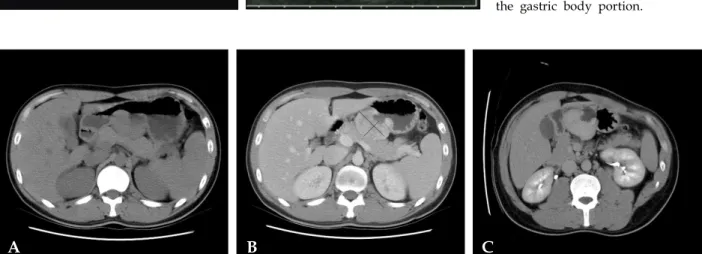
An abdominal CT scan demonstrated about 5.5
× 3.2 cm sized, well-defined and large exophyti- cally growing mass arising from the posterior aspect of the lesser curvature side in the gastric body. The mass showed a large ulceration at the gastric luminal side and a cavity in the central portion communicating with the gastric lumen.
Solitary Fibrous Tumor Arising from Stomach: CT Findings
Sung Hee Park,
1Myeong-Jin Kim,
1,2Jieun Kwon,
3Jong-pil Park,
3Mi-Suk Park,
1Joon Seok Lim,
1Joo Hee Kim,
1and Ki Whang Kim
11