Case Report pISSN: 1011-8942 eISSN: 2092-9382
Korean J Ophthalmol 2014;28(2):194-196 http://dx.doi.org/10.3341/kjo.2014.28.2.194
© 2014 The Korean Ophthalmological Society
This is an Open Access article distributed under the terms of the Creative Commons Attribution Non-Commercial License (http://creativecommons.org/licenses /by-nc/3.0/) which permits unrestricted non-commercial use, distribution, and reproduction in any medium, provided the original work is properly cited.
194
A Case of Ocular Myasthenia Gravis Presenting as Double Depressor Palsy
Kwanbok Lee1, Ungsoo Samuel Kim1,2
1Department of Ophthalmology, Kim’s Eye Hospital, Seoul, Korea
2Department of Ophthalmology, Konyang University College of Medicine, Daejeon, Korea
Double depressor palsy (DDP), which is a rare condition, refers to simultaneous paralysis of the inferior rectus mus- cle and superior oblique muscle of the same eye. This con- dition may occur because of congenital disorders, cerebro- vascular disease, trauma or superior rectus contracture after strabismus surgery [1]. To the best of our knowledge, DDP caused by ocular myasthenia gravis (OMG) has not been reported to date. Therefore, we present a case of DDP caused by OMG and review the related literature.
Case Report
A 65-year-old man who had been experiencing diplopia in front and down gaze for 15 days visited our hospital. His
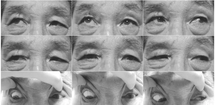
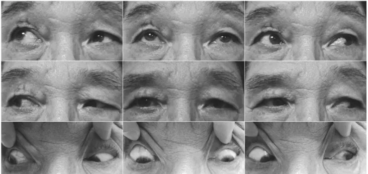
medical history revealed no contributing factors such as diabetes mellitus, hypertension, and cerebral ischemic at- tack. Ophthalmic examinations revealed normal vision in both eyes and no abnormal pupillary reflex. Fourteen prism diopter hypertropia was noted in the patient’s left eye and limitation of depression was found in the adduction, pri- mary gaze, and abduction (Fig. 1). Forced duction test revealed no restriction. Brain magnetic resonance imaging showed no remarkable findings. Two weeks after the first visit, the patient complained of ptosis in the left eye. An ice test was performed and the ptosis was resolved after the test (Fig. 2). Then, anti-acetylcholine receptor binding antibody levels were checked and found to be slightly ele- vated (0.416 nmol/L). We prescribed methylprednisolone per os 24 mg for 2 weeks, and his symptoms improved after 2 weeks. Three weeks after medication, the patient showed an ortho result in the alternate prism cover test, normal oc- ular movements, and complete resolution of diplopia (Fig. 3).
A 65-year-old man who had been experiencing diplopia in front and down gaze for 15 days visited our hospital. Hypertropia was noted in the patient’s left eye, and limitation of depression was found in the adduction, primary gaze, and abduction. Brain magnetic resonance imaging showed no remarkable findings. Two weeks after the first visit, the patient complained of ptosis in the left eye. An ice test was performed and the ptosis was resolved after the test. Then, anti-acetylcholine receptor binding antibody levels were checked and found to be slightly elevated. We prescribed methylprednisolone per os 24 mg for 2 weeks, and his symptoms improved after the 2-week treatment. Five weeks after his first visit, the patient showed an ortho result in the alternate prism cover test and normal ocular movements. This may be the first case in which ocular myasthenia gravis presented as double depressor palsy, and in such cases, the possibility of ocular myasthenia gravis should be considered to rule out double depressor palsy.
Key Words: Double depressor palsy, Myasthenia gravis, Strabismus
Received: February 11, 2014 Accepted: February 21, 2014
Corresponding Author: Ungsoo Samuel Kim, MD. Department of Oph- thalmology, Kim’s Eye Hospital, Konyang University College of Med- icine, #136 Yeongsin-ro, Yeongdeungpo-gu, Seoul 150-034, Korea. Tel:
82-2-1577-2639, Fax: 82-2-2677-9214, E-mail: [email protected]
195 K Lee, et al. Ocular Myasthenia Gravis as Double Depressor Palsy
Discussion
Although congenital DDP patients usually do not complain of diplopia, acquired cases have diplopia commonly. DDP is a very rare condition. The relatively low incidence of this condition can be attributed to the fact that the inferior rec- tus muscle is supplied by the inferior branch of the 3rd cranial nerve while the superior oblique muscle is supplied by the 4th cranial nerve [2]. Etiology of DDP is not clear and it results from various conditions such as a primary paralysis of the inferior rectus muscle (congenital or acquired), a primary supranuclear palsy of depression, and secondary dysfunction of the inferior rectus due to ipsilateral superior rectus contracture [3]. To define the pathogenesis, a careful history taking is needed to rule out secondary causes including orbital wall fracture, thyroid orbitopathy and previous vertical rectus muscle surgery.
OMG is an autoimmune disorder that leads to ptosis and/
or diplopia that is caused by weakness of the extraocular eye muscles, levator palpebrae superioris, and orbicularis oculi, without dysfunction of other muscles. A history of variable and fatigable ocular muscle weakness in the pres- ence of normal pupillary function should raise the index of suspicion for OMG [2]. The ice test is a convenient, specif- ic, and relatively sensitive technique for diagnosing myas-
thenia gravis [4]. In the present case, the ice test proved useful in diagnosing OMG.
In conclusion, this may be the first case in which OMG presented as DDP, and in such cases, the possibility of OMG should be considered to rule out DDP.
Conflict of Interest
No potential conflict of interest relevant to this article was reported.
Fig. 1. A limitation of depression was found in adduction, primary gaze and abduction in the left eye.
A
B
Fig. 2. (A) Left eye’s ptosis was noted. (B) Five minutes after application of ice pack, ptosis improved.
196
Korean J Ophthalmol Vol.28, No.2, 2014
References
1. Nayak BK, Menon V, Prakash P. Acquired double depres- sor palsy. Indian J Ophthalmol 1983;31:77-8.
2. Vaphiades MS, Bhatti MT, Lesser RL. Ocular myasthenia gravis. Curr Opin Ophthalmol 2012;23:537-42.
3. Jacobs L, Anderson PJ, Bender MB. The lesions producing paralysis of downward but not upward gaze. Arch Neurol 1973;28:319-23.
4. Golnik KC, Pena R, Lee AG, Eggenberger ER. An ice test for the diagnosis of myasthenia gravis. Ophthalmology 1999;106:1282-6.
Fig. 3. Three weeks after oral steroid medication administration, the limitation of depression was completely resolved.