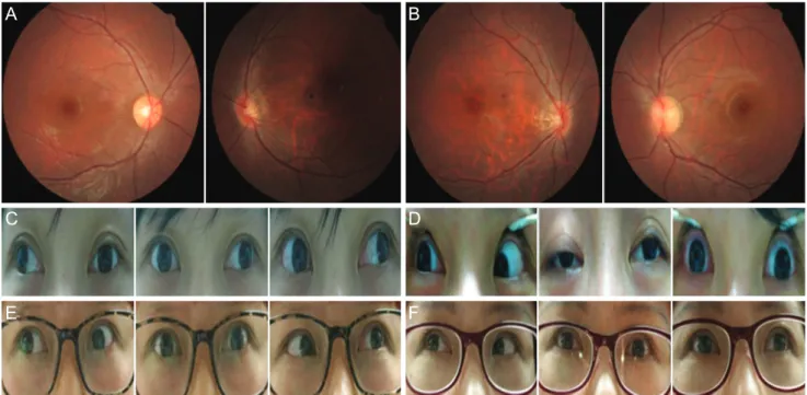
Ocular Myasthenia Gravis in Monozygotic Twins with Mirror-image Myopic Anisometropia
2
0
0
전체 글
(2)
수치
관련 문서
Phase profile of conventional DOE Obtained diffraction image. Optical vortices appear in the diffraction image generated by
Taubin, “The QBIC Project: Querying Image by Content using Color, Texture and Shape,” Proceedings of SPIE Storage and Retrieval for Image and Video Databases, pp.. Pala,
To develop a prolonged ocular drug delivery system with less frequency dosing, nanoparticles covalently bonded with levofloxacin in contact lenses will be a
load and differential output have a superior p p high frequency response over differential amplifiers with current mirror load and. amplifiers with current mirror load
* Symmetry operation symmetry element reflection mirror plane rotation rotation axis inversion inversion center..
Photonic crystals containing rugate structure result in a mirror with high reflectivity in a specific narrow spectral region and are prepared by applying
Postoperative Postoperative Postoperative Postoperative Magnetic Magnetic Magnetic Magnetic resonance resonance resonance image resonance image image image shows
With uses of a techniques named mola that pursues modern and Korean image by overlapping of cloth with various colors, and a technique named slash that
