Vol. 21, No. 1, 2009 53
Received June 2, 2008, Accepted for publication July 24, 2008
*This study was supported by a grant from the Korea Health 21 R&D Project of the Korea Ministry of Health and Welfare (Grant 01-PJ3- PG6-01GN12-0001). N.K. was supported by a Brain Korea 21 Re- search Fellowship from the Korea Ministry of Education and Human Resources.
Reprint request to: Jeung Hoon Lee, M.D., Department of Dermatology, College of Medicine, Chungnam National University, 640, Daesa- dong, Jung-gu, Daejeon 301-040, Korea. Tel: 82-42-280-7707, Fax:
82-42-280-5098, E-mail: [email protected]
Ann Dermatol (Seoul) Vol. 21, No. 1, 2009
CASE REPORT
A Case of Isolated Plexiform Neurofibroma in a Patient with Myasthenia Gravis
Seung Ju Back, M.D., Dae Hun Kim, M.D., Nari Kim, M.S., Young Lee, M.D., Young Joon Seo, M.D., Jang Kyu Park, M.D., Jeung Hoon Lee, M.D.
Department of Dermatology, College of Medicine, Chungnam National University, Daejeon, Korea
We report a case of an isolated plexiform neurofibroma occurring in a patient with myasthenia gravis. A 48-year-old man presented with asymptomatic skin-colored nodules on the tip of his 4th finger. Microscopically, a plexiform neurofibroma was identified located in the dermis that appeared to originate from small superficial nerves. He had a 20-year history of treated myasthenia gravis; otherwise, his personal and family histories were unremarkable. Given that myasthenia gravis is a disorder of the peripheral nerves, plexiform neurofibromas could be associated with my- asthenia gravis. However, the development of an isolated plexiform neurofibroma in a case of myasthenia gravis has not yet been reported. The occurrence of a neurofibromas in a patient with myasthenia gravis suggests a link in the pathogenesis of these two diseases. (Ann Dermatol (Seoul) 21(1) 53∼55, 2009)
-Keywords-
Isolated, Myasthenia gravis, Plexiform neurofibroma
INTRODUCTION
Plexiform neurofibromas, which are histologically similar to discrete neurofibromas, are benign peripheral nerve sheath tumors that involve one or more nerve fascicles
often arising from the branches of a major nerve1. In this tumor, the nerve is converted into a convoluted mass which has been likened to a 'bag of worms'. The plexiform neurofibroma is considered to be pathognomic of neurofibromatosis 1 (NF1)1,2. However, a few cases of isolated plexiform neurofibroma that were not associated with NF1 have been reported.
Myasthenia gravis is a relatively rare autoimmune disorder of the peripheral nerves associated with pemphigus, alopecia areata and psoriasis3-5. The development of an isolated plexiform neurofibroma in myasthenia gravis has not been reported.
Thus, we report a rare case of an isolated plexiform neurofibroma in a patient with a history of myasthenia gravis, and discuss the possible pathogenetic relationship between the two.
CASE REPORT
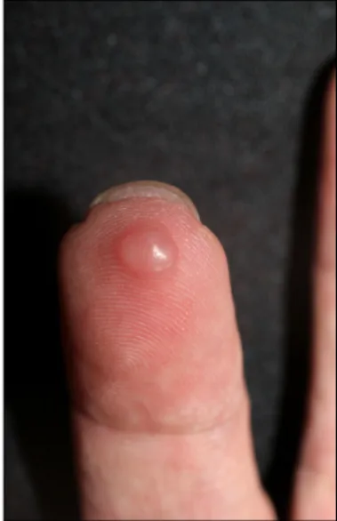
A 48-year-old man presented with an asymptomatic skin-colored nodule on the tip of his right 4th finger that had been enlarging slowly for approximately 3 years. One year earlier, it had been excised with a CO2 laser, but had since recurred. Examination disclosed a 0.7×0.5 cm rubbery nodule (Fig. 1). It was not associated with any pain, tenderness, or ulceration. Other than a 20 year history of treated myasthenia gravis, the medical evalua- tion of this patient was unremarkable and there was no evidence of other skin tumors or pigmented lesions. His family history was negative for similar skin disaeses and other hereditary conditions. The lesion was completely excised.
Histopathological examination showed an unremarkable epidermis with slight hyperkeratosis. The dermis con- tained a circumscribed, lobular proliferation of spindle cells in a loose fibular background distorting much of the
SJ Back, et al
54 Ann Dermatol (Seoul)
Fig. 1. 0.7×0.5 cm sized rubbery nodule on right fourth finger tip.
dermis (Fig. 2A). The tumor was well circumscribed and surrounded by concentric bands of collagen in an 'onion
skin pattern'. It consisted of faintly eosinophilic, thin, collagen bundles, myxoid changes, and occasional blood vessels (Fig. 2B). The cell nuclei were spindle- or S-shaped and quite uniform in size. The nuclear chromatin was bland without significant polymorphism or increased mitotic activity. A fairly large proportion of the tumor cells were immunopositive for S-100 protein (Fig. 2C).
DISCUSSION
We have described an isolated plexiform neurofibroma that developed in a man with myasthenia gravis but no stigmata of von Recklinghausen's disease. Plexiform neurofibromas consisting of a heterogeneous mix of va- rious cell types, including Schwann cells and fibroblasts6,7, are reported only in patients with von Recklinghausen's disease, and are considered to be pathognomic for NF11,2. Enzinger and Weiss8, while acknowledging the extremely high association with von Recklinghausen's disease, were somewhat more dubious about this relationship when the neurofibromas involved small nerves in the subcutis.
Fig. 2. (A) Well circumscribed numerous nerve fascicles with irregular configurations embedded in cellular matrices (H&E stain, ×40). (B) Spindle shaped cells with wavy collagen fibrils in myxoid stroma (H&E stain, ×100). (C) Numerous cells of the fascicles present intensive positivity for S-100 protein (S100 stain, ×200).
A Case of Isolated Plexiform Neurofibroma in a Patient with Myasthenia Gravis
Vol. 21, No. 1, 2009 55 Moreover, several cases of isolated plexiform neurofi-
broma have recently been observed in otherwise healthy patients with no family history of NF or other anomalies, suggesting that the presence of a plexiform neurofibroma does not always indicate associated NF or structural anomalies9-11. In our case, the tumor presented on the right 4th finger tip as a small rubbery nodule that was located superficially on the dermis. It involved a small superficial nerve, and was similar to recently described cases in which an isolated plexiform neurofibroma oc- curred as a superficial form involving small nerves in the dermis10,11. The patient had no personal or family history of other abnormalities suggestive of NF, but had been treated for myasthenia gravis since 1989.
Myasthenia gravis is a relatively rare autoimmune disorder of peripheral nerves in which antibodies form against acetylcholine nicotinic postsynaptic receptors at the myoneural junction12. It is characterized by weakness and rapid fatigue of the muscles under voluntary control. It may cause double vision, drooping eyelids, difficulties with speech, chewing, swallowing and breathing, as well as limb weakness. Myasthenia gravis is associated with other autoimmune diseases, such as thyroiditis, rheumatoid arthritis, systemic lupus erythematosus and thymoma3,13. It is also associated with skin diseases such as pemphigus, alopecia areata, vitiligo and psoriasis3-,5,14.
There has been one other reported case of neurofibroma associated with myasthenia gravis. Waugh15 reported the development of myasthenia gravis in a case of multiple NF. However, no neurofibroma developing in a case of myasthenia gravis has been reported. The relationship between the diseases may be coincidental, but is worthy of further investigation due to the fact both developed from peripheral nerves.
In myasthenia gravis, autoantibodies block the post- synaptic receptor at the myoneural junction, preventing signal transduction which results in inflammation around the junction. This inflammation tends to be worse in areas of the body subject to frequent trauma. This inflammatory process enhances cell proliferation and, ultimately, the proliferation of nerve cells occurs. Based on this hypothesis, a clear mechanism for the development of the neurofibroma on our patient's fingertip is evident.
This case seems to confirm the premise that the superficial
form of plexiform neurofibroma, involving small nerves in the dermis or subcutis, is not necessarily pathognomic for von Recklinghausen's disease, as previously reported10,11. Furthermore, we have reported this case because of the rare occurrence of plexiform neurofibroma in myasthenia gravis.
REFERENCES
1. Robert L, Joel C. The neurofibromatoses. In: Wolff K, Goldsmith LA, Katz SI, Gilchrest BA, Paller AS, Leffell DJ, editors. Fitzpatrick's dermatology in general medicine. 7th ed. New York: McGraw-Hill, 2008:1332-1334.
2. Legius E, Descheemaeker MJ, Fryns JP, Van den Berghe H.
Neurofibromatosis type 1. Genet Couns 1994;5:225-241.
3. Kubota A, Komiyama A, Hasegawa O. Myasthenia gravis and alopecia areata. Neurology 1997;48:774-775.
4. Younus J, Ahmed AR. The relationship of pemphigus to neoplasia. J Am Acad Dermatol 1990;23:498-502.
5. Kwan SY, Lin JH, Su MS. Coexistence of epilepsy, myasthenia gravis and psoriasis vulgaris. Zhonghua Yi Xue Za Zhi (Taipei) 2000;63:153-157.
6. Korf BR. Plexiform neurofibromas. Am J Med Genet 1999;
89:31-37.
7. Crawford AH, Schorry EK. Neurofibromatosis update. J Pediatr Orthop 2006;26:413-423.
8. Enzinger FM, Weiss SW. Soft tissue tumors. 2nd ed. St Louis:
Mosby, 1988:748-749.
9. Aloi FG, Massobrio R. Solitary plexiform neurofibroma.
Dermatologica 1989;179:84-86.
10. Fisher DA, Chu P, McCalmont T. Solitary plexiform neuro- fibroma is not pathognomonic of von Recklinghausen's neurofibromatosis: a report of a case. Int J Dermatol 1997;36:439-442.
11. Lee HJ, Koh BK, Ha SJ, Kim JW. A case of isolated plexiform neurofibroma. Ann Dermatol 2000;12:271-274.
12. Younger DS, Worrall BB, Penn AS. Myasthenia gravis: his- torical perspective and overview. Neurology 1997;48(Supp 5):S1-S7.
13. Lovelace RE, Younger DS. Myasthenia gravis with thymoma.
Neurology 1997;48(Supp 5):S76-S81.
14. Tan RS. Ulcerative colitis, myasthenia gravis, atypical lichen planus, alopecia areata, vitiligo. Proc R Soc Med 1974;67:
195-196.
15. Waugh WH. The development of myasthenia gravis in a case of multiple neurofibromatosis. South Med J 1953;46:
1132-1134.